Note: Descriptions are shown in the official language in which they were submitted.
~ WO 95/26158 PCTIUS95/03514
2186382
METHOD AND APPARATUS FOR DIAGNOSING ERECTILE DYSFUNCTION
Technical Field
This invention relates generally to the
diagnosis of erectile dysfunction. More particularly,
the invention relates to a novel noninvasive procedure
for diagnosing erectile dysfunction, particularly
vasculogenic erectile dysfunction, by measuring various
penile hemodynamic parameters subsequent to transurethral
administration of a selected vasoactive agent. The
invention additionally relates to kits for carrying out
the diagnostic method, and to methods of treatment
deriving from the diagnosis.
Background
Impotence is the consistent inability to
achieve or sustain an erection of sufficient .~igidity for
sexual intercourse. It has recently been estimated that
approximately 10 million American men are impotent (R.
Shabsigh et al., "Evaluation of Erectile Impotence,"
Urolovv 32:83-90 (1988); W.L. Furlow, "Prevalence of
Impotence in the United States," Med. Aspects Hum. Sex
19:13-6 (1985)). Impotence is recognized to be an age-
dependent disorder, with an incidence of 1.9 percent at
40 years of age and 25 percent at 65 years of age (A. C.
Kinsey et al., "Age and Sexual Outlet," in Sexual
Behavior in the Human Male, A.C. Kinsey et al., eds.,
Philadelphia, PA: W.B. Saunders, 218-262 (1948)). In
1985 in the United States, impotence accounted for more
' 35 than several hundred thousand outpatient visits to
-1-
WO 95126158
~1~65~2
PCT/US95103514 ,~
physicians (National Center for Health Statistics,
National Hospital Discharge Survey, 1985, Bethesda, MD,
Department of Health and Human Services, 1989 DHHS
publication no. 87-1751). Depending on the nature and
cause of the problem, treatments include psychosexual
therapy, hormonal therapy, administration of vasodilators
such as nitroglycerin and a-adrenergic blocking agents
("a-blockers"), oral administration of other
pharmaceutical agents, vascular surgery, implanted penile
prostheses, vacuum constriction devices and external aids
such as penile splints to support the penis or penile
constricting rings to alter the flow of blood through the
penis.
A number of causes of impotence have been
identified, including vasculogenic, neurogenic,
endocrinologic and psychogenic. Impotence can also be a
side effect of various classes of therapeutic drugs, or
can be associated with various diseases, including
diabetes, multiple sclerosis and sickle cell anemia.
Impotence resulting from any one of these causes can be
exacerbated by additional factors such as cigarette
smoking, a poor diet, or the like.
Vasculogenic impotence occurs either as a
result of arterial occlusion--the obstruction of adequate
blood flow to the penile arteries necessary for erection
--or as a result of cavernovenous leakage, i.e., excess
venal outflow. As explained by Krane et al., "Medical
Progress: Impotence," The New Encrland Journal of Medicine
321(24):1628-1639 (1989), alteration in the flow of blood
to and from the penis is believed to be the most frequent
organic cause of impotence.
Current methods of diagnosing vasculogenic
impotence or other vasculogenic erectile disorders
involve measurement of penile hemodynamics after inducing
an erection by direct injection of a vasoactive agent
-2-
TWO 95126158 PCT/US95/03514
~186~$~
into the corporal cavernosum. For example, T. I-Sheng
Hwang et al., "Impotence Evaluated by the Use of
Prostaglandin E1," The Journal of Uroloav 141:1357-1359
(1989), describes a method for diagnosing impotence using
intracavernous injection of prostaglandin E1, followed by
subcutaneous injection of 133xenon to enable hemodynamic
evaluation of penile vascularity. Reference may also be
had to R. Virag et al., "Intracavernous Injection of
Papaverine as a Diagnostic and Therapeutic Method in
Erectile Failure," Anctiology - Journal of Vascular
Diseases, February 1984, pp. 79-87, who describe a method
for diagnosing erectile failure involving intracavernous
injection of papaverine and measurement of subsequent
arterial changes using Doppler ultrasound.
Such diagnostic techniques, involving injection
of vasoactive agents using a hypodermic needle, are
painful and frequently unacceptable to patients. In
addition, intracorporeal injections have been associated
with priapism, development of fibrosis at the injection
site and hematomas.
There is accordingly a need in the art for a
noninvasive method of diagnosing erectile dysfunction,
particularly vasculogenic erectile dysfunction. As used
herein, the term "vasculogenic erectile dysfunction" is
used to refer not only to vasculogenic impotence, but
also to Peyronie's syndrome, a condition characterized by
fibrosis of the cavernous tissue and associated painful
and distorted penile erection. The term is also used to
refer to erectile dysfunction resulting from local
vascularized injury or vasculogenic changes.
Accordingly, the method of the invention is
useful to diagnose vasculogenic erectile dysfunction,
i.e., to determine whether or not a patient's impotence
is due to vasculogenic causes. Unlike the diagnostic
-3-
WO 95/26158 PCTIUS95I03514
21 X36382
methods of the prior art, the present technique is
noninvasive, fast, cost-effective, and easy to perform.
Summary of the Invention
Accordingly, it is a primary object of the
present invention to address the aforementioned need in .
the art, by providing a novel technique for diagnosing
erectile dysfunction, i.e., for determining whether or
not a patient's erectile dysfunction is due to
l0 vasculogenic causes.
It is another object of the invention to
provide a method for diagnosing vasculogenic erectile
dysfunction which involves transurethral administration
of a vasodilating agent followed by measurement of penile
hemodynamics.
It is still another object of the invention to
provide such a method wherein the measurement of penile
hemodynamics is conducted using ultrasound or nuclear
magnetic resonance ("NMR") spectroscopy.
It is yet another object of the invention to
provide such a method wherein the vasculogenic erectile
dysfunction is impotence or Peyronie's syndrome.
It is a further object of the invention to
provide such a method wherein the hemodynamic parameters
which are measured include cavernosal artery peak
systolic velocity, cavernosal artery end diastolic
velocity, maximum arterial dilation, and pressure.
It is still a further object of the invention
to provide such a method wherein the vasodilating agent
is a nitrate (e. g., nitroglycerin, isosorbide dinitrate,
or nitric oxide compounds), a short-acting a-blocker, an
ergot alkaloid, or a prostaglandin.
Additional objects, advantages and novel
features of the invention will be set forth in part in
the description which follows, and in part will become
-4-
CA 02186382 2000-10-03
77971-2
apparent to those skilled in the art upon examination of the
following, or may be learned by practice of the invention.
In one aspect the invention provides a method for
diagnosing erectile dysfunction in a male individual,
comprising: (a) transurethrally administered to the individual
an agent in an amount sufficient to induce erection of the
penis; (b) after erection has been induced, conducting penile
hemodynamic measurements, wherein the hemodynamic measurements
include evaluation of cavernosal artery peak systolic velocity,
evaluation of cavernosal artery end diastolic velocity or both;
and (c) determining from the results in step whether penile
vascular insufficiency exists, such that it may be concluded
that the erectile dysfunction is or is not a result of
vasculogenic factors. The hemodynamic parameters such as
cavernosal artery peak systolic velocity (PSV) cavernosal artery
end diastolic velocity (EDV), and maximum arterial dilation are
evaluated, preferably using duplex ultrasonography, although
other techniques may be used as well. Pressure may also be
measured, using a simple cuffing technique or a corpus
cavernosagram. Based on the results of the hemodynamic
evaluation, a determination is made as to whether there is
penile vascular insufficiency; this will generally be the case
when the measured PSV, EDV, maximum arterial dilation and/or
pressure are below certain predetermined values.
In another aspect of the invention, a method is
provided for treating erectile dysfunction. The method involves
conducting the aforementioned diagnostic procedure, determining
whether the patient's erectile dysfunction is due to
vasculogenic causes, and treating the patient in a manner that
comports with the diagnosis.
5
CA 02186382 2000-10-03
77971-2
In a further aspect of the invention, a kit is
provided for conducting the diagnostic method. Generally, the
kit will include at minimum the drug to be administered, a
device for administering the drug transurethrally, a sealed
container housing the drug and device prior to use, and written
instructions for carrying out the diagnostic method. The kit
may include a means for administering the drug at different
doses, or
5a
WO 95!26158 PCT/US95/03514
2186382
it may include different drugs. Measurement instruments
may be included in the kit as well.
Brief Description of the Drawings
Figure 1 is a cross-sectional view of one
embodiment of a device which may be used in conjunction ,
with the present invention, to administer a drug
transurethrally.
Figure 2 is a cross-sectional view of a second
embodiment of such a device.
Figure 3 is an exploded view of a penile insert
which may be used in conjunction with the present
invention.
Figure 4 is a side view, partly in section, of
an inserter/container assembly for introducing a drug
into the urethra.
Figure 5 is a top view of the
inserter/container of Figure 4.
Figure 6 is a side view of another inserter
construction for introducing a drug into the urethra.
Figure 7 is a cross-section through the
inserter of Figure 6 in the filling position.
Figure 8 is a cross-section through the
inserter of Figure 6 in the loaded position.
Figure 9 is an exploded view of a further
embodiment of a device which may be used in conjunction
with the present method.
Figures 10 through 13 are graphs illustrating a
comparison of a diagnostic method using transurethral
administration of a vasodilating agent with the
diagnostic method of the prior art, wherein the
vasodilating agent is administered by intracorporeal
injection. '
'
-6-
~",~WO 95/26158 PCT/US95/03514
2186382
Detailed Description of the Invention
Before describing the present invention in
detail, it is to be understood that this invention is not
limited to particular drugs, transurethral delivery
systems or equipment for conducting penile hemodynamic
measurements, as such may, of course, vary. It is also
to be understood that the terminology used herein is for
the purpose of describing particular embodiments only,
and is not intended to be limiting.
It must be noted that, as used in this
specification and the appended claims, the singular forms
"a", "an" and "the" include plural referents unless the
context clearly dictates otherwise. Thus, for example,
reference to "a vasodilating agent" or "vasodilator"
includes a mixture of two or more such agents, reference
to "a transurethral permeation enhancer" includes
mixtures of two or more enhancers; and the like.
In describing and claiming the present
invention, the following terminology will be used in
accordance with the definitions set out below.
The terms "transurethral" or "intraurethral" as
used to specify the mode of administration of vasodilator
are used interchangeably to refer to delivery of the drug
into the urethra such that drug contacts and passes
through the wall of the urethra and enters into the
bloodstream.
"Penetration enhancement" or "permeation
enhancement" as used herein relates to an increase in the
permeability of the skin or mucosal tissue to a selected
pharmacologically active agent, i.e., so as to increase
the rate at which the drug permeates through the skin and
enters the bloodstream. "Transurethral permeation
enhancers" increase the permeability of the urethral wall
to drugs administered as described herein.
WO 95/26158 PCT/US95/03514 f
218b382
"Carriers" or "vehicles" as used herein refer
to carrier materials suitable for transurethral drug
administration, and include any such materials known in
the art, e.g., any liquid, gel, solvent, liquid diluent,
solubilizer, or the like, which is nontoxic and which
does not interact with other components of the '
. composition in a deleterious manner.
In order to carry out the method of the
invention, a selected vasodilating agent is initially
administered transurethrally to induce an erection.
Suitable vasodilators include, but are not limited to:
nitrates such as nitroglycerin and isosorbide dinitrate;
long and short acting a-blockers such as
phenoxybenzamine, dibenamine, doxazosin, terazosin,
phentolamine, tolazoline, prazosin and trimazosin; ergot
alkaloids such as ergotamine and ergotamine analogs,
e.g., acetergamine, brazergoline, bromerguride,
cianergoline, delorgotrile, disulergine, ergonovine
maleate, ergotamine tartrate, etisulergine, lergotrile,
lysergide, mesulergine, metergoline, metergotamine,
nicergoline, pergolide, propisergide, proterguride and
terguride; antihypertensive agents such as diazoxide,
hydralazine and minoxidil; chlorpromazine; haloperidol;
yohimbine; naturally occurring prostaglandins such as
PGE1, PGAl, PGBl, PGFla, 19-hydroxy-PGA1, 19-hydroxy-PGB1,
PGE2, PGA2, PGB2, 19-hydroxy-PGA2, 19-hydroxy-PGB2, PGE3,
and PGF3a; semisynthetic or synthetic derivatives of
natural prostaglandins, including carboprost
tromethamine, dinoprost tromethamine, dinoprostone,
gemeprost, metenoprost, sulprostone and tiaprost;
vasoactive intestinal peptides; and any other agent which
is capable of producing an erection when administered
transurethrally. For example, dopamine agonists such as
apomorphine and bromocriptine, and opioid antagonists
such as naltrexone have been reported to induce erection
-g-
z~8s382
and they may also be useful in conjunction with this
invention. See S. Lal et al., "Apomorphine: Clinical Studies
on Erectile Impotence and Yawning," Pro4. Neuropsycho-
pharmacology 13:329-339 (1989) and A. Fabbri et al.,
"Endorphins in Male Impotence, Evidence for Naltrexone
St imulat ion of Erect 1 le Act ivit y in Pat lent Therapy, "
Psvchoneuroendocrinology 14(89):103-111.
Additionally, simultaneous administration of two or
more vasodilating agents may be desirable and may in some
cases exhibit a synergistic effect.
The vasodilator will typically be administered in a
pharmaceutical composition containing one or more selected
carriers or excipients, as noted above. Examples of suitable
carriers for use herein include water, silicone, waxes,
petroleum belly, polyethylene glycol, propylene glycol,
liposomes, sugars such as mannitol and lactose, and a variety
of other materials. The composition may also include
transurethral permeation enhancers, e.g., dimethylsulfoxide
(DMSO), dimethyl formamide (DMF), N,N-dimethylacetamide (DMA),
decylmethylsulfoxide (ClOMSO), polyethylene glycol monolaurate
(PEGML), glycerol monolaurate, lecithin, the 1-substituted
azacycloheptan-2-ones, particularly 1-n-
dodecylcyclazacycloheptan-2-one (available under the trademark
Azone~ from Nelson Research & Development Co., Irvine, CA),
alcohols, or the like.
The amount of vasodilating agent administered is
selected such that it is sufficient to induce an erection.
9
74972-1
i y
18638 2
As explained in co-pending Canadian patent application Serial
No. 2,040,914, entitled "Treatment of Erectile Dysfunction"
(published internationally as W091/16021), transurethral
administration of vasodilator can be carried out in a number
of different ways. For example, the drug can be introduced
into the urethra from
9a
74972-1
WO 95126158 ~ PCTIUS95/03514 ~..,~
21 a63~z
a flexible tube, squeeze bottle, pump or aerosol spray.
The agent may also be contained in coatings, pellets or
suppositories which are absorbed, melted or bioeroded in
the urethra. In certain embodiments which are
illustrated in Figures 1 and 3, the agent is included in
a coating on the exterior surface of a penile insert.
Referring now to Figure 1, a penile insert 1
comprises a shaft portion 2 which is sized to be easily
and comfortably inserted into the male urethra. It is
preferable, however, that the end of shaft 2 is provided
with an enlarged terminal portion 3 configured to prevent
complete insertion into the urethra and to facilitate
removal of the device after the agent has been delivered.
The internal end of shaft portion 2 is preferably
provided with a rounded, blunted end to prevent
discomfort on insertion and is typically from about 3 to
5 millimeters in diameter~and from about 2 to 12
centimeters in length.
The insert itself may be made from any
pharmacologically acceptable material and although it may
. be rigid, it is preferred that the device be relatively
soft and flexible for purposes of comfort, merely having
sufficient rigidity to facilitate insertion. For this
purpose, various pharmaceutically acceptable natural or
synthetic rubber or polymeric materials may be used, such
as natural rubber, silicone rubber, ethylene vinyl
acetate (EVA) copolymers, polyethylene, polypropylene,
polycarbonate, polyester, polyurethane and
polyisobutylene polymers.
Although the vasoactive agent and any carriers,
enhancers, or the like may be dispersed throughout the
body of the insert 1, it is preferable that the agent be
concentrated on the urethra-contacting surfaces of the
device in order to permit rapid absorption of the agent
and any carrier, enhancer, or the like. As shown in '
-10-
TWO 95126158 PCTIUS95I03514
2186382
Figure 1, the shaft portion 2 of the insert 1 is provided
with an agent-containing coating 4 which comprises the
desired agent dose and, if used, a permeation enhancer,
- dispersed throughout a rapidly releasing carrier. The
coating 4 may be applied to the insert by means of dip
coating in an appropriate agent-containing bath, spray
coating, heat melt coating, evaporation of a fixed volume
of a solution or suspension of the agent in a volatile
vehicle or by co-extrusion of an agent-containing layer
onto the surface of shaft 2, for example.
To facilitate insertion, coating 4 preferably
has lubricating properties and may contain materials such
as polyethylene glycol ("PEG"), propylene glycol, or
hydroxy alkyl celluloses, for example, which are or
become slippery upon insertion into the urethra.
Materials such as glycerol monolaurate, polyethylene
glycol monolaurate, and glycerol monolaurate, for
example, may combine permeation enhancing properties with
lubricating properties.
To facilitate adherence of the drug coatings to
the penile insert, the surfaces to which the coatings are
applied may be slightly roughened. Also, to provide a
visual indication of complete agent release, the coating,
instead of being clear and transparent, can be selected
to provide a different visual appearance from that of the
uncoated insert. This can be accomplished with the use
of dyes or pigments or can be a property of the agent or
coating material itself.
In use, the device is to be inserted into the
urethra up to the terminal portion 3 and either
maintained in place until the agent is absorbed. With
shorter devices (about 2-5 cm in length), the device 1
would be inserted into the urethra up to portion 3 and
then, while compressing the penis around shaft 2, gently
but firmly rotated and reciprocated to wipe all the
-il-
WO 95/26158 PCT/US95/03514 ~
~1~~3~~
agent-containing material from the surface of the device
prior to removal.
When the agent dose is formed from a water-
soluble material such as PEG, it is also preferable that -
the patient urinate shortly before administration of the
dose. The residual urine in the urethra causes the dose
to dissolve more rapidly producing more rapid drug
absorption.
Referring now to Figure 2, a combination insert
10 is shown in which the insert 11 is provided with a
tapered agent-carrying shaft portion 12 which terminates
in a plug portion 13 which may also be provided with
sealing ridges 13a. Plug element 13 terminates in cap
portion 14 which may be larger than plug 13 and
preferably of a square or other polygonal configuration
to make it easy to rotate insert 11 for removal from its
container 15. Container 15 is generally tubular in shape
closed at one end and of sufficient length to receive the
insert up to contact with cap 14. The interior diameter
of container 15 and the exterior diameter of plug 13 with
sealing ridges 13a are selected to provide a sliding seal
that is sufficient to prevent insert i from falling out
of the container and the passage of contaminants into the
container while permitting removal of the insert with the
application of a reasonable force on cap 14.
Referring now to Figure 3, another embodiment
of the invention is shown in which the penile insert 20
comprises a shaft portion 22 adapted to be receive within
the male urethra and a terminal portion 23 in the form of
a tubular cap adapted to enclose the glans and, if more
agent delivering surface is required, some portion of the
shaft of penis 17. The body-contacting surface of insert
20 is provided with an agent-containing coating 24
similar to that described with respect to Figure 1 which
coating is applied to the shaft 22 and such other portion
-12-
TWO 95/26158 PCT/US95/03514
2186382
of the interior of the terminal portion 23 as is desired.
The embodiment of Figure 3 may be used with respect to
less potent agents which require an administration rate
- greater than can be obtained directly through the
urethra. Thus, the portion of the coating 24 which
- contacts the glans and the shaft of the penis also
provides for the administration of the agent directly
through the skin of the penis in addition to the
transurethral administration.
In use, the device would be inserted into the
urethra 16 and in contact with the skin of penis 17 and
maintained in place until all the agent has been released
from coating 14.
In the practice of this invention it is
desirable that the entire dose of drug be reproducibly
deposited in contact with the urethra at the desired
location within the urethra. Because the coatings on the
inserts of Figures 1-3 are in contact with the urethra
during the insertion and removal procedure, it is
possible that some of the coating may be deposited at
nonoptimum locations or that all of the coating may not
be removed prior to withdrawal of the insert. In order
to obtain a more precise control of the dose administered
and the site of application, the dose can be contained
within the insert where it is protected from contact with
the urethra during insertion and means can be provided to
positively displace the entire dose from the insert into
the urethra at the desired depth of application.
Referring now to Figures 4 and 5, another
embodiment of this invention is shown for use when the
agent is contained in an ointment, paste, suppository,
cream or gel formulation of the type described above
rather than as a coating on the shaft of an inserter.
The dosage inserter/container 25 comprises a container 26
closed at one end and receiving inserter 27 in the other
-13-
WO 95/Z6158 PCT/US95103514 .~'1,
2186382
end. Although container 26 can be cylindrical in
configuration, it is preferred to form container 26 into
a more volume efficient flattened configuration such as
elliptical or rectangular because there is no need to -
maintain a large clearance between the exterior of
inserter 27 and the interior of container 26, to prevent
inadvertent removal of any coating on inserter 27.
Inserter 27 comprises a shaft portion 28 having an
external configuration similar to that of the inserter
shown in Figures 1 and 2 but provided with a longitudinal
bore which receives the piston portion 29 of plunger 30,
the agent-containing dose 31 in the form of an ointment,
paste, suppository, cream or gel having sufficient
viscosity to enable it to remain, without spillage,
within the cavity formed between the tip of piston 29 and
the end of the bore. The bore may communicate with the
urethra through the single outlet shown in Figure 4
through which the dose is ejected by movement of piston
29. Alternatively, the end of the inserter could be
provided with a multiplicity of small holes distributed
about the tip through which the dose could be extruded in
small streams into contact with the urethra.
Preferably, means are provided to prevent
unintentional activation of plunger 30 which in its
simplest form could be a frangible bead or bond which
resists relative motion of plunger 30 with respect to
shaft portion 28 until a predetermined force is applied.
A more positive means is illustrated in Figures 4 and 5
wherein shaft portion 28 terminates in a plug portion 32
configured to form a sliding seal with the interior of
container 26. The plug portion 32 terminates in cap
portion 33 provided with receptacle means 34 configures
to receive.plunger 30 when plunger 30 is in a first
position and to be incapable of receiving plunger 30 when
in a second position and being of sufficient depth to
-14-
',.TWO 95!26158 PG"T/US95/03514
allow displacement of piston 29 over sufficient travel to
fully displace dose 31 from the inserter. In Figures 4
and 5 the receptacle 34 is shown as a slot across cap 33,
- Plunger 30 is mounted transverse to slot 34 and
maintained in this first position by a frangible bond 35.
- Cap 33 is likewise sealed to container 26 by a similar
frangible bond 36. These frangible bonds can be formed
by any suitable technique which include adhesive bonding,
heat or sonic welding or the application of some form of
"shrink wrap" material, for example.
This configuration is readily adaptable to
automated filling together with precise control of the
quantity of dose 31 and provides for positive
administration of the desired quantity of agent at the
desired site of application.
- Prior to use the device is protected from
inadvertent d~.splacement of dose 31 by means of the
frangible seal 35 and inadvertent removal of the inserter
by means of frangible seal 36. In use, frangible seal 35
would be broken by rotating plunger 30 from its first
position to a second position where it is in alignment
with receptacle 34 and frangible seal 36 would be broken
to remove the inserter 27 from container 26. The
inserter would then be placed into the urethra to the
depth of plug 32 and plunger 30 depressed into receptacle
34 to completely eject dose 31 into the urethra at the
desired point of application. The inserter 27 would then
be removed leaving the dose 31 within the urethra.
The materials used to form the
inserter/container 25 are the same as those which can be
used in fabricating the devices of Figures 1 and 2 for
example and when these materials are thermoplastic the
formation of the frangible bonds 35 and 36 by sonic
fusion is a preferred technique.
-15-
WO 95/26158 PCT/US95103514 r"~
2186382
In order to assure the complete displacement of
dose 31 by piston 29, relatively precise tolerances must
be maintained with respect to the internal and external
diameters of the bore through shaft portion 28 and of the
piston 29, respectively. Figures 6, 7 and 8 describe a
device in which manufacturing tolerances can be more
relaxed while still maintaining positive displacement of
the entire dose to the urethra at the desired position.
Inserter 40 of Figures 6, 7 and 8 comprises a sleeve 41
which is preferably slightly thinner or otherwise
weakened about the periphery of its distal end 42 such
that this portion is more flexible than the remainder of
sleeve 41 so that it will deform preferentially at this
location. Sleeve 41 is also preferably provided with a
thickened terminal portion 43. Sleeve 41 is sized to be.
received within the male urethra and preferably
terminates at a shoulder 44 on handle 45, shoulder 44
being of sufficient diameter to prevent insertion into
the urethra. Sleeve 41 may be formed as a unit with
handle 45 or it may be formed separately and bonded or
otherwise attached to shoulder 44. Handle 45 is provided
with a central bore having a diameter corresponding to
the interior of sleeve 41 and piston 46 is slidably
received within sleeve 41 and handle 45. The distal end
of piston 46 is firmly connected to the interior portion
of the end portion 43 of sleeve 41. When piston 46 is
moved to a position where it completely fills sleeve 42,
the inserter has the configuration shown in Figure 6.
However, when piston 46 is withdrawn slightly from the
position of Figure 6, the end 43 of sleeve 41 will be
withdrawn with the piston to form a cup-shaped cavity
into which a suppository 47, preferably spherical,
comprising the agent dose can be received. Upon further
withdrawal to the position shown in Figure 8, the
peripheral portion 42 of the end of the sleeve 41 will
-16-
TWO 95126158 PC"TlUS95/03514
2186382
have been withdrawn by piston 46 into the sleeve 41
causing it to surround and envelop suppository 47.
In operation, the spherical suppositories 47
. are fabricated in the frozen condition by any of the
conventional techniques used for the manufacture of
spherical granules of predetermined size. Equipment for
manufacturing small spherical particles is known to the
art and includes rotary processing, multiple-step
extrusion and spheronization equipment. Suitable
equipment is available, for example, from Niro-Aeromatic,
Inc. of Columbia, Maryland.
To load the inserter, piston 46 is moved to the
position shown in Figure 7 and the frozen suppository 47
deposited in the cup shaped receptacle so formed. The
piston 46 would then be withdrawn to the position shown
in Figure 8 completely enclosing and enveloping the
suppository 47 within the retroverted tip 42 of the
sleeve. In use, the loaded inserter would be inserted
into the male urethra until shoulder 44 abuts the meatus
and plunger 46 moved forward to the position shown in
Figure 6 thereby releasing suppository 47 from the tip of
the inserter and depositing at the desired depth.
Inserter 40 may be made from any of the
materials described in connection with the embodiments of
Figures 1-4 and may be provided with means for preventing
inadvertent actuation as described with respect to
Figures 4 and 5. For example, after the inserter is
loaded with the suppository as shown in Figure 8, sonic
bonds could be formed between the handle 45 and the
piston 46 or a cap-like structure similar to that of .
Figures 4 and 5 could be employed.
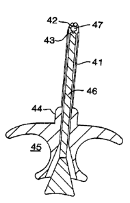
Figure 9 illustrates another embodiment of the
invention, wherein a transurethral drug delivery device
is shown generally at 48. The device comprises a
transurethral inserter 49 having an easily graspable
-17-
WO 95/26158 PCT/US95103514 ~"~
a
2188382
segment 50 that has opposing symmetrically concave
surfaces 51 and 52 adapted to be held by two fingers.
Drug is contained within shaft 53, which is sized to fit
within the urethra. A longitudinal plunger, the tip of
which is seen at 54, is slidably insertable into the
longitudinal bore contained within shaft 53. To extrude
drug into the urethra, shaft 53 is inserted into the
urethra, and plunger tip 54 is pushed into segment 50.
The inserter 49 is then removed. Prior to use, and
during storage, the device is capped with elongate cap 55
which fits snugly over flange 56 at the proximal end of
shaft 53. The cap 55 is provided with a series of
parallel ridges 57 to facilitate gripping of the cap and
removal from inserter 49.
Although in the configurations shown in Figures
4-9 are preferred configurations, other inserter/
container configurations can be used and any mechanism by
which a predetermined quantity of drug can be introduced
from the inserter at a predetermined depth in the urethra
is suitable for use with this invention. The
aforementioned devices can either be manufactured under
sterile conditions, thereby eliminating the need for
post-manufacturing sterilization, or they can be
manufactured under non-sterile conditions and then
subsequently sterilized by any suitable technique, e.g.,
radiation sterilization. The devices can be manufactured
by typical plastic forming and coating processes known in
the art, including molding extrusion, heat forming, dip
coating, and the like.
After administration of the vasodilator,
various penile hemodynamic parameters are measured,
typically including cavernosal artery peak systolic
velocity, cavernosal artery end diastolic velocities,
maximum arterial dilation, and pressure. Based on these
-18-
'~ 18638 ~
measurements, a determination is made as to whether penile
vascular insufficiency is present.
It will be appreciated by those skilled in the art
that any number of devices can be used to conduct the
aforementioned measurements, providing that the desired level
of accuracy is achieved. Duplex ultrasonography is the
preferred mode of evaluating the penile hemodynamic parameters
of interest. However, other types of techniques and equipment
may be used as well, e.g., NMR spectroscopy, pressure cuffs,
corpus cavernosograms, angiography, NPT (nocturnal penile
tumescence) "Rigiscans," magnetic resonance imaging (MRI),
computer aided tomography (CAT), pulsoximeters, and the like.
Examples of duplex ultrasonography devices which can
be used in conjunction with the present method include those
described in U.S. Patent Nos. 4,334,543 to Fehr, 4,485,821 to
Iinuma, and 4,612,937 to Miller. Suitable devices are
available from a number of manufacturers, including, for
example, Advanced Technology Laboratories (Bothell,
Washington) and Siemens Quantum (Issaquah, Washington). In
general, an apparatus is used which includes a transducing
means for emitting ultrasound pulses, a means for receiving
ultrasound reflected from the blood, and a detector circuit
for determining the frequency difference between the
transmitted and received ultrasound. The velocity of blood is
then determined from the frequency difference. As explained
in U.S. Patent No. 4,612,937, individual velocity estimator
signals are produced at a predetermined one of a plurality of
points along each beam directions these estimator signals are
19
74972-1
~~~s~s
then coupled to a display, preferably a color display in which
the brightness of color is proportional to the magnitude of
blood flow velocity.
Based on the hemodynamic parameters measured using
the aforementioned ultrasonography technique, a diagnosis can
be made as to penile vascular sufficiency. Generally, if the
measured PSV is less than about 50 cm/sec, more typically less
than about 35 cm/sec, vascular inflow is insufficient, and a
diagnosis of arterial insufficiency may be made.
Alternatively, or additionally, if the measured EDV is greater
than 0 cm/sec, more typically greater than about 5 cm/sec, a
diagnosis of venous leakage may be made. In either case, the
cause of erectile dysfunction is presumed to be vasculogenic,
and the patient is then treated accordingly. A preferred mode
of treating vasculogenic impotence involves the therapeutic
regimen described in co-pending Canadian patent application
Serial No. 2,040,914. Briefly, the mode of treatment
comprises transurethrally administering a therapeutic
composition containing a unit dosage of a vasodilating agent
(suitable vasodilators are as identified earlier herein) and a
dispersant which is selected so as to dissolve, melt or
bioerode within the urethra to release the drug.
Administration is repeated, incrementally increasing dosage if
necessary, until the desired results are obtained.
If the diagnosis is that the patient's erectile
dysfunction is due to other than vasculogenic causes,
treatment will proceed accordingly depending on the factors
involved, e.g., psychogenic, neurogenic, or the like.
74972-1
~" !~
286382
The invention also encompasses a kit for conducting
the diagnostic method. The kit contains the drug to be
administered, a device for administering the drug
transurethrally (e.g., as shown in the Figures herein), a
sealed container housing the drug and device prior to use, and
written instructions for carrying out
20a
74972-1
TWO 95/26158 PCTIUS95/03514
21 c~63g2
the diagnostic method. The kit may include a means for
administering the drug at different doses, or it may
include different drugs, or a combination thereof. (That
is, if the drug initially administered is not effective
in inducing an erection, incrementally higher doses of
drug can be used, or different drugs may be administered,
until an erection is induced which is sufficient to
enable measurement of the desired penile hemodynamic
parameters.) Instruments for conducting the evaluation
l0 of one or more penile hemodynamic parameters may be
included in the kit as well, e.g., a pressure cuff or the
like.
As exemplified below, the method of the
invention is as effective in providing diagnoses of
vasculogenic erectile dysfunction as known methods which
involve injection of the vasodilator. However, in
contrast to these known methods, the present invention
enables use of a noninvasive technique which avoids the
pain of injection and the subsequent discomfort, and
which involves easily used, inexpensive equipment.
It is to be understood that while the invention
has been described in conjunction with the preferred
specific embodiments thereof, that the foregoing
description as well as the examples which follow are
intended to illustrate and not limit the scope of the
invention. Other aspects, advantages and modifications
within the scope of the invention will be apparent to
those skilled in the art to which the invention pertains.
-21-
WO 95/26158 PCT/US95/03514 t"'
2186582
Example 1
Ten patients (20-57 yrs) with vasculogenic (8)
or psychogenic (2) impotence underwent color-flow duplex
'ultrasound evaluation employing intracorporeal injection
("ICI") of prostaglandin El (alprostadil, obtained from
Upjohn; 10 ~,g), and Doppler Model No. ATL UM-9 (Advanced
Technology Laboratories, Bothell, Washington). Within 60
days, duplex ultrasound was repeated employing
intraurethral administration of alprostadil using the
drug administration device of Figure 9. Patients, after
voiding, administered the medication by inserting a 3 cm
x 4 mm applicator that contains 500 ~cg Alprostadil, into
the urethra. Erection occurred within minutes after
application. Hemodynamic parameters examined at
baseline, 5 and 15 minutes included: 1) cavernosal
artery peak systolic velocity ("PSV") at baseline, 2)
cavernosal artery end diastolic velocity ("EDV"), and 3)
maximum arterial dilation.
The overall mean PSV following ICI was at 5
min: 35.4 +/- 13.8 cm/s (R) and 36.2 +/- 19.7 cm/s (L);
at 15 min: 36.3 +/- 16.3 (R) and 37.1 +/- 17.8 cms (L).
The overall mean PSV following intraurethral
administration was at 5 min: 37.0 +/- 14.4 cm/s (R) and
29.5 +/-19.7 cm/s (L); at 15 min 32.8 +/- 13.8 cms/(R)
and 35.8 +/- 21.0 cm/s (L). There was no statistically
significant difference between the responses to the two
different modes of delivery on either cavernosal artery
(t-test). In addition, within each individual the right
and left cavernosal arterial response to ICI and
intraurethral administration did not statistically differ
at either 5 or 15 minutes (t-test). The EDV responses
were identical. The maximum arterial dilation following
ICI was 0.080 +/- 0.026 cm (R) and 0.078 +/- 0.012 cm
(L). The maximum arterial dilation following MUSE was
-22-
TWO 95!26158 PCTlUS95/03514
218682
0.078 +/- 0.030 cm (R) and 0.072 +/- 0.027 cm (L). The
dilation response did not differ significantly (t-test).
Additionally, the dilation responses to ICI versus
- intraurethral administration did not differ significantly
within each individual (t-test). The most dramatic
difference with intraurethral administration was observed
in the cross-sectional demonstration of a diffuse
arterial dilation in the extracorporal and
periospongiosal arteries that often formed the origin for
collateral intracorporal arterial inflow. Results are
summarized in Table 1 and illustrated in graph form in
Figures 10 through 13 (wherein the shaded bar represents
the results obtained after injection of alprostadil while
the unshaded bar represents the results obtained after
intraurethral administration of alprostadil).
It may be concluded that transurethral
administration effectively produced intracorporal smooth
muscle relaxation comparable to intracavernosal injection
of alprostadil (10 ug). This is demonstrated by arterial
and veno-occlusive hemodynamic measurements of arterial
dilation, arterial PSV increases, and EDV decreases
(veno-occlusion). Furthermore, arterial dilation
following transurethral administration appeared to be
more diffuse compared to ICI.
This noninvasive method of delivering a
vasodilating agent such as alprostadil can accordingly
provide an equally effective and far more acceptable
alternative to intracavernosal injection for patients
undergoing duplex ultrasound evaluation for the diagnosis
of erectile dysfunction.
-23-
WO 95126158 PCTIUS95103514 ~"~
2186382
w o 0 0 ~ 000
N O O N If1l~O
M C~ ri
00 00 00 tf1d'N
O O N ~-iN N
't'1
~ O O 10 t~ u11f1
N1 M
N N rlri
w ro ro o ~n a,
~ of o 00
1 O r~ N
d' M d'~O
~i'N rlrl
G tf110 n tI1
u1 ..~ r~ r~ U
* rl r~ o th
w ro ~ 01 O O 1G
cn~ ~ ,n ~ o 0
w
'~ ~""~ 0 0 0 0 ~ o,
d~ C~ O N
l~ t0 O O
ro
N
2 0 ~ w +' w +'w
~w
~ a ~ a
'
. .
~
a c~ ~x
~ ~ i
a +~
0
'Cf U
O
ro o~
rl
Ul ~.I O ..
O N w
O +~
i ro w
-
a ro *
x b
ro a~
o ~ w w
a ro ro b
-24-
~,.,, WO 95/26158 PCTIUS95103514
2186.382
Example 2
The procedure of Example 1 is repeated, except
that PGE2 is substituted for~alprostadil. Substantially
the same result will be obtained, i.e., that the
diagnostic method involving transurethral administration
of PGE2 followed by hemodynamic evaluation will be at
least as effective as the diagnostic method which
involves intracavernosal injection of PGE2, without trr
associated pain and discomfort. Hemodynamic evaluation
may be conducted as in Example 1, using duplex
ultrasonography, or it may be carried out using other
techniques, e.g., NMR, angiography, or the like.
Example 3
The procedure of Example 1 is repeated, except
that phentolamine is substituted for alprostadil.
Substantially the same result will be obtained, i.e.,
that the diagnostic method involving transurethral
administration of phentolamine followed by hemodynamic
evaluation will be at least as effective as the
diagnostic method which involves intracavernosal
injection of phentolamine, without the associated pain
and discomfort. Hemodynamic evaluation may be conducted
as in Example 1, using duplex ultrasonography, or it may
be carried out using other techniques, e.g., NMR,
angiography, or the like.
Example 4
The procedure of Example 1 is repeated, except
that a combination of PGEl and prazosin is substituted
for alprostadil. Substantially the same result will be
obtained, i.e., the diagnostic method involving
transurethral administration of PGE1 and prazosin
followed by hemodynamic evaluation will be at least as
effective as the diagnostic method involving
-25-
WO 95/26158 PCT/US95103514 ~
2186382
intracavernosal injection of PGE1 and prazosin, without
the associated pain and discomfort. Hemodynamic
evaluation may be conducted as in Example 1, using duplex
ultrasonography, or it may be carried out using other
techniques, e.g., NMR, angiography, or the like.
Example 5
The procedure of Example 1 is repeated, except
that a combination of PGE2 and prazosin is substituted
for alprostadil. Substantially the same result will be
obtained, i.e., the diagnostic method involving
transurethral administration of PGE2 and prazosin
followed by hemodynamic evaluation will be at least as
effective as the diagnostic method which involves
intracavernosal injection of PGE2 and prazosin, without
the associated pain and discomfort. Hemodynamic
evaluation may be conducted as in Example 1, using duplex
ultrasonography, or it may be carried out using other
techniques, e.g., NMR, angiography, or the like.
Example 6
The procedure of Example 1 is repeated, except
that a the device of Figure 9 is replaced with a simple
shaft coated with alprostadil (10 fig). Substantially the
same result will be obtained, i.e., the diagnostic method
involving transurethral administration of drug followed
by hemodynamic evaluation will be at least.as effective
as the diagnostic method which involves intracavernosal
injection, without the associated pain and discomfort.
Hemodynamic evaluation may be conducted as in Example 1,
using duplex ultrasonography, or it may be carried out
using other techniques, e.g., NMR, angiography, or the
like.
-26-
A..~WO 95!26158 PCT/US95103514
2186382
Example 7
The procedure of Example 1 is repeated, except
that a the device of Figure 9 is replaced with a pellet
comprising a polyethylene glycol/alprostadil melt.
Substantially the same result will be obtained, i.e., the
diagnostic method involving transurethral administration
of drug followed by hemodynamic evaluation will be at
least as effective as the diagnostic method which
involves intracavernosal injection, without the
associated pain and discomfort. Hemodynamic evaluation
may be conducted as in Example 1, using duplex
ultrasonography, or it may be carried out using other
techniques, e.g., NMR, angiography, or the like.
20
30
-27-