Note: Descriptions are shown in the official language in which they were submitted.
W0 95/~668~ 2 1 ~ 6 8 8 ~ PCi~ll595103'i45
DEVICE FOR TREATING CONSTRICTION IN A BODILY CONDUIT
BACgGROUND OF 1~ INVENTION
1. Field of the Invention
The present invention relates to the field of treating
a stenosis which would occur in various blood vessels and other
bodily conduits as well as to the field of angioplasty.
Additionally, the present invention is directed to the field of
treating cancer which would occur in various body conduits or
ducts, as well as to the field of brachytherapy.
2. Descri~tion of the Prior Art
Various techniques have been developed to treat many
different conduits in the body when these conduits have become
reduced in size due to the existence of a stenosis or have been
completely occluded. These techniques include introducing a
deflated balloon catheter to the site of the stenosls or
occlusion, inflating the balloon one or more times to eliminate
the size of the stenosis, deflating the balloon and then removing
the balloon catheter from the treatment site.
With respect to the vascular pathways, angioplasty is
used to open an artery or blood vessel in the region where the
stenosis or the occlusion has occurred. A typical angioplasty
procedure consists of making a small incision through the body
and into a blood vessel and then maneuvering a guide wire through
the vascular system to a point beyond the stenosis or occlusion.
A hollow catheter with a deflatable balloon near its distal end
is threaded over the guide wire and advanced to the point of
stenosis or occlusion. The balloon is then inflated and deflated
several times to widen the constricted area, and is then
withdrawn from the body.
Unfortunately, although the angioplasty procedure does
markedly reduce the area of stenosis or occlusion, many patients
exhibit a reoccurrence of the stenosis within a few months of the
original procedure.
Although the original stenosis occurs by means of the
build up of plaque over a relatively long period of time, experi-
mentation has lead many to believe that the reoccurrence of the
W095/26681 2 1 ~ 6 ~ 8 9 PCT~SgS/036~5
stenosis after the original angioplasty procedure is unrelated
to the cause of the original stenosis. It is believed that the
inflation of the balloon catheter used in the angioplasty
procedure or the placement of a stent in the area of the stenosis
causes irritation to the blood vessel. This irritation produces
a mechanism of action called hyperplasia, inducing the inner
layer of the blood vessel cells to rapidly reproduce, thereby
causing restenosis. It has been proposed that if the blood
vessel is irradiated at the point of the stenosis with a
radioactive dose, the mechanism that causes hyperplasia would be
destroyed without harming the blood vessel itself.
During this procedure, it is important to precisely
control the amount of radiation which is directed to the blood
vessel wall, since too much radiation could actually induce
hyperplasia as well as destroying a portion of the blood vessel,
making it possible for an aneurism or rupture to occur. U.S.
Patent 5,213,561 issued to Weinstein et al and U.S. Patent
5,199,939 issued to Dake et al, as well as PCT Application
PCT/US92/07447 to Shefer et al, describe various methods and
apparatus for introducing radiation to the site of a stenosis to
endeavor to prevent restenosis.
The Weinstein et al patent describes a method and
apparatus for preventing restenosis after angioplasty. A balloon
catheter transported by a conventional guide wire is delivered
to the location of the stenosis. Particles or crystals of
radioactive material are embedded or mounted on a tube provided
inside the balloon catheter. A retractable radiation shielding
sleeve is slidable along the tube to cover the source of
radioactive material. Upon completion of the angioplasty, the
shielding sleeve is retracted and the area of the stenosis is
irradiated. Although this apparatus does introduce radiation to
the point of the stenosis, the retractable shielding surrounding
the source of radioactive material makes this catheter bulky and
unwieldy to use. In this regard, it is very doubtful that a
catheter system this bulky would fit into the smaller branches
or vessels of the heart. It is also doubtful that a catheter
WO9S/26681 2 1 8 6 8 8 9 PCT~SgS/036~s
this bulky and stiff couid be maneuvered through the tighter
bends and turns in many of the vessels.
An additional embodiment of the Weinstein et al patent
illustrates a stent which is made of or coated with a radioactive
material such as iridium 192. Since the radioactive material is
provided on the outer surface of the stent, it is very difficult
to precisely administer the proper dosage of radiation to prevent
hyperplasia without administering a level of radiation which
would actually induce hyperplasia or other deleterious effects
to the blood vessel.
The PCT application illustrates a method and apparatus
for restenosis treatment by applying a radioactive dose to the
stenosed region after reduction of the region by angioplasty or
other means. As shown in FIG. 4, an angioplasty balloon is
expanded in the vicinity of a lesion site and radioactive
elements provided on the exterior surface of the balloon are
forced into contact with the region. Therefore, similar to the
Weinstein et al patent, the presence of the radioactive material
on the exterior of the catheter would make it very difficult to
apply the precise amount of radiation to the region of interest.
Additionally, both the PCT application as well as the patent to
Weinstein describe balloon catheters which do not allow the blood
within the vessel to flow during inflation of the balloon.
The patent to Dake et al shows a radioactive catheter
for preventing restenosis after angioplasty. However, this
patent merely indicates that an elongated flexible catheter is
transported to the area of the original stenosis after a balloon
catheter has been withdrawn, thereby lengthening the time to
A~ml n~ ster the entire procedure.
SU~RY OF 'L~ih' INV~ITION
These and other deficiencies of the prior art are
addressed by the present invention which is directed to a method
and apparatus for treating the location of a stenosis in a blood
vessel or other hollo~ conduit in the body by inflating and
deflating a balloon catheter one or more times. A source of
radiation is then advanced through the catheter to the site of
21 86~9
the stenosis, centered within the blood vessel, and the site is
then treated for a period of time with radiation. Once the
treatment is completed, both the radiation source and the balloon
catheter are withdrawn.
According to the teachings of the present invention,
a radiopaque guide wire is inserted into the body through a small
incision and is then introduced into a blood vessel or similar
conduit. Once in place, a catheter having a ribbed balloon
attached near the distal end thereof is threaded over the guide
wire and is also advanced to the location of treatment. The
interior of the catheter is provided with an elastic membrane,
one-way valve or other similar device for sealing the distal end
of the catheter, but allowing the guide wire to pass there-
through. The guide wire is then removed and the ribbed balloon
lS is inflated one or more times to reduce the size of the stenosis,
while allowing blood to flow around the site of the stenosis to
greatly decrease the patient's risk of a myocardial infarction
or heart attack. A radioactive source is advanced into position
through the balloon catheter to the site of the original
stenosis. With the balloon inflated, the balloon catheter and
the radioactive source are correctly centered within the blood
vessel to administer a precise dose to the original area of the
stenosis. After a period of time in which the original site of
the stenosis is irradiated from the radioactive source, both the
radioactive source and the balloon catheter are then removed from
the blood vessel and the body of the patient.
Contrast dye, helpful in locating the position of the
catheter within a body vessel is injected therein by a conduit
provided on the exterior surface of the catheter or through the
guide wire itself, after the core of the guide wire has been
removed.
W095/26681 2 1 ~ 6 8 8 9 PCT~S9sl0364s
BRIEF DESCRIPTION OF THE DRAWINGS
The above and other objects, features and advantages
of the present inventicn will become apparent from the following
description and the appended claims, taken in conjunction witn
5the accompanying drawings, in which:
FIG. 1 is a side view of a ribbed balloon catheter
according to the present invention;
FIG. 2 is a side view of a second embodiment of the
10ribbed balloon catheter according to the present invention;
FIG. 3 is a transverse cross-sectional view of the
ribbed balloon catheter of the present invention taken along
lines 3-3 of FIG. 2;
FIG. 4 is a longitl~; n~l sectional view of the ribbed
15balloon catheter of the present invention showing the radioactive
source within the balloon catheter;
FIG. 5 is a longit~ nAl sectional view of the present
invention showing the guide wire and the one-way valve;
FIGS. 6-9 are end views of the elastic membrane shown
20in FIG. 4 with or without the guide wire inserted therethrough;
FIG. 10 is a front view of the one-way valve shown in
FIG. 5;
FIG. 11 is a side view of the one-way valve with the
guide wire passing therethrough;
25FIG. 12 is a front view of the one-way valve showing
-the smaller opening behind the flap;
FIG. 13 is a side view of a removable core guide wire
inserted into the body;
FIG. 14 is a side view of the guide wire shown in FIG.
3013 after the core has been removed;
FIG. 15 is a side view of the guide wire shown in FIG.
14 after a Luer-Lock has been attached thereto; and
FIG. 16 is a side view of a catheter for the treatment
of cancer within a vessel, duct or airway according to yet
35another embodiment of the present invention.
W095/266~1 2 1 8 6 8 8 ~ PCT/USg51036~5
DETATT.F~n DESCRIPI ION OF THE PRE~RRED EMBODIMENI S
Although the present invention can be used to treat
blockages in many body conduits, for ease of explanation, the
present invention will be discussed with respect to a stenosis
provided in a blood vessel.
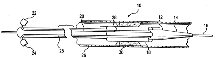
FIGS. 1, 2 and 3 illustrate the catheter 10 of the
present invention after it has been inserted into the body and
moved to the site of a stenosis 30 in a blood vessel 26. The
catheter itself consists of a hollow, generally cylindrical
member 12 which is constructed from a fairly flexible material
such as polyethylene glycol so that it can be easily maneuvered
within the body and travel over a guide wire 16 which was
initially maneuvered in the blood vessel to a position beyond the
actual site of the stenosis. The interior of the catheter can
be made of or coated with a friction reducing material, such as
TEFLON (PTFE) to aid in the passing of the guide wire and the
radioactive sources to the treatment site. The catheter itself
is slightly tapered at its distal end 14 to facilitate movement
through blood vessels or similar conduits or ducts. Both the
guide wire 16 and the catheter 12 should be of sufficient length
to travel to the site of occlusion or constriction in various
conduits and certainly should be long enough to reach the heart.
A ribbed balloon 18 surrounds a portion of the outer surface of
the catheter 12 and contains a number of ribbed pleats. When
these pleats are inflated by a syringe 24 injecting air into a
conduit 25 extending along the exterior surface of the catheter
12 to the balloon 18, the size of the stenosis would be reduced
as well as allowing the catheter to be properly centered when a
radioactive source is introduced to the original site of the
stenosis.
A second syringe 22 is also attached to the catheter
12 for injecting contrast dye into the blood vessel to aid in the
proper location of the catheter. This contrast dye would travel
through a conduit 20 also provided on the exterior surface of the
catheter to a site 28 near the proximal end of the balloon 18
(see FIG. 1) or could extend to a point 32 beyond the distal end
of the balloon 18 (see FIG. 2).
WO95/26681 21 ~b889 PCT~S95/036~5
Alternatively, contrast dye can be introduced to the
site of the stenosis by injecting the contrast dye directly into
the interior of the catheter 12. This is accomplished utilizing
a guide wire provided with a removable core, the operation cf
which will be subsequently explained.
Since the ribbed balloon 18 would inflate in a
symmetrical pattern, blood would be allowed to profuse at various
locations 34 during both the angioplasty procedure as well as the
radiation treatment. This flow of blood would greatly decrease
the incidence of a myocardial infarction or a heart attack and
would allow the angioplasty procedure as well as the radiation
treatment to be performed as iong as needed without completely
blocking the flow of blood through the vessel.
Since the catheter of the present invention would act
as a conduit to allow a radiation source to be introduced to the
site of the original stenosis, it is important that the catheter
should be sealed at a point proximate to its distal end, while
allowing a guide wire to exit the distal end of the catheter 12.
Consequently, the present invention utilizes an elastic membrane
40 shown in FIGS. 4, and 6-9 to perform this function. This
membrane can be constructed of any biocompatible material 44 that
will expand large enough to allow the guide wire 16 to pass
therethrough and then contract to form a closed seal when the
guide wire is removed.
FIG. 6 illustrates the elastic membrane which is
completely sealed prior to the guide wire passing through this
membrane. FIG. 7 illustrates the membrane with a small hole 46
forming in the middle thereof which would allow the guide wire
to pass therethrough as shown in FIG. 8. FIG. 9 illustrates the
elastic membrane 40 immediately after the guide wire 16 has been
removed.
As shown in FIG. 4, more than one elastic membrane 40
can be utilized to insure that the catheter is sealed after the
guide wire 16 is removed. Regardless of whether a single
membrane or a plurality of membranes are used, the membrane is
placed in the interior of the catheter 12 at a location beyond
the ribbed balloon 18, in such a manner as to effectively seal
wo 95/26681 2 1 ~3 6 8 8 9 Pcr/uss~/036~5
the catheter from the blood vessel. Filters 42 can be provided
between each of these menbranes for wiping the guide wire as it
travels through the balloon catheter 12. Because the guide wire
16 extends into the blood vessel, and is then removed from the
catheter 12 after the catheter has been maneuvered to the correct
location, it is important that blood or other liquids not be
introduced into the sealed portion of the catheter since this
would inhibit the proper placement of the radioactive source.
The filtered material 42 can be constructed from any biocom-
patible material that freely allows the guide wire 16 to pass
therethrough as well as wiping the guide wire as it is withdrawn
from the catheter 12. Cotton or angel foam have been found to
be particularly efficacious for this purpose.
An alternative embodiment in which a one-way valve 48
is used with, or in place of the elastic membrane 40 is shown in
FIGS. 5, 10, 11 and 12. The one-way valve 48 is placed in the
interior of the catheter beyond the ribbed balloon 18. The one-
way valve is provided with a relatively large flap 50 which is
considerably larger than the hole 54 which it covers. A tension
hinge 52 insures that the flap r.om~;ns in the closed position
during the absence of the guide wire 16. In use, as shown in
FIG. 5, the guide wire 16 advances in the direction shown by
arrow 56 and the catheter advances in the direction shown by
arrow 58. In this instance, as the guide wire passes through the
relatively small hole 54, it pushes against the flap, causing the
flap to rise and allow passage of the guide wire therethrough.
As illustrated in FIG. 5, since the hole 54 is much smaller than
the size of the flap 50, the flap can only move in the clockwise
direction and not in the counterclockwise direction. A "funnel-
shaped" entry port 52 assists in allowing the guide wire 16 to
pass through the hole 54. If the one-way valve is used in
conjunction with at least one of the elastic membranes 40 shown
in FIG. 4, filter material 42 can be provided between these two
sealing members.
FIGS. 13-15 demonstrate a removable core guide wire 64
which can be used instead of the guide wire 16 illustrated in
FIGS. 1 and 2. The guide wire 64 is provided with a flexible
wo ss/266~l - 2 1 8 6 8 8 9 PCT/US95/036-~5
outer housing 58 which can be constructed from such a material
as nitinol. The removable core guide is provided within the
outer housing 58 and includes a soft, flexible, rounded tapered
end leader 54 extending beyond one end 61 of the outer housing
58. A slightly oversized cap 56 is provided over the second end
63 of the outer housing 58 to allow the removable core to be
removed from the outer housing with the guide wire has been
properly positioned within the blood vessel. Once the core is
removed, the guide wire would only include the hollow outer
housing 58 as well as a series of external threads 60 on the end
of the guide wire extending out of the patient's body.- This
threading would allow a Luer-Lock 62 or similar device to be
screwed onto the outer housing 58 so that a syringe can inject
contrast dye into the catheter. The removable core can be
constructed from Teflon, nitinol or any springy, soft biocompati-
ble material. If the removable guide wire as illustrated in
FIGS. 13-15 is employed, the conduit 20 shown in FIGS. 1 and 2
used to deliver contrast dye to the vicinity of the stenosis is
not needed.
The balloon catheter of the present invention as
described can be utilized in the following manner to treat a
stenosis as well as to prevent reoccurrence of the stenosis.
Once the site of a stenosis is determined by appropriate
gnostic procedures, a small incision is made in the body and,
assuming that an angioplasty procedure is necessitated, into a
vessel. The guide wire 16 is then maneuvered into the vascular
pathway and is imaged under fluoroscopy while being advanced
through the blood vessel pass the area of stenosis. The catheter
12, with the balloon 18 being deflated, is threaded over the
guide wire 16 and it is also advanced such that the balloon 18
is maneuvered to the area of the stenosis. Contrast dye is
injected either through the external ports 28, 32 or the
specially designed removable core guide wire illustrated in FIGS.
13, 14 and 15. The contrast dye enters the vascular pathway
causing the blood vessel to become temporarily opaque and
allowing it to be imaged under fluoroscopy.
WO9S/26681 2 i 8 6 8 8 9 PCT~S95/036~5
Since the contrast media is quickly absorbed by the
body, multiple injections of contrast dye are possible. An
opaque marker can be applied to one or both ends of the ribbed
balloon 18 allowing it to be imaged under fluoroscopy. Once the
ribbed balloon is verified to be in position, the balloon is
inflated, the guide wire is withdrawn from the body, and the
angioplasty procedure commences.
At this point, the balloon 18 is inflated and deflated
one or more times to widen the constricted area. When the
balloon is deflated, contrast dye can be injected again to verify
the widening of the prior constricted area. The balloon is then
inflated to hold the catheter in place for the radioactive
treatment.
One or more radioactive sources 38 are provided on, or
inside the distal end of a flexible member 36 which is advanced
through the interior of the catheter 12 until it reaches the
proper location (see FIG. 4). The radioactive source treats the
area of the original stenosis for a specific period of time. The
time that the source r~m~-n~ inside the catheter depends upon the
strength of the radioactive source and the distance between the
source and the inner blood vessel walls. Examples of gamma type
radiation sources which can be utilized in this procedure would
be cesium 137, cobalt 60, iodine 125, iodine 131, cobalt 57,
iridium 192, gold 198, palladium 103, etc. Typically, treatment
times could last between approximately four minutes to approxi-
mately thirty minutes or longer. Since iridium 192 has a well-
defined energy level with a strength of 1-2 Curies, it is
particularly well-suited to treat the area of the original
stenosis at the prescribed distance. In this instance, treatment
times would be in the range of 5 to 10 minutes. After treatment
with the radiation source has been completed, both the radiation
source and the catheter, with the balloon deflated, are then
removed from the body.
Since the radiation source can have a deleterious
effect on the body if it is not precisely positioned with respect
to the area of treatment, the present invention insures that the
radiation source is positioned in the center of the vessel at a
WO95/2668l 2 1 8 6 ~ 8 9 PCT~SgS/036~5
1 1
predetermined distance from the area o' treatment. Thls is
accomplished by in'lating the ribbed balloon 18 when the.
radiation source is delivered to the proper location. Addition-
ally, for safe measure, the balloon 18 can be inflated at all
S times when the radiation source is being delivered to the site
of the treatment. The positioning of the radiation source with
respect to the area of treatment is crucial since next to the
radiation source, it is possible to receive thousands of Rads or
centiGrays, units of measurement of radiation dose. This dosage
would drop to only a few hundred Rads or centiGrays approximately
10 ~;11i~eters away from the source.
Although the present invention has been explained with
respect to an angioplasty procedure, it is noted that this
treatment could be conducted in virtually any conduit of the body
with or without the inclusion of radiation treatment. This
catheter can also be used to treat cancer in various areas of the
body, such as the common bile duct, the bladder, the liver, the
lungs, etc. employing the same balloon catheter shown in FIGS.
1-15.
There are many instances in the body where cancer
invades around and into a vessel or airway. Treating and
controlling the invasion of the cancer is difficult since a
sealed prior art catheter having a removable backbone wire on its
inside was used to try to access the cancerous area. Since the
hollow duct of a vessel or other conduit includes many turns and
bend inside the body, the cancerous area could not be reached due
to the stiffness of the catheter and the fact that the backbone
wire was unable to negotiate the turns. If the backbone wire was
removed, the catheter would bunch up and advancement would not
be possible. The balloon catheter of the present invention
avoids these problems since a flexible guide wire is easily
maneuvered into position and the closed-end catheter is advanced
over this guide wire giving access to the cancerous area.
With this in mind, the following procedure can be
utilized to treat a cancerous area with radiation utilizing the
catheter, guide wire and sealing means illustrated in FIGS. 1-15:
The radiopaque guide wire 16 is maneuvered into position either
WO 95126681 2 1 8 6 8 8 9 PCT~US95/036~5
through a body orifice leading into the hollow duct or an opening
created into the hollow duct by means of a small incision or
puncture. The radiopaque nature of this guide wire allows X-rays
to be used to properly position the guide wire beyond the tumor
or cancerous site, which in many ways, is similar in appearance
to the stenosis 30 of FIG. 1. The catheter system 10 is then
threaded over the guide wire 16 and advanced into position. A
radioptic marking on the ribbed balloon 18 makes it easy to
position the catheter utilizing X-rays. To further confirm
position of the catheter, a contrast dye may be injected through
either of the external ports 28, 32 or through the removable
guide wire illustrated in FIGS; 13-15. The balloon catheter is
then inflated and the guide wire is removed. The inflation of
the balloon is especially valuable if the tumor has invaded the
duct or is causing extrinsic compression from outside the duct.
This inflation will give temporary relief from the constriction,
allowing greater passing of bodily fluids. A radioactive source
or sources 38 contained on the end or inside the end of the
flexible drive member 36 (FIG. 4) is advanced inside the catheter
to align with the tumor or cancerous area. After a specified
time, the radiation and catheter are removed from the body.
The catheter apparatus including the flexible membrane
or the one~way valve is very important since, once the guide wire
is removed, the system becomes closed, thereby not allowing the
radioactive source or sources to advance out the end of a
catheter and into the body if they become detached from the drive
member 36. Furthermore, similar to the previously described
embodiments, the inflated ribbed balloon allows body fluids to
pass around the catheter. For example, when treating the bile
duct, the catheter does not allow passage of the bile, cholecys-
titis can develop due to the back up of bile into the liver and
cause liver dysfunction. Additionally, when treating the airway
of the lung, if the catheter does not allow mucus or air to pass,
atelectasis (collapsing of the lobe or the lung) or obstructive
pneumonia can develop. This is a very harmful situation to the
patient since the patient's lung capacity has already been
compromised due to the presence of the cancer.
~ WO95/26681 2 1 8 6 8 8 9 PCT~Sss/036~5
Similar to the previously described embodiments, the
use of the inflated balloon catheter 18 is helpful in centering
the radioactive source or sources inside the hollow duct. Since
radiation emission observes the inverse square law, it is quite
important that the radioactive source be properly centered
because in areas of the body where the walls of the vessels are
extremely radiosensitive, such as the bile duct, great harm can
be caused to the patient if the source is not centered and kept
from the vessel wall. Too much radiation for a period of time
in an area proximate to the vessel wall can cause severe
hemorrhaging or radiation necrosis.
FIG. 16 illustrates a catheter and guide wire combina-
tion previously described, with the exception that a ribbed
balloon or other means does not surround a portion of the
exterior surface of the catheter. This catheter system is
important since, in instances where a cancerous site 70 has
invaded the vessel or duct wall 72 to a great extent, it would
be very difficult if not impossible to maneuver a catheter having
a ribbed balloon to the cancerous site. Once the guide wire 16
is removed, the radioactive source or sources is maneuvered in
place in a manner similar to the above-described procedures
relating to the treatment of stenosis or cancer.
Although preferred forms of the present invention have
been herein disclosed, it is to be understood that the present
2S disclosure is made by way of example and that variation of
posture without departing from the scope of the hereinafter
claimed subject matter.