Note: Descriptions are shown in the official language in which they were submitted.
WO 95128947 r~ 'C
~1 87 45q
--1--
p~.T~l~Rl~RTT.T~Rl? PEPTTnRC FOR INCREASING
,- BLOOD-OCUI~R BARRIER PRRMBABILITY
Ba~;h~ ~ vulld of the Invention
The blood-ocular barrier (BOB) I?rovides a ahield for
5 the ocular tissues and fluids, prevelltin~ the transport of
certain molecules f rom the plasma into the eye . This is
generally a f avored f eature of the BOB, except when the
transport of therapeutic or diagnostic agent6 to the eye is
desired. The ~30B i8 comprised of ~ril 1 Ary endothelial
10 cells rr-nnect.od by tight junctions . ~1 th~u~h alterations
in the p~ -hi 1 ;ty of the barrier may c.~rur in certain
~1; c~ c, as well as from trauma, aurger~ or certain
rhArm-col~gic agents, it is generally not sufficient to
permit adequate quantities of a theL cl~euLic agent into the
15 eye.
Currently, age~ts are administered for delivery to the
eye by a number of methods ; nrl ~ ; n~ systemic
administration (intravenous), local application of an
agent-cont~in;ng solution, rl~ ~ of a porous component
20 in contact with the eye for sustained release of an agent,
or insertion of an implant c~n~;n;n~ an agent. These
delivery methods are often ;n~A~ te due to the limited
ability of the agent itself to penetrate the BOB. In other
cases, agents are administered by direct inj ection into the
25 eye. This means is inadequate due to a high level of
discomfort, risk of injury, and expense of treatment.
The need exists for an effective and non-invasive
means for delivering adequate ~l~n~;t;~ of a therapeutic
or diagnostic agent into the eye an~ across the BOB in
30 order to provide better treatment and diagnosis of ocular
diseases .
WO 95/28947 P~.l/uw.,.~ n
~7459
r~ of the Invention
The present invention pertains to a method of
increasing the p~ -hil; ty of the blood-ocular barrier of
a host to a molecule present in the host' 8 bloodstream.
The method comprises administration to the host of an ~-
ef f ective amount of a p~ -h; 1; 7er peptide wherein the
peptide co~prises bradykinin or an analogue of bradykinin.
In a preferred ~ ' '' - t, the bradykinin analogue,
referred to herein as A-7, has the core sequence
Arginine-Proline-~Iy-lL~ yl-L~line-Glycine-ThienylAlAn;n~-Seri
ne-Proline-4-Me-Tyrosine- (CEI2NH)Arginine (SEQ. I.D. NO. 1)
from N-terminus to c-t~rm; nll~ where CH2NH denotes a reduced
peptide bond between the 4-Me-tyrosine and arginine amino
acids. Conformational analogues of the A-7 sequence are
lS also preferred peL, -h;l;~r8 useful in this invention
provided they have the p~ L Ly of increasing the
peL -h; l; ty of the BOB .
The I leclll F' to be delivered to the eye can be an
endogenous molecule residing in the bloodstream or an
e~uy~ u8 molecule that is co-administered se~n~;Ally or
simultAn~ ly with the peL --h;1;7~r peptide.
An advantage of the present invention is that it
provides a practical means of ;nrreA~;n~ the p~ -h; 1 itY
of the blood-ocular barrier to a co-administered molecule
or drug of theLa~u-ic, prophylactic or diagnostic value.
The p~LI =h; 1; 7~.r peptide can be administered
intravascularly (illL~ ,us or intraarterial), or by any
route that permits it to enter the bl~odYLL~,,I, of the host.
In contrast to methods wherein a drug is directly inj ected
into the eye, or introduced by way of a component placed in
contact with the eye for sustained release, intravascular
administration is significantly less traumatic, causes less
discomfort to the patient and is unlikely to necessitate
anaesth~sia .
W0 95128947 r~ 4rcn
~ 2~ 8745q
--3--
Finally, the invention pertains to methods for
delivery of a tlleLc~u~ic or diagnostic agent into the eye
of a patient in need of such 1.~ ~ ' comprising
co-administering an ef f ective amount of a p~l ~hil; 7er
5 peptide and an agent to be delivered to the eye in order to
effect the p, --hil ~ ty of the blood-ocular barrier to the
agent of interest.
Brief Descrition of the Drawincs
Figure 1 is a diagram of a human eye.
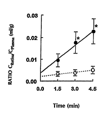
Figure 2 is a graph showing the relative ~nr~ntration
of sucro~e delivered to the retina in the presence (-) and
absence (o) of A-7 as a function of time.
Figure 3 is a graph showing the relative ~n~ntration
of sucrose delivered to the vitreous humor in the presence
15 (-) and absence (o) of A-7 as a function of time.
Figure 4 is a graph showing the relative cr~nr~ntration
of sucrose delivered to the aqueous humor in the presence
(-) and absence (o) of A-7 as a function of time.
Figure 5 is a graph showing the relative c~n~-~ntration
20 of sucrose de~ivered to the lens in the presence (-) and
absence (o) of A-7 as a function of time.
Figure 6 is a graph showing the relative c~n~~~ontration
of sucrose delivered to the cornea in the presence ~-) and
absence (o) of A-7 as a flln~t;nn of time.
25 Detailed De3crition of the Invention
In general, the BOB can be separated into the
blood-aqueous barrier (BAB) and the blood-retinal barrier
(BRB). These barriers divide the eye into three chambers
w0 95/28947 r~ S - ?
~ ~7 459 ~
--4--
(anterior, posterior and vitreou8) cnntR;n;ng two humors,
the aqueous humor and the vitreous humor (Figure 1). The
RntPr; rr chamber is anterior to the iri5 and the posterior
chamber extends anteriorly f rom the vitreous body and
comprises the ciliary proce85es, len5 and posterior part of
the iris. The vitreous chamber i5 bounded po5teriorly by
the retina and anteriorly by the pars plana and cnntR;n~
the vitreous humor.
The cells of the anterior chamber barrier are somewhat
different from those in the posterior chamber. The barrier
in the anterior chamber i5 made up of va5cular epithelium,
b~r membrane and iri5 5troma. The posterior chamber
barrier is made up of vascular endothelium, hR~
' ~.,e, stroma and two layer5 of ciliary epithelium (Cole
et al, 1984, In Davson, H. (ed.~ The Eye. Vegetative
Physiology and Bi~rhPm; ~try New York, Academic Press, vol .
la, p 269; Cunha-Vaz, 1979, Surv. Opthalmol. 23 :279) . All
of these cells appear to be cnnnPrted by tight jllnrtinnA~
The barrier protecting the vitreous chamber consists
of the ciliary epithelium, the retinal pigment epithelium
and the endothelial layer that line5 retinal blood vessels.
These cells are also cormected by tight jllnrt;onl~
However, the retinal ve55el5 have even tighter tight
junctions than the DAB.
A8 used herein, the term "blood-ocular barrier" means
the blood-aqueous barrier or the blood-retinal barrier,
alone or irl combination, as well as any other system of
tight junction5 withi~ the ocular compartments. The terms
"blood-retinal barrier'~ a~d "blood-Yitreous barrier~ can be
3 0 used interchangeably to de5cribe the barrier protecting the
vitreous chamber.
The present invention relate5 to a method for
increasing the p~ h; 1; ty of the blood-ocular barrier of
a host to a molecule pre5ent in the ho5t ' 5 blood8tream . By
increasing the pe~ - h; 1 ity of the blood-ocular barrier to
. .
WO 95/28947 P~
~\ 21 ~745q
--5--
a molecule of interest, the molecule more readily leaves
the bloodstream and enters the interstitial fluids of the
eye. The increase in blood-ocular barrier peL -h; l; ty to
a molecule of interest in the presence of the pell --h; 1; 7~r
5 peptide provides accessibility of the molecule to the eye
in higher relative rrnr~ntrations than in the absence of
the ~:e- -h; 1; 7er peptide.
For purposes of this invention, a, ~ is a
peLl -h; l; ~er of the blood-ocular barrier w~en it
10 signif icantly increases the pe -h; 1; ty o- the
blood-ocular barrier to a molecule of interest. This
effect may operate through a recepto~^ v-';~t~d event. The
preferred pe ~h;1;7~rq are peptides, peptoids, or
p~pti ,~ t ;cs like bradykinin or bradykinin analogues.
15 Bradykinin is a naturally occurring peptide comprised of
nine amino acids having the following geqn~nre
Arginine-Proline-Proline-Glycine-Phelly1 ~1 ~n; n~-Serine-
Proline-Phenyl l1~n;n~-Arginine (SEQ. I.D. N0. 2)
(~ehninger, A.~., 1975, Biochemist~, p. 97). An analogue
20 i8 a structural derivatïve of a pare:nt . _ ~. Analogues
of bradykinin can be ~_ _ ~lq which are derivatives of the
number and/or sequence of~ amino acidq in the bradykinin
structure r- ; r,noc~ above which have a similar or ~nh~nr~d
effect on r~ -h;l;ty of the blood-ocular barrier.
25 ~Ir~;~;r~t;nr~ of the bradykinin molecule can be made by
rh~ng;n~ or modifying peptide bonds, adding C-t~rm;n~1 or
N-terminal .o,~t-~nqirnq, etc.
Particularly pref erred bradykinin analogues which are
p~ --hil; 7ers of blood-ocular barrier permeability are
30 peptide A-7 and conf~rr~t;~n~l analogues of A-7. A-7 has
the following linear amino acid sequence from N-terminus to
C-tf~m; nll~
Arginine-Proline-Hydroxyproline-Glycine-Thienyl~ n;n~-
Serine-Proline-~-Me-Tyrosine- ~CH2NH) Arginine (SEQ. ID
35 N~. 1) .
wo ssn894~ r~ ,n
21 8745~ ~
--6--
The peptide A-7 differs from a conv~nt;nn~l linear amino
acid sequence in the following ways: the fifth amino acid
is thienyl~1~n;n~ which is similar to phenyl~1~n;n~ but
where a thienyl group has replaced the phenyl group; the
5 eighth amino acid is tyrosine which has been substituted
with a methyl group at the 4 position; and the peptide bond
between the eighth and ninth amino acids has been replaced
with a reduced peptide bond isotere, i.e. CH2NH. Peptide,
peptoid and peptidomimetic analogues of A-7 are also part
10 of this invention provided they allow the proper
conformation ln a~ueous ~ol~lt; nn 80 they effect an increase
in pe~ h; 1 ity of the blood-ocular barrier to molecules of
interest. These compositions are termed "confnrr-t;nn~l
analogues ~ of this ~
The preferred peL h;l;7er A-7 differs from
bradykinin iI~ the following respects: at the third amino
acid, hydroxyproline replaces proline; at the fifth amino
acid, thienylAl~n;np replace5 pheny1~1An;n~; at the eighth
amino acid, 4-Me-tyrosine replaces pheny1A1~n;nP; and
20 between the eighth and ni~th amino acids, a reduced peptide
bond replaces a conv~nt;nn~1 peptide bond.
Characteristic feature5 of the peL -h; 1; 7~r A-7 or
conforr-t;nn21 analogues of this invention are important
for the ~ -h;1;7~r A-7 or conforn~~ti~n~1 ~n~lo~l~ to
25 allow the proper conform-t;nn to effect an increase in the
~eL, - -h; l; ty of the blood-ocular barrier to a molecule of
interest. Further and more ~1~t~;1ed descriptions of
r ~;f;c~t;nn~ that can be made to bradykinin, analogues of
bradykinin and peLI h;l;7~r A-7 are provided in U.S.
30 Patents No. 5,112,596 and No. 5,268,164 assigned to the
same assignee, the te~r~;n~R of which are hereby
incorporated by ref erence .
The invention relates to a method ~or increasing the
peL, -h; ] i ty of the blood-ocular barrier of a host to a
35 molecule present in the host' 8 bloodstream. The host can
wo 95/28947 P~~ 'Q
2~ 87459 _7_
be any organism which possesses an eye, including mammals,
such as humans and domestic animals (e.g., dogs, cats,
- cows, sheep, goats or horses), as well as animals ;nt~nrl~
for experimental purposes (e.g., guinea pigs, rats, mice,
5 rabbits).
The molecule in the host ' 8 bloodstream can be
~uye~uus to the host. For example, it can be an agent
which ha~ a therapeutic effect on an ocular disease or
disorde~ Examples of ocular diseases and disorders
10 include ~:iral infections, such as AIDS- associated
cytomegalovirus (CMV) retinitis, bacterial infections or
~n~npthAlm;tis (bacterial or fungal infection caused by
trauma or surgery), cystoid macular degeneration, diabetic
ret;nnpathy, ;nfl tinnl or tumors (e.g.,
15 ret; nnhl A~toma)
Classes of thera~-utic agents which can be used in
this invention include Ant;h;ot;r~, antiviral agents,
anti-;nfl tnry agents, and chemotlleL~euLic agents.
Examples of antibiotics include cPrhAlospnring, p~n;r;ll;n~
20 and qn;nnl ;nl~ Examples of antiviral agents include
ganciclovir and foscarnet. Examples of anti-;nfli tnry
agents include ketorolac, 11e~YLV~11 or any non-steroidal
anti-;nfl ory drug (NSAID). Examples of chemo-
therapeutic agents include riqrhopl~t; - and cisplatin. The
25 theLcl~ueuLic agents can also include the prodrug form of an
agent which is r -~iqhol; 7ed to the active form of a drug
following administration. The molecules in the host's
bloodstream can also be a diagnostic agent, such as an
imaging or contrast agent or a dye. Examples of diagnostic
30 imaging agents include substances that are labeled with
rAtl;~Artivity, such as 68Gallium for positron emission
t , L c.~hy (PET) scanning, gadolinium based agents for
magnetic r~onAnr~ imaging (MRI), and 99Tc-DTPA for Single
Photon r~ ; nn Computed T~ (SPECT) scanning.
35 Examples of dyes include fluorescein and indocyanine green.
.
wo95/28947 r~u~,_'C~
2~3745q
--8--
The invention further pertains to a method of treating
cyt.~ _ lovirus retinitis in the eye of a patient,
comprising administering a therapeutically effective amount
of an antiviral agent and an effective amount of a
p~ ~h;1;7~r peptide, wherein said permeabilizer peptide .
ia bradykinin or a bradykinin analogue, and said
pe, ~-h;1;7~r peptide is effective for increasing
peL --h;l ;ty of a blood-ocular barrier to said antiviral
agent .
In addition, the method pertains to the treatment of
ret;n~hl~ctoma, comprising administering to a patient a
therapeutically effective amount of a chemot~ c
agent and an effective amount of a p~ h; l; ~ r peptide,
wherein said p~ -hil; 7~r peptide is bradykinin or a
bradykinin ;~n::llo~l~, and said p~ -h;1;7~r peptide ia
effective for increasing peL -h; l; ty of a blood-ocular
barrier to said chemo~h~ ; c agent .
The administration of an ~-~yt~ JuS molecule to the
host's bloodstream can be parenterally by sllh~-lltAn~ollc,
intravascular, preferably intravenous or intraarterial, by
intl cclll ~r injection, by oral administration, by
eyedrops, or by any route that delivers a molecule to the
bloodstream of the host. The form in which the molecule is
administered (e.g., g~ lt;c~n, l~;-ln, tablet, capsule,
etc. ) will depend, at least in part, on the route by which
it is administered .
The administration of the _~:-J~ llc molecule to the
host ' s bloodstream and the administration of the
peL, -~h; l; zer peptide can occur simult~nPollcly or
~e~l~nt;~l~y in time. For example, a therapeutic drug can
be administered orally in a tablet form while the
intravascular administration of the peL -h; 1; 7~r is
performed some time later. This allows time for the drug
to be :~hg~rhe~ in the gastrointestinal tract and taken up
by the bloodstream before the pel -hil; 7er ~is given to
.
wo 95/28947 F~ ''C
21 8745~
g
increase the permeability of the BOB to the drug. On the
other hand, the pe -hil; 7~r can be administered before or
- at the same time as an il~Lr~velluus injection of a drug.
Thus, the term nr~A~' n;~:trationn iE used herein to mean
5 that the peLI --h; 1; 70r peptide and the ~uy~ uuS molecule
will be administered at times that will achieve sign;f;r3nt
c.tions in the blood for producing the simultaneous
effects of increasing the p~ --h;l;ty of the blood-ocular
barrier to the ~ molecule and allowing the maximum
10 passage of the e,wy~, uUs r~ 1 ~cl~l e from the blood to the
interstitial ~ ~ of the eye.
In addition, the -1PC111~ to be delivered to the eye
via the bloodstream can be ~ to the host. That
is, it can be a h; ol o~ l product that is naturally
15 synthF~; 7~1 and produced by the host. Examples of auch
biological products include augara, such as glucose, and
amall peptides, such as ~nkF-rh~l;n~ and thyroid 8t; l~t;
hormone releasing factor.
An effective amount of bradykinin or a bradykinin
20 analogue is that amount which will sign;f;r~ntly increase
the blood-ocular barrier peL, -h; l; ty to the molecule of
interest. In other words, it will increase the
pe~ h; 1 ~ ty of the blood-ocular barrier to allow
suf f icient quantities of a molecule of interest to pass
25 from the blood to an ocular ~ , - i t to exert a
therapeutic or prophylactic effect or allow diagnostic
procedures. The effective amount will be determined on an
individual basis and will be based, at leaat in part, on
c~n~irl~ration of the individual's size, the specific
30 diaease, the aeverity of the symptoma to be treated, the
reault sought, the ~p~-; f i c bradykinin analogue, t~ie
variation of individuals' affinity binding of bra~ kinin
receptors, etc Thus, the effective amount can be
determined by one of ordinary skill in the art employing
WO 9!5128947 r~ ",,~ . r~
2~ 87459 -lo-
such factors and using no more than routine
exper; - tAt; on .
One or more pP --h;l; 7Pr peptides can be administered
to a host in a suitable pharmaceutically acceptahle
5 carrier, any of a number of w_ich are known to one of skill
in the art. The actual amounts and rnnl~Pntrations of
peL ~hi 1; 7Pr peptides in the compositions can be readily
ascertained by a person of skill in the art.
The increase in pe~, - -h; 1; ty of the blood-ocular
10 barrier in response to a p~r, -h; 1; 7Pr peptide relates not
only to the quantity of -1PCI11P~ passing from the blood
into the ocular c, ~R, but also, to the type of
molecule .
The invention i5 further illustrated by the following
15 specific 1 PR, .
r le 1 - Effect Qf ~-7 on u~take of l4C-sucrose into
O~lll Ar, -rtmentS
Adult Hartley guinea pigs of either_sex ~2~0-300 g)
were used. Pe:ll -h;l; ~r A-7 was prepared by the method
20 described in U S. Patent Number 5,268,164, assigned to the
same assignee and hereby ine~ulc.ted by reference.
14C-sucrose (560 mCi mmol~l, New England Nuclear, Boston,
MA) is a well accepted model molecule and was used to
IJ ~ te pe~ -hi l i ~tinn of the blood-ocular barrier by
25 peLI --h; l; ~Pr A-7 .
Animals were anaesthPt~ ~ed with 6 mg/kg xylazine
(Rompun~, Mohay Corp., Shawnee, KS) and 30 mg/kg kPtAm;np
(Vetacetl, Abeco Co., Inc., Fort Dodge, IA) before
surgically P~oR; ng the neck vessels . The vascular eye
30 perfusion (VEP) technique used here and previously
described by ZlokoviC et al (1992, Exp. Eye Res., 54:471)
allows for the study of uptake of agents in to the eye
while protecting the eye f rom systemic metabolic changes .
The terhn;~r1P is briefly summarized here. A fine
wo gs/28947 2 ~ 8 7 4 5 9
polyethylene catheter ~nnn~oct~l to the extracu ~ ~uOLeal
perfusion 6ystem by silicon tubinga was in5erted into the
-~ right common carotid artery. T ~ t~ly after the start
of the perfusion, the contralateral carotid artery was
5 ligated and both jugular veins cut to allow free drainage
of the perfusate.
The perfusion medium consisted of 2096 washed sheep red
blood cells (R3C) suspended in mock plasma of the following
composition (in mM): 123 NaCl, 4 KCl, 2.5 CaCl2.H2O, 25
10 NaHCO3, 1.2 KH2PO4, 1.8 MgCl2.6H2O and 5.5 D-glucose. The
perfusion medium was gassed with 96~ 2 and 4~ CO2 and
warmed to 37.6C, and was pumped from a reservoir through a
water bath using a Rainin Rabbit peristaltic pump (Rainin
Instruments, Woburn, MA) . All tubing was low gas p~ --hle
15 silicon, and all metal was medical grade stainless steel.
The t -r~t~lre and perfusion ~res~ure were ~n~;n~ u~ly
recorded, and the acid-base status rle~uellLly monitored.
Perfusion pres8ure was kept 81ightly above the animal ' 8
blood ~lés~u~c: to ol;m;n~te any possihle ingress from the
2 0 systemic circl l l ~ t i ~n .
Perfusion medium was delivered to the eye with or
without the addition of permeabilizer A-7 (total dose of
l~Lg/kg) by ~ nt;nll~llc 5 minute arterial infusions at a rate
of 0 . 2 ml min~l using a Harvard syringe pump (Harvard
25 Apparatus, South Natick, MA). Isotopically labeled
[l4C]-sucrose was introduced into the perfusion circuit
after the 5 minute infusion at a rate of 0.4 to 0.6
Ci/ml/min, over periods ranging from 1.5 min to 4.5 min.
In all experiments, the perfusion was terminated by
3 0 severing the right common carotid artery and decapitating
the animal.
AB large a sample a5 possible (30-40 ~Ll) of the
aqueous humor was removed with a 0.5cc U-100 insulin
syringe using 28G 1/2 microfine IV neêdle (no dead-space),
35 immediately after the perfusion was terminated. The eyes
~, .
W0 95/28947 1 ~
2187~59
--12--
were enucleated and the lenses rapidly excised via a
lateral approach 1.5 mm posteriorly to the limbus. The
lenses were blotted on filter paper to remove any adhering
aqueous humor and to avoid epithelial c nnt~m; n~t; on by
5 aqueou8 r~;o~ot;vity. Corneas were dissected
circumf erentially about 0 . 5 mm anteriorly to the limbus .
Af ter removal of the anterior segment of the eye, the
vitreous body was dissected from the retinal surface.
Retinas were scooped from the lying epithelium. All
10 tissues were blotted after ~;~Rect;nn to m;n;m;~e
nnnt:-m;n;lt;nrl by adjacent tis8ue layers and/or fluids.
Sample8 of plasma, a~aueous humor, lens, cornea, retina and
posterior vitreous were treated with 2 ml Beckman Tissue
SQ1 llh; l; 7~r ~BTS) -450, and 16 ml of 8cintillant (Beckman5 Ready Organic, Fullerton, CA). R~;o~ct;vity was
rm;n~d in a Beckman LS-7500 liquid ~rint;ll~tion
spectrometer.
The data in Figures 2-6 and Tables I and II are
e,~ ed as ratios or perC~n~a~R of tracer cnnrPntration8
20 in different ocular fluids and tissues over that in plasma
f or a 4 . 5 minute time period in the presence or absence of
A-7 (Zlokovic, supra). ~When data are expressed as
percentages the ratios were multiplied by 100 . ) The
following equations were used to calculate ratios:
25 C~queou8 or Cvitreous/CplaBma = (DPM/ml aqueous or
vitreous) / (DPM/ml plasma)
Clens or CCornea or Cretina/Cplz~B~a = (DPM/g lens or cornea or
retina) / (DPM/ml plasma)
Shaded areas in the Figures indicate the range of error in
3 0 control samples .
The unidir~t;nn~l rates of tr:~nRpnrt (KIN) of
[l'iC]-sucrose from the blood into the eye compartments in
the presence and absence of permeabilizer A-7 were
-' -
Wo gS/28947 } ~lr~ ?
~1 87459 -13-
r:-lc~ tf-d for the interval between 3.0 and 4.5 minutes
after addition of [14C]-sucrose. ~Atll~ t;r~l treatment
was ~ased on p-lhl; ~h~d theoretical tr~nRp~nrt model (8)
(Davson and Matchett, 1953, J. Physiol. 122:11-32;
5 DiMattio, 1989, Inveat. OFh~h~ l. Vis. Sci. 30:2320-2330
and Exp. Eye Res. 49:873-885.), and the initial step of
solute exchange kinetics, i.e., the unidirect~onal
compa, ~l transport rates were /~t;r -t~d as previously
reported (Zlokovic, supra). Multiple-time uptake series
10 were performed, and the unidirect;rn~l tL.'.l~ULL rate
constant, KIN~ and initial volume of tracer distribution,
VIt were r;~l C111 ~ted using the following equations where T
is the perfusion time:
KIx(plasma-aqueou8 or vitreou8) ~ [C~gueoun or
15 vitrcou8/cplan~a] T + VI (aqueous or vitreous/plasma)
KIN(plasma-lens~ cornea or retina)~[Clerm, cornea or
r~tina~CP1Z~nma] T + VI (len8, cornea or retina/plasma)
The respective KIN and VI values were grArh;c~lly estimated
by linear regression analysis as a slope and ordinate
20 intercept of the line corr~pnn~;n~ to the best linear fit
to exp~ l data points for the time period when there
was no significant departure from linearity in studied
~ .
WO 95/~8947 P~ LI.. r ~
21 87459 ~ --
--14--
5~ ~ 6 ' ~ ",
3 ~ o ' 3 o + o . 3 + ~ 8
p~o O ~ ~ O _ ' t~
.q
E ~, O ~ " o e ~
a ¢ ~
o cq +l - - +I Z + Z o z C O q
t~ t O , _ ~,, ~ _
` ~ ` ',
E z O - z + + ~ +l, Z - D~
g' C . O O- O O ~ O
t~ ~ ¢ ~ ~ t¢ ~ ~ ¢ ~ ~
~':
o ~ ~ ~
t'
Wo gsl28947 P~
2~ 87459
Results were ~ CI by analysis of variance (ANOVA),
and multiple comparisons were corrected by the 80nferroni
method (Posner, 1986, F~l~ 'i " ~1 # of Biostatistic8,
Duxbury Press, Boston, MA); p~O . 05 was taken to be
5 statist:ically significant. Results are presented as means
+ SE.
Table I summarizes the uptake of [14C]-sucrose into
dif~erent ocular fluids and tissues a~ter 1.5, 3.0 and 4.5
minutes of VEP in the presence and absence of A-7 (llLg/kg).
10 The amount of [l4C]-sucrose in each, _ i at the
t; ~L-in~ is expressed as the percent of the plasma
[l4C]-sucrose c~ tion that appears in the ocular
(w/w; mean plus range). The unidirectional
transport rate (KI~) calculated for the 3.0 minute to 4.5
15 minute time period is also shown in Table I.
The retinal uptake of 8ucrose was increased by 3 to
4 . 5 times .~ d to vehicle treated control animals from
1. 5 to 4 . 5 minutes of VEP ( Figure 2 ) . An increase of
sucrose uptake into the vitreous was also seen after 4.5
20 minutes of VEP. In contrast to the retina, A-7 did not
affect sucrose uptake by vitreous at earlier time points
(Figure 3). The delayed effect of A-7 on the
blood-vitreous barrier can pos5ibly be P~li~; ned by a lag
time due to the initial sucrose dif fusion across retinal
25 layers before reaching the vitreous, as well as by a
relatively slow diffu8ion rate of sucrose across vitreous
gel, as eP denced by the uptake values that were one order
of magnitude less than in the retina . A signif icant
increase in blood-aclueous barrier permeability was obtained
30 after 4.5 minutes of VEP (Figure 4) that was also
accn~r~n; ~d by increased uptake of sucrose in the lens
(Figure 5) and cornea (Figure 6).
The kinetics of sucrose entry into the retina and
lens in the presence and absence of A- 7 is illustrated in
35 Figures 2 and 5. A-7 produced a marked increase in the
wo 95/28947 . ~~ 04~C
21 87459 -16-
initial linear slope of sucrose uptake in both tissues.
The KIN valuea for sucrose in the retina and lens were 6 . 7
and 6.5 times higher, respectively, in the presence of A-7 --
(Table I). There was no initial increase in aqueous or
vitreous pe~, -h;l;ty to sUcrose within the first 3 minutes
of VEP (Table I). However, an apparent KIN estimated
between 3 and 4.5 minutes for aqueous pe~, -Ah;l;ty in the
presence of A-7 was 9.13 + 3.66 in comparison to 1.13 +
o . 34 ILl/min/g in control animals . The control KIN value in
vitreou~ waa; ~Rllr~hl~ due to extremely low uptake
values, while an estimate of 0 . 84 ~ll/min/g in the presence
of A-7 was made on the basis of increase in vitreous uptake
of sucrose between 3 and 4 . 5 minutes of VEP. In the
cornea, the KIN values estimated between 3 and 4 . 5 minutes
were 2.26 + 0.75 for control subjects and 16.20 ~ 3.42
/~l/min/g for A-7 group. The differences in KIN values for
aqueous, vitreous and cornea p~ -h; l; ty in the presence
and absence of A-7 were highly sign;f;r ~nt (p ~ 0.01) .
r le 2 - Rffect of A-7 on Phvsioloqical Functions
Increasing doses of A-7 in perfusion medium were
delivered to the guinea pig eye by -~nt;nllnl-~ 5 minute
arterial ;nf~ n~ as described above. Within the first
minute of infusion following administration of A-7 at 0, 1,
3 and 10 ~g/kg body weight, the animals were tested for
body weight, respiration rate, blood pressure (mm Hg) and
heart rate. The result5 are shown in Table II. At 1 ~g/kg
A-7, the dosage used in the studies described herein, no
significant changes in the designated parameters were
ob~rved.
W095128947 2 1 8 7459 ~"~ rGn
--17--
Table II Effect of A-7 on Physiologic~l Functions
A-7 Body Weigh~ Respirationsl Blood Hear~ Rate
(llg/kg Body (g) Minute Pressure (Beats Per
Weight) (mm Hg) Minute)
Con~ol (0) 283 ~7 85 200
(1) 283 26 75 170
(3) 283 44 90 200
(10) 255 58 1~O 1~ ''
.
.. ..
W0 9sl28947 2 1 8 7 4 5 9 P~
--18 -
r le 3 - Effect of A-7 on u~take of r3H]-qanciclovir
The --thn~nl ngy of these experiments is similar to
that of Example l described above. In this case,
[3H]-~:In~ lnvir (22Ci mmol~l~ New England Nuclear, Boston,
5 MA) was administered fol 1 ~wi n~ a 5 minute arterial infusion
of A-7, over periods ranging from l-S to 4.5 minutes. The
ocular COmpdL f3 were tl; ~eet~ as described above and
analyzed for uptake of [3H]-ganciclovir.
Results are 3hown in Table III. Reti~al uptake
lO increased two-fold ~ -- ed to vehicle control from 1.5 to
4.5 minutes of VEP. A si~n1f;~-Ant (1.5-2-fold) increase in
uptake of ~nr; cl nvir in to the lens also occurred over the
same time period. A-7 did not substantially increase
~ in~1 rlnvir uptake into other . , i ~ within the
15 measured time periods.
The KI~ values for the retina and lens, which were
c~ lated for the 0 to 3.0 min time period, were 2.6 and
1.3 t~ higher, r~!pegt13ely, 1n the p~e~ence ~f A 7
.
wo 95/28947
2 ~ 1 7 4 5 9 ~ A :''Q
~ E --` ~ . ` ~
1 . ~ ~1 Z ~ ~ ' ~ ~ Z
E -- g _ , ~ -- v ~ `
L~ ; O L~ ~o ` ~
3 ~ e C V~ V, Z +l ~ e ' ' z
:C ~;; E
. _
¢& ~L-& ~L`,~
C ~ , ,, o
~.
WO 951Z8947 r~ I'C'!
21 87459
--20-
F le 4 - Effect of A-7 on u~take of 3H-qanciclovir into
th~ ret;n~ and l~nA followinq i~travenous administration of
~:Z
The methr~ ogy of the5e experiments is similar to
that of Example 1 except for the following modifications. `.
In this case, [3H]-~nri~ vir (22 Ci mmol~l, Moravek,
Brea, CA) was injected into the right jugular vein instead
of the common carotid artery. [3H]-ganciclovir was
introduced following a five minute intravenous
administration of A-7, and the animals were sacrificed
af ter 1. 5 or 4 . 5 minutes . The lenses and retinaa were
removed as described in Example 1.
Results are shown in Table IV. Retinal uptake of
[3H] -ganciclovir~ was increased more than 2-fold above
vehicle treated controls animals (p=0 . 001) . A si~ni ~; rAnt
(1.8 to 2-fold) i~crea5e in uptake into the lens alEo
occurred over the same time period (p=0 . 015) .
WO 95128947 P~
21 874~
-21-
2 7' ~' ~r' æ
Z ~ $ 11
!~ R ~ , a
-H ~
~ æ ll
~ a
3 a
w09sl28947 P~~ ,r'~
21 87459
-22-
Eauivalents
Those skilled in the art will recognize, or be able to
ascertain using no more than routine exprr; ' ~t; r~n, many
equivale~ts to the Eper; f j r ,~ ; ' s of the invention
S described sper; f; ~1 ly herein. Such er~uivalents are
; n~Pnfl~ to be ~n~ e~ in the scope of the following
claims .
WO 95/28947 r. ~ t :S60
2~87459
8EQUENOE LISTL~
( 1 ) GENERAL L~ ~TnN
( i ) APPLICANT:
(A) NAME: ALRERMES, INC.
(B) STREET: 64 8idney Street
(C) CIT~: Cam~ridge
(D) STATE/PROVINCE: '~
(E) COUNTRY: 1~. S .A.
(F) POSTAL CODE/ZIP: 02139
(G) TELEPRONE: (617) 494-0171
(I) TELEFAX: (617) 494-9263
(ii) TITLE OF INV~;TION: .T5:Pl~ PEPTIDES FOR T~r~P7~qTr~r,
BLOOD-OCtlLAR BARRIER TTy
(iii) N~ER OF SEQllENCES: 2
(iV) ~ ADDRESS:
(A) ADDRESSEE: }iamiIton, Brook, Smith & Reynolds, P.C.
(B) STREET: Two Militia Drive
(C) CITY: Lexington
(D) STATE: P~
( E ) COUNTRY: IJ . S . A .
(F) ZIP: 02173
(v) CQMPUTER READABLE FORM:
(A) '~EDIUM TYPE: Floppy disk
(B) COMPUTER: IBM PC ~hl.~
(C) OPERATING SYSTEM: PC-DOS/MS-DOS
(D) SOFTWARE: PatentIn Release #1.0, ~ersion ~1.25
(vi) Ct;RRENT APPLICATION DATA:
(A) APPLICATION NUMBER:
(B) FILING DATE:
(C) CL~s5Lr~
WO 95/28947 r~ Q
21 87459
(vii) PRIOR I~PPLIC~TION. DATA:
(A) APPLICATION NO.: 08/306,873
~B) FILING DATE: Séptember 12, 1994 ~,
(vii) PRIOR APPLICATION DATA:
~A) APPLICATION NO.: 08/232, 526
~B) FILING DATE: April 22, 1994
~Viii) ATTORNEY/AGENT lh=l -- :
~A) NAME: David E. Brook
~B) RT.'~:T~R~JnN NUMBE.~: 22,592
(C) REFER~NOE/DOC}~ET N~3ER: AL~C92-08A PCT
(iX) l~T. 1~.1 1 I(IN .L~r~
(A) TELEPEIONE: 617-861-6240
(B) TELEFAX: 617-861-9540
(2) lhrl _ FOR SEQ ID NO:1:
(i) SEQUENCE t~TT.Z~
(A) LENGT~i: 9 amino acid~
(B) TYPE: amino acid
(D) TOPOLOGY: linear
(ii) MOLEWLE TYPE: peptide
(iii) n~ T: NO
(vi) ORIGINAL SODRCE:
(C) INDIVIDUAL ISOLATE: Synrh~.~i7~.
( ix) FEATURE:
(A) NAME/XEY: Modified-~ite
(B) LOCATION: 3
(D) OT}IER INFORMATION: /laoel= other
/note= nl-y~ y~.,line"
wo 95l2
89~7 P~ .'C''5~`
,~ 21 ~7$59
--2s--
(ix) FEA=:
(A) NAME/KEY: Modified-s$te
~- (3) LOCATION: S
(D) OTHER lNr~ : /label= other
/note= `' thienylalanine n
( ix) FEATURE:
(A) =/KEY: Modified-site
(B) LOCATION: 8
(D) OTE~ER lNr~ : /lahel~ other
/note~ "8~h~t; t~ t is a 4-methyl group~
( ix ) FEATURE:
(A) NAME/KEY: Modified-site
(B) LocAT}rJN: 8..9
(D) OTEER lNr' lUh: /label= other
/note= "reduced peptide bond~'
(xi) SEQllENCB Llr;bwLl~lluN: SEQ ID NO:1:
Arg Pro Xaa Gly Xaa Ser Pro Tyr Arg
(2) IN-FORMATION FOR SEQ ID NO:2:
(i) SEQUENCE ~
(A) LENGTE: 9 amino acids
(B) TYPE: amino acid
(D~ OPOLOGY: linear
(ii) MOLECULE TYPE: peptide
(iii ) ~y~(J . .1 ~ T - NO
(X) prTl~T.TrDTTI'170 lNrl lUN:
(A) ADTEIORS: A. L . Lehninger
(B) TITLE:
(C) JOVRNAL: R; rm~h~m; el~ry
WO 95/28947 I~ 5
21 ~7459
-26-
(D) VOLOME:
~E) ISS~lE:
(F) PAGES: 97
(G) DATE: 1975
(xi) SEQUENCE L)l!;~ Kl~LLU~: SEQ ID NO:2:
Arg Pro Pro Gly Phe Ser Pro Phe Arg