Note: Descriptions are shown in the official language in which they were submitted.
21 S 7 ~5~
ET: 1718
CANADA
SURGICAL RETRACTOR
BACKGROUND
Technical Field
This application relates to a surgical
retractor, and more particularly, to a surgical retractor
which facilitates the harvesting of veins.
Background of the Related Art
In certain surgical procedures, it is necessary
to remove an artery or vein from the patient. For
example, in coronary artery bypass surgery (CABG), to re-
route the blood flow to or from the heart to bypass a
blockage in the coronary artery, an artery or vein is
harvested from the patient and connected to the coronary
artery to enable the unobstructed flow of blood.
In certain instances, e.g. when only a short
graft is required, the mammary artery can be harvested
and used for CABG. However, the mammary artery is
oftentimes of insufficient length. Therefore, the
patient's saphenous vein is most often utilized. The
saphenous vein runs the length of the leg and is about
1/4 to 1 inch below the skin. The most common method of
removing the saphenous vein currently performed. by
surgeons involves making an incision in the patient's leg
extending the length of the section of the vein to be
removed. Frequently, this requires an incision running
~'1~7~5~
2
the entire length of the leg, from the ankle to the
groin, which can be over 40 inches in length. Once the
leg is opened in this manner, the surgeon, utilizing a
light source supported on headgear or a headband,
dissects the vein from the surrounding tissue and ligates
and severs the vein from its numerous branches along its
length. The vein is then cut at both ends and removed
from the patient for use as a graft attached to the
coronary artery or aorta. After removal of the vein, the
leg incision is sutured.
Such formation of a large leg incision has many
disadvantages. It is time consuming, complicates the
procedure, creates a large scar, and increases the risk
of infection and skin necrosis. It also adds to the
expense of the procedure by requiring additional surgeon
time to close the leg incision. Moreover, it increases
the patient's discomfort and prolongs the patient's
recovery time. In fact, the recovery time from the leg
incision can take even longer than the recovery time from
the chest incision from the heart surgery.
The need for a less invasive method and
instrumentation to remove the saphenous vein is
recognized in the field. For example, in U.K. Patent No.
2,082,459, an apparatus is disclosed for harvesting the
saphenous vein utilizing two small incisions. A center
rod is inserted into the lumen of the vein, and the
tubular body having a series of cutting blades is
217852
3
introduced over the center rod and passed along the vein
to cut the tributaries and fatty tissue around the vein.
U.S. Patent No. 4,793,346 to Mindlich discloses an
apparatus which has a pair of knife blades extending from
an elongate plastic tube. The tube has an inner diameter
larger than the outer diameter of the vein. In use, the
tube is inserted through an incision, and guided over the
vein by a flexible guide which is inserted through the
vein. The tube is rotated as it is advanced so that the
knife blades can sever the vein branches. Electrically
conductive wires are coupled to the knife blades to
cauterize the severed end of the branches. U.S. Patent
No. 5,373,840 to Knighton discloses an endoscope and
method for vein removal under visualization. A
dissecting tool is inserted through one of the endoscope
channels to separate the blood vessel from the connective
tissue and a forceps is inserted through a second channel
to hold the vessel during the procedure. The endoscope
is inserted through a small incision and the dissecting
tool is advanced along the vein. When a side branch is
encountered, the dissection tool is removed and a
ligating-cutting tool is inserted through the channel to
sever the side branch.
Each of the instruments of the prior art
described above are complex and expensive. Furthermore,
they require the procedure to be performed in a tight
working space as the vein is not separated from the
CA 02187852 2005-O1-07
4
surrounding tissue and the instruments are wedged between the
vein and the tissue.
It would be advantageous to provide an apparatus
which could minimally invasively separate the skin (and
subcutaneous tissue) from the vein to enable dissecting and
ligating instrumentation to be inserted through small incisions
to facilitate removal of the saphenous vein. It would also be
advantageous to equip such apparatus with illumination
capabilities to enable the surgeon to better visualize the vein
as it is dissected. This would not only eliminate the need for
the surgeon to wear cumbersome head gear, but would avoid the
expense involved with the use of an endoscope as well as avoid
the additional time required for the constant withdrawal and
reinsertion of the instruments through the endoscope's working
channels.
SUMMARY
In accordance with an embodiment of the present
invention there is provided a surgical apparatus for retracting
tissue comprising a base adapted to lie on the patient's skin,
a handle slidably mounted with respect to the base, a tissue
retracting blade extending from the handle, and a locking
member cooperating with the handle and movable from at least
a first position to a second position to retain the tissue
retracting blade in a selected position.
In accordance with another embodiment of the present
invention there is provided a surgical apparatus for retracting
tissue comprising: a) a base having a neck portion and a pair
of legs spaced apart to define a space therebetween, the legs
configured for positioning on the patient's skin and the neck
portion defining an opening; b) an elongated shaft positioned
in the opening and the space and slidable with respect to the
base; c) a retracting member extending from the shaft and
configured for insertion into the patient' s body; and d) a grip
CA 02187852 2005-O1-07
4a
portion extending from the shaft for moving the retracting
member away from the patient.
In accordance with yet a further embodiment of the
present invention there is provided a surgical apparatus for
retracting tissue comprising: a) a base having a pair of
stationary legs spaced apart at a fixed distance; b) an
elongated shaft movably mounted with respect to the base; c)
a tissue retracting blade extending from the shaft and movable
upon movement of the elongated shaft; and d) means mounted with
respect to the base or the elongated shaft for enabling
illumination of the surgical site.
The present application discloses, in preferred
embodiments, a retractor which advantageously increases the
working space to facilitate minimally invasive harvesting of
the vein from the patient. More specifically, the retractor
lifts the skin and subcutaneous tissue away from the saphenous
vein to improve visibility and enable dissecting and ligating
instruments to more easily access the vein.
The retractor has a base adapted to lie on the
patient's skin, a handle slidably mounted with respect to
JO
~1~~~52
the base, a tissue retracting blade extending from the
handle, and a locking member cooperating with the handle
and movable to retain the tissue retracting blade in a
selected position. The locking member preferably
5 comprises a rotatable knob which engages threads on a
shaft portion of the handle. The apparatus may include
means for enabling illumination of the surgical site.
A method for accessing the saphenous vein to
facilitate harvesting the vein is also disclosed
comprising the steps of, making a small incision in the
leg of the patient, positioning a retractor on the
patient's leg such that a retractor blade extends into
the incision and a base portion lies on the surface of
the patient's leg, and pulling the retractor blade away
~15 from the patient to lift the tissue away from the
underlying saphenous vein.
BRIEF DESCRIPTION OF THE DRAWINGS
Various embodiments are described herein with
reference to the drawings, wherein
FIG. 1 is a perspective view of a first
embodiment of the surgical retractor;
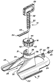
FIG. 2 is an exploded perspective view of the
retractor of FIG 1;
FIG. 3 is a cross-sectional view taken along
lines 3-3 of FIG. 1 showing the handle and retractor
blade in the initial position;
z I sls~z
6
FIG. 4 is a perspective view showing the
surgical retractor positioned in a first incision in the
patient's left leg and the retractor handle in the
initial position;
FIG. 5 is a perspective view similar to Fig. 4
showing the handle and retractor blade in the deployed
position to separate the vein from the skin and
subcutaneous tissue from the vein;
FIG. 5A is a cross-sectional view taken along
lines 5A-5A of Fig. 5 showing the handle and retractor
blade in the deployed position;
FIG. 6 is a perspective view similar to FIG. 5
showing rotation of the locking knob into the locking
position to retain the handle and retractor blade in the
selected position;
FIG. 7 is a perspective view illustrating the
surgical retractor positioned in a second incision,
oriented in the opposite direction of that of FIG. 4 and
showing the handle and retractor blade in the initial
position;
FIG. 8 is a perspective view similar to FIG. 7
illustrating the handle and retractor blade in the
deployed position to separate the skin and subcutaneous
tissue from the vein;
FIG. 9 is a perspective view of a surgical kit
for harvesting the saphenous vein which includes the
surgical retractor of Fig. 1;
218782
FIG. 10 is a perspective view of an alternate
embodiment of the surgical retractor;
FIG. 11 is an exploded perspective view of the
retractor of Fig. 10;
FIG. 12 is a cross-sectional view of the
surgical retractor taken along lines 12-12 of Fig. 10
showing the handle and retractor blade in the initial
position; and
FIG. 13 is a cross-sectional view taken along
lines 13-13 of Fig. 12 showing the keyway for orienting
the handle with respect to the base of the retractor.
DETAILED DESCRIPTION OF PREFERRED EMBODIMENTS
Referring now to the drawings wherein like
reference numerals identify similar or identical parts
throughout the views, and particularly to FIGS. 1-3, the
surgical retractor of the present application is
identified generally by reference numeral 10. Retractor
10 includes a handle 14 having a hook portion or
retractor blade 53 for engaging the tissue to be
retracted, a base 12 for supporting handle l4, and a
locking knob 16 for retaining the handle 14 and retractor
blade 53 in the selected position.
In short, the retractor blade 53 is placed
inside a skin incision and is manually pulled upwardly by
pulling on handle 14 to lift the tissue layers. This
separates the lifted tissue from the underlying structure
CA 02187852 2005-O1-07
8
to improve access for performing the surgical procedure
as described below.
Base 12 of retractor 10 has a pair of spaced
apart legs 22, 23, extending from walls 32, 34,
respectively, which curve upwardly and outwardly away
from the center of the base 12. The skin engaging bottom
surfaces 20, 21 of legs 22,23, respectively, are adapted
to lie on the patient's skin. Walls 32, 34 are spaced
apart to form gap 25 to allow access to the surgical site
with the necessary instrumentation.
Extension 24 has a support 28 with an aperture
27 formed therein configured to receive a conventional
adapter 26 for mounting an optical fiber 29~to illuminate
the surgical site. Alternatively, a light pipe can be
provided to guide light from a conventional light source
such as Storz Coldlight Fountain*. Thus, the optical
fiber or light pipe provides a means for enabling
illumination of the surgical site as an alternative to
the headgear currently worn by surgeons. It should be
understood that the means for enabling illumination
could alternately be positioned at other locations with
respect to the base and the handle as long as it performs
the desired function.
Walls 32, 34, as shown, extend downwardly and
outwardly from neck portion 31 and are integrally formed
with the respective leg 22, 23. A U-shaped outer wall 36
and a U-shaped inner wall 38 are formed on neck portion
*trade-mark
21~78~2
9
31 and are configured to receive the locking knob 16 as
best seen in Figure 2. The opening in the U-shaped walls
36, 38 allows the handle 14 to be mounted to the base 12.
Handle 14 is slidably mounted with respect to
base 12 and has a shaft 50 dimensioned for slidable
reception in the opening in inner wall 38. Shaft 50 has
an integral retractor blade 53 extending substantially
perpendicular thereto, terminating in atraumatic tip 54.
Although shown as integral, it is also contemplated that
the retractor blade can be a separate element attached to
shaft 50. A plurality of external threads 52 are formed
along the length of shaft 50 to engage the internal
threads on locking knob 16 as will be described below.
Handle grip 56 is illustrated with an opening for the
user's fingers to facilitate grasping. It should be
appreciated that alternate grips can be utilized.
With continued reference to Figs. 1-3, locking
knob 16 has an axial opening 44 to receive shaft 50 of
handle 14 and internal threads which engage the external
threads 52 of handle 14. Cylindrical flange -42 is seated
in the space between the inner and outer walls 38, 36 and
bottom surface 43 rests on the upper surface of inner and
outer walls 38, 36 when the locking knob 16 is in the
locking position.
In use, retractor portion (blade) 53 is
inserted through an incision formed in the patient and
the base 12 is placed on the patient's skin. Handle grip
218~8~2
56 is grasped by the user and the handle 14 is pulled
upwardly away from the patient. This causes the blade 53
to lift the patient's skin and a portion of the
subcutaneous tissue. When the handle 14, i.e. the
5 retractor blade 53, is in the desired position, locking
knob 16 is rotated clockwise to slide it towards base 12
until flange 42 is seated in the space between the outer
and inner walls 36, 38, and lower surface 43 rests on
walls 36, 38, thereby locking handle 14 in position.
10 This frees the surgeon's hands as it effectively retains
the tissue in the lifted position without the surgeon
having to hold the handle 14. When the surgeon desires
to release the retractor blade 53, locking knob 16 is
rotated counterclockwise, thereby releasing the locking
engagement of locking knob 16 and shaft 50 to allow the
handle 14 to be lowered to return the tissue to the
initial portion. The retractor blade 53 can then be
removed from the incision.
It should be noted that if controlled
progressive lifting of the retractor blade 53 is desired,
initially the locking knob 16 can be placed in the
lowermost position, i.e. flange 42 positioned between U-
shaped walls 36,38 and lower surface 43 resting atop
walls 38, 36. Rotation of knob 16 clockwise will then
progressively move retractor blade 53 upwardly to lift
the skin.
2187~~2
11
By way of example, the retractor of the present
invention will be described in conjunction with saphenous
vein harvesting as illustrated in Figs. 4-8, although
other uses of the retractor are possible. The retractor
10 advantageously enables the saphenous vein V to be
harvested by requiring only several (e. g. four), small
incisions in the leg, each about 40 mm, as contrasted
with a longitudinal incision running the length of the
leg. As shown, four incisions A1, A2, A3 and A4 are made
in the leg, two above the knee and two below the knee.
The retractor 10 is inserted into each incision to
separate the surrounding tissue from the vein to improve
access to the vein and increase the working space. More
specifically, it lifts the tissue away from the vein to
enable the vein to be dissected and ligated along the
extent of its length which is accessible by the surgical
instruments inserted through the incision. The retractor
10 is inserted in each incision in two directions (e. g.
Fig. 4 and Fig. 7) so the vein can be accessed in both
directions through each incision.
More particularly, as shown in Figure 4, the
retractor blade 53 of retractor 10 is placed through
incision A1 in the leg with the engaging surfaces of legs
22, 23 of the base 12 resting on the patient's skin.
Optical fiber 29 illuminates the surgical site. Handle
grip 56 is pulled upwardly in the direction of the arrow
of Figure 5 to lift retractor blade 53, thereby lifting
CA 02187852 2005-O1-07
12
the skin and a portion of the subcutaneous tissue away
from the saphenous vein V (see also Fig. 5A) . When the
skin and subcutaneous tissue have been lifted to a
desired degree to provide a sufficient gap for
visualization and access to the branches of the vein,
locking knob 16 is rotated clockwise as shown in Figure 6
to abut inner and outer walls 38, 36 to secure the handle
shaft 50 in position. This locks the retractor blade 53
in position so the surgeon can release the handle 14 and
free his hands for the procedure, with the blade 53
maintaining the working gap between the tissue and the
vein.
If more controlled progressive lifting of the
tissue is desired as described above, the locking knob 14
can initially be seated on the upper surface of inner and
outer walls 38, 36 and rotated clockwise to progressively
lift the retractor blade 53.
Once the tissue is lifted, a dissecting and
ligating instrument are inserted through the gap 25 in
the base 12 to legate and dissect the branches from the
vein. As illustrated, this dissects and ligates the
branches to the left of the incision as viewed in Fig. 4.
On example of instruments which can be used are the Auto
Suture ENDO SHEARS* instrument and Auto Suture PREMIUM
SURGICLIP* clip applier. A conventional retractor such
as GELPI manufactured by George Tiemann Co., can be
inserted through gap 25 to achieve lateral spreading of
*trade-mark
21878~Z
13
the tissue adjacent the vein. The light guide which is
supported by support 28 illuminates the surgical site as
the tissue is retracted as well as during dissection and
litigation of the vein. After the branches of the vein
are dissected within the reach of the instruments, the
locking knob 16 is rotated counterclockwise to release
the handle shaft 50 and allow the skin and subcutaneous
tissue to return to its non-lifted (initial) position.
The retractor 10 is then, in the same incision,
reoriented 180° from the original position. The
dissecting and ligating instruments can then be inserted
again through gap 25 to separate the portion of the vein
from the branches on the other side of the incision ,
i.e. to the right of the incision. As is apparent, this
enables the portion of the vein to the right and the left
of the incision to be dissected, limited by the reach of
the instruments.
When the vein is severed in both directions
through the first incision, the retractor 10 is then
placed in the second incision A2. The skin and a portion
of the subcutaneous tissue is lifted away from the vein
as described above and the instruments are inserted to
ligate and dissect the branches from the vein. Retractor
10 is then reoriented 180° in the incision A2 to ligate
the portion of the vein extending in the other direction.
Figure 8 illustrates the retractor 10 positioned in the
second incision oriented in the opposite direction from
2~878~~
14
that shown in Figures 4-6. The retractor 10 is placed in
each of the four incisions, oriented in both directions
to access the vein in two directions. This enables
access to the entire length of the vein through only four
small incisions. Note that the extent the vein can be
accessed in each direction through each incision is
limited by the length of the ligating and dissecting
instruments.
After all the branches are severed, the
saphenous vein V is severed at both ends and removed from
the leg through the incisions for use as a bypass graft.
One way to remove the vein is to pull a portion of the
vein up through incision A1, followed by pulling the vein
portion around A1 through the incision A2, followed by A3
and finally through A4.
It should be appreciated that not only can more
than four incisions be made, but fewer incisions can be
utilized if a smaller section of the vein is desired or
if longer instruments can be provided. Also, the order
of insertion and orientation of the retractor 10 in each
incision is not limited to the order discussed above.
The retractor 10 can be provided in a sterile
package which includes the instrumentation for removing
the vein from the leg. The kit, as shown by way of
example in Fig. 9, includes a clip applier for ligating
the branches of the vein, a dissector for severing the
branches, and a grasper for holding the vein during
CA 02187852 2005-O1-07
dissection and ligation. An Auto Suture* ENDO GRASP*,
ENDO SHEARS*, and PREMIUM SURGICLIP* instrument are shown,
designated by reference numerals 75, 85 and 95,
respectively. Clearly, other combinations of instruments
can be included in the kit. As illustrated, recesses
conforming to the shape of the instruments are formed in
tray 70 with accompanying shaped recesses formed in cover
72.
Note that the retractor 10 can be packaged
10 fully assembled or packaged with the three elements, i.e.
the handle, base, and locking knob, separated for quick
assembly by the user.
An alternate embodiment of the retractor is
illustrated in Figs. 10-13. Retractor 100 includes a
base 112, a handle 114 and a locking knob 116. The base
112 functions in a similar manner as base 14, i.e., it
rests on the patient's skin and mounts handle portion
114. However, as shown, it is different in configuration
as, for example, extension 24 has been eliminated and
walls 132 and 134 are angled at edges 135, 136
respectively.
The handle 114 has a hook portion or retractor
blade 153 which progressively decreases in width towards
the distal end to reduce the stress on the blade. A
plurality of external threads 152 formed on shaft 150
engage the internal threads formed on locking knob 116.
A pair of longitudinal grooves 151 (only one of which is
*trade-mark
CA 02187852 2005-O1-07
16
shown) are formed along the length of the handle shaft
150 to create a projecting surface 154 which sits within
the keyway (recess) 139 in the U-shaped inner wall 138 of
neck portion 131 of base 112. This alignment of the
projecting surface 154 and recess 139 ensure that the
retractor blade 153 is oriented in the correct position
during use and prevents rotation of handle portion 114.
On the portion of the handle shaft 150 opposite
the projecting surface 154, (180° apart), is an elongated
recess 158 configured to receive a light guide 170. As
shown, the light guide 170 is in the form of a plastic
tube which snaps into the elongated recess 158 and
extends around the radiused portion 155 of retractor
blade 153, terminating at distal tip 172 underneath
retractor blade 153. The proximal end 174 of light guide
170 protrudes through opening 118 in handle grip 156 for
connection to a conventional light source, such as Storz
Coldlight Fountain*. Thus, the light guide 170 provides
means for enabling illumination for the surgical site.
It should be appreciated that the means for enabling
illumination can be positioned at other parts of the
. handle portion 114 or the base 112. For example, the
tube 170 can be attached to the outside of the shaft 150.
Also, although the means is disclosed as a light guide
which cooperates with an independent light .source, it is
also contemplated that an illumination means which
*trade-mark
2187852
17
contains a light source can be included as part of the
retractor.
The rotating knob 116 is similar to the
rotating knob 16 of the first embodiment of Figs. 1-3
except that instead of the flange 42, portion 139 of
inner wall 138 extends upwardly to mount the locking knob
116. Locking knob 116 functions in an identical manner
as locking knob 16 to retain the handle 114 and retractor
blade 153 in the selected position.
The surgical retractor 100 is used in the
identical fashion as retractor 10 described in Figs. 4-8.
The retractor 100 can also be packaged as a kit in the
same manner as described above with respect to the first
embodiment.
The retractor 10 or 100 can optionally be
offered with retractor blades of different
configurations. For example, the retractor can be
packaged as a kit including two or more handles having
retractor blades of different sizes.
It will be understood that various
modifications may be made to the embodiments disclosed
herein. For example, different shaped handles can be
provided. Also the instrument can be entirely disposable
or the entire instrument or parts thereof can be
sterilized and reusable. Therefore, the above
description should not be construed as limiting but as
merely exemplifications of preferred embodiments. Those
218 7852
18
skilled in the art will envision other modifications
within the scope and spirit of the claims appended
thereto.