Note: Descriptions are shown in the official language in which they were submitted.
W095/30385 218935~ l~l" '-- I
6TEN-T AND NETEIOD OF NARING T~I3 SANE
FIELD OF T}IE lNV~LlUN
The present invention relates to a stent to be
5 deployed inside a cavity of an animal for --;ntA;n;n~
patency of the cavity. This invention also relates to a
method of making and positioniny such a stent in the
body cavity of an animal.
BACRGROllND OF TEIE lNvh~lL~
In many diseaseg, a body cavity, such as a
passageway or channel (e.g. blood vessel, esophagus,
bronchus, etc. ~ through which a body fluid or other
substance (e.g., air in bronchus) flows, may collapse or
15 be narrowed to be substantially restricted. A
passageway can be 50 restricted that fluid flow in the
passageway is ~l;m;n; ~hf~fl or blocked. For example, in
coronary artery disease, the coronary artery of a mammal
is narrowed or restricted such that blood f low through
20 the artery is rl;r;n;qh~d Even after balloon
angioplasty procedures, such as percutaneous
tr~n~ min~1 angioplasty, in which a blood vessel is
dilated by means of a balloon catheter to flatten plaque
against the artery wall, in many cases, restenosis
25 occurs soon afterwards and the vessel becomes restricted
again. Following perCutAn~ollc balloon angioplasty, the
arterial wall ~ develops tears As a result,
f laps of the inner layer of the arterial wall may hang
loosely in the blood vessel, causing obstruction to
30 blood flow and requiring emergency bypass surgery.
There is a need for a means to -~-;ntA;n patency of :: ~ -
collapsing body cavities or blood vessels and to prevent
the renarrowing of the vessel after angioplasty.
Stents can be used to provide mechanical
35 support to --;nt~;n the patency of blood vessels.
Similarly, the patency of body cavities and passageways
such as urethra, bile duct, esophagus, ureters,aorta,
etc., can also be r-;ntA;n~d by stents. Stents of
various shapes and designs have been l]t; 1; 70d for such
Wo 95l30385 2 1 8 .9 3 ~ ".,~ s 1 ~
purposes. For example, U.S . Patent No. 4, 886, 062
(Wiktor) discloses an intravascula~ radially ~ n~lAhle
stent and method of i~?lAn -nt the3~eof-: The stent
disclosed by Wiktor comprises a~wire wound into a
5 r~nt;nlloug helix along the length of the stent. The
stent is made of a low-memory metal and is radially
expanded by ;nflAt;n~ a ~thrt,~r balloon, which exerts a
force on the stent. U.S. Patent No. 4,969,458 (Wiktor)
also discloses a stent made of low-memory metal,
10 ~YpAntl~hle radially to a larger diameter by the action
of inflation of a balloon. U.$. Patent No. 5,133,731
(Wiktor) discloses yet another stent made of low-memory
material. The stent has a cylindrical body coiled from
a generally r~nt;n~ us wire with a deformable zig-zag
15 structure . Means f or preventing the stent ~ 8 body f rom
stretching along its longitudinal axis are also present
in the stent.
Becau5e stent5 made with low-memory materials
rer~,uire -h~n;~i~l force to expand the stent, such as a
20 force exerted by the inflation of a balloon, their use
can result in trauma to the body caused by the imprecise
control of the expansion of the stent or the balloon.
Moreover, stents made with low memory material may have
a tendency to be compressed into a smaller ~; i t~ by
25 the radial,-~inwardly directed force exerted by the body
tissue on the stent. Self-~n~i;ng stents have been
developed to obviate the use of ~t~rn~ll 1 y applied
mechanical orce for their expansion. For example, IJ.S.
Patent No. 4,830,003 (Wolff) discloses a cylindrical
30 stent for preventing arterial closure and restenosis.
The stent is made of biocompatible metal wires welded
together ~n pairs at alternate ends with each pa~r of ~ '
w~res bent into a V-section. The stent is compressed
and loaded ~nto an outer catheter, posit~oned in a
35 selected location and released for self-expans~on by an
inner catheter. ~.S. Patent No. 5,104,404 (Wolff)
discloses an art~culated stent made up of a number of
.. . .. , .. , , ,, ~ _ _ _
WO 9!il3038!; ~ 1 8 ~ 3~ ~ P~ v3$11
~ . :
individual stent segments~ A number of wires are welded
together to form a segment, and adjacent stent segments
are connected together to provide a hinge action. In a
similar fashion, U.S. Patent No. 5,035,706 (Gianturco)
5 discloses a self-~An-l;n~ stent formed of stainless
steel wire arranged in a closed zig-zag configuration
including an endless series of straight sections joined
at their ends by bends. The bends of at least one end
of the stent are formed into eyes f or connection with
10 eye8 at one end of a similarly constructed stent. The
stents are compre8sible into reduced diameter size for
insertion into and removal from a body passageway.
Because self-P~An~l;ng stents, such as those
made from 8tainless steel, once ~Antl.~d, cannot be ~-
15 deformed unless an ~lr~rn;ll force is applied thereto,
such stents generally cannot be removed from the body
cavity once they are deployed therein . Theref ore,
stents that can be brought back to a smaller shape and
size after expansion within a body cavity have been
2 0 developed to enable removal af ter deployment . For
example, U.S. Patent No. 5,037,427 (Harada et al.)
discloses a method of implanting a stent and removing
same from a tubular organ. The stent is formed of a
two-way shape-memory alloy and expands or shrinks in the
25 radial direction, in accordance with changes in
temperature. Also, U.S. Patent No. 5,147,370 (M_Nc.",a
et al. ) discloses a coil stent constructed from a
nitinol alloy. ~owever, because such stents expand by
the heat of the body, there can be a ri5k that such a
3 0 stent would expand bef ore it is properly deployed or
positioned in the desired location.
U.S. Patent No. 5,026,377 (Burton) discloses a
stent pl A~--' instrument and method for deployment or
retraction of a self -f~YrAnt1; n~ stent in a body canal .
35 The instrument comprises an elongated tubular outer
sleeve having disposed therein an elongated core which
is movable relative to the sleeve and has a grip member
Wo gs/303~5
210935g
for releasably holding a self~ n~n~ stent within the
outer sleeYe U.S. Patent No. 5,078,720 (Burton)
discloses yet another stent pl~c: '_ instrument and
method for the ~ t of a aelf-expanding stent in a
5 body canal. The inatrument comprisea an elongated inner
tube having an outer tube dispoaed along ita axia for
carrying and retaining a aelf -~ n~l; ng stent and an
aLL~ y. t for r~ ; n~ the 8tent, in combination with
at least one of: (a) a location member for positioning
10 and fixing the instrument so that the stent is released
at a desired ~ ocation in the body canal, and (b) a
memher for r~ c;n~ the stent in a retrograde manner.
The stenta diacloaed by Burton in the two patents are
wire-mesh-type stents.
sur~aRY OF T~IE ~\/~l
The present invention provides a stent having
an elongated (e.g., generally cylindrical) body which
includes a ~1llr~l ;ty of generally closed windinga (or
20 loops) and strips inter~ nn~-t;ng the windings such that
the stent is prevented from stretching longitl~;nzllly
(or along its axis). The cylindrical body is
conatructed from a aingle piece of material 8uch as a
wire. The stent is self-~n~hl~ from a first,
25 radially-constrained, lln~ n~P~l geometry to a aecond,
radially-unconatrained, F-~n~ geometry. The stripa
can be interconnected to form an aligned, longitudinally
oriented apine which helpa to prevent longitudinal
stretching of the stent, thus --~ntA;n;n~ the geometry
30 thereof. The windings can further have curves (or
waves) which, for example, can have a generally
sinusoidal appearance. The stent of present invention,
being capable of self-expanaion, is effective for
supporting and ~-intFl;n;n~ patency of a body cavity,
35 auch aa a paasageway (e.g., artery, aorta, bile duct,
urethra) through which a fluid flows. Such a stent can
_ _
WO 95/30385 21 8 9 3 ~ ~ r 5/lJ~. . Al
fl,
be implanted in a body cavity of an animal, such as a
mammal, 1n~11It8;n~ humans.
In another aspect, the present invention
provides a stent comprising a eelf-P~n~l~hle
5 cylindrical body f ormed f rom a continuous wire . The
cylindrical body i8 a coil having successive windings of
wire wherein each of the windings (or loops) is an
essentially closed, complex loop . The term " complex
loop" refers to a loop that has curves (or waves) or
10 structures such a6 o-shaped eyelet9 on the loop. The
stent is formed such that it i9 prevented from
stretching longitudinally (or axially) by portions of
wire interconnecting adj acent windings . When compresaed
and put under radial pressure by a radial constraint,
15 such as a sheath of a catheter, which prevents the stent
from ~ n~1lng radially outward, the stent has a first
diameter reflecting the dimensions of the constraint.
When the radial constraint is removed, the stent can
self-expand from the first radially-constrained,
20 llnP~n~f~d diameter to a second, radially-unconstrained,
P~rln~n~l~(l diameter. The stent can be wound such that any
two adjacent windings (for example, a first and second
successive windings), are connected and restrained from
stretching longitudinally by a portion (or strip) of
25 wire interposed between the first and second successive
windings and intertwining with a portion of one of said
two adj acent windings . The portion of the wire
interposed between the two successive windings can be
connected to a f irst end or end portion of the f irst
3 0 winding and an end or end portion of the second winding
and intertwined with a second end portion of the f irst
winding to prevent stretching longitudinally. The
intertwining of the wire interposed between successive
windings can be aligned to result in a generally
35 straight longitudinal spine (or cord) in the stent. The
stent can be formed from a single, continuous wire into
such a coil having successive windings.
W095/30385 ~ ' 0~
21893~4~
The present invention also provides a method
of making a stent. ~ The method includes a step of
winding a wire on a cylindrical mandrel to form a self-
n~lin~ coil of successive windings (or loops) such
5 that each of the windings having curves and that the
coil i8 prevented from stretching longitudinally by
portions of ~he wire connecting the successive windings.
Spikes can be used on the mandrel for the wire to be
wound thereupon. The wire can be wound such that the
10 portion of wire connecting any adj acent first and second
successive windings is connected to a f irst end portion
of the f irst winding and an end portion of the second
winding and intertwined with a second end portion of the
f irst winding.
The mandrel can be disassemblable (or capable
of being taken apart) to enable a o~med or wound stent
to be removed therefrom without distortion. Such a
disassemblable mandrel can contain a elongated,
preferably generally cylindrical body with spikes
20 disposed thereon for a wire to be wound to form the
stent. The cylindrical body can contain digagg~ hlf.
longitudinal layers. In another aspect, the spikes can
be movably affixed on the cylindrical body, for example,
by screwing into the cylindrical body.
The stent can be made with a flexible
material, for example, a shape-memory material such as
nitinol. In the preferred embodiment, the stent is made
of "superelastic" ~nitinol, which is an alloy c~nt~;n;ns
equal parts of nickel and titanium. A stent made with
such a superelastic nitinol, upon ~nne~l ;n~ at an
elevated temperature, gains memory of the shape in which
it is ~nnP~ (i . e., a preprogrammed shape) . If
deformed into other shapes, upon release, the stent will
spring back to its p~ yl ~1 shape. The method of
making the stent can include ;3nne~l ;ng the wound coil on
the mandrel at an elevated temperature above room
temperature, preferably at above 5000C, more preferably
_ . _ _ _ _ _ _ . _ _ .. _ . ... . . . _ _ _ _ . _ _
WO 95/30385 2 1 8 9 ~ ` P~
about 500C, for a determined period, for example, about
3 0 minutes .
Also provided by the present invention is a
system for positioning (or deploying) in a body cavity a
5 self ~ r~n~hl ~ stent . This system includes an
instrument for r~ ~r~m~nt (or deployment) of a self -
~lr~ntl;n~ gtent and a self-~r~n~l;nrJ stent releasably
held by the instrument. In this aystem, the stent
;nrlll~lP~ a self-f~ n~hle/ cylindrical body formed by a
10 rt~nt;nll~lus wire. The cylindrical body is a coil of
successive windings each having curves and the stent is
prevented from stretching in its longitudinal axis. The
stent is self-~r~n~hle from a first radially-
constrained, l~n~r~n~f3 diameter to a second, radially-
15 unconstrained, ~l~rAn~ d diameter.
The instrument can include an elongatedtubular outer sheath (or sleeve) having a first end and
a second end, such as a conventional catheter, for
radially constraining the stent proximate the distal end
20 of the instrument, and an elongated core or pusher
device having a f irst end and a second end movably
disposed within the lumen of the sheath. As used
herein, the term.. "proximal" means the end or part
nearest to the operator of the instrument and the term
2~ "distal" means the end or part farthest from the
operator. The stent can be self-~l~r~n~hle such that
it self-expands and r~nt~rtcl or rests on the body tissue
or wall in the body cavity when the sheath is moved
longit~ ; ni~l l y away from the distal end o~ the core,
30 thus releasing the radial constraining by the sheath on
the stent.
The stent of the present invention has many
superior characteristics which render it highly useful
as scaffolding support to m-;nt~;n patency of body
35 cavities and passageways. Because the stent of the
present invention can be made from a single r~mt;nll~us
wire, compared to prior art stents, the manufacturing ~:
W0 95/30385 ~ r ~ . c or 11 ~
~ .
process of the stent of the present invention is greatly
simplified and the amount of waste material resulting
from ~nllf~-turing is greatly reduced, thereby reducing
the cost of.production.
Furthermore, the stent of the present
invention overcomes many of the 8hortcomings of the
prior art stents. For example, the zig-zag stents have
many ends of wire (or wire ends), which are welded to
other wire ends. With a large number of wire ends, as
is present in a multiple-wired stent, special effort may
be needed tQ shield these wire-ends or prevent them from
protruding into tissue of the body. Such effort is
labor-intensive. Likewise, the wire-mesh stents also
have multiple wires and thus impose similar risk
associated with multiple wire ends. In the present
invention, using one continuous wire to make a stent
reduces the number of wire ends, thus greatly lowering
the risk of causing irritation or injury to body tissue.
Such a single-wired stent has only two wire ends, whioh
can easily be shielded or curved radially inward to
avoid irrlt:~t;n~ or injuring body tissue.
With multiple-wired steuts such as the zig-zag
type stent or the ~wire-mesh stent, because of the
interlocking or intermeshing of the wires, the stents
are not very compressible and flexible. The wire-mesh
stent tends to become compres8ed (or narrow) radially at
the bend when it is flexed and lengthen9 longitl-~l;n~l ly
when compressed radially. In contrast, the stent of the
present invention ~ is advantageous in that it can be
flexed without causing significant radial compression.
Moreover, it can be compressed radially without
longitudinal t~;r~ n:~l change. Further, using a
single piece of material (such as a c nnt;nl1nus wire) to
make a stent affords the advantage that a stent of any
desirable length can be made without having to join
sections o~ =wire together.
_ _ _ _ _ .
WO9~/30385 21 8~3S4 ` ` : -
Often, balloon inflation is needed to expand
prior art coil stents or wire-mesh stents fully to the
- desired diameter. The present invention can be 8elf-
ntl1ng such that no external force, such as that
5 provided by an i n~ ; ng balloon, is needed to fully
expand the diameter of the stent . Such self -~n~1 ng
nature of the stent of the present invention obviates
~ h~ftU...C and possibly trauma-causing procedures such
as balloon inflation.
Furthermore, unlike the coil or helical stents
that do not have means for constraining longitudinal
extension and tend to 5tretch under longitudinally
directed f orces ( such as the f orce caused by f lowing
fluid or movement of the vessel contacting the stent),
the stent of the present invention has constraint means
80 that the stent does not extend lo~gitll~1n~l1y from
the ~ ntl~rl f orm .
Becau8e the deployment of thermoelastic shape-
memory stents generally re~uire ice-cold saline to
r-;nt~;n the stent in the soft or shrunken form or
saline of relative hot temperature to expand the stent,
the use of self ~ n~i n~ stents obviates such
cumbersome procedures. Although the stent of the
present invention can be made of thermoelastic shape-
memory material to render it soft and shrunken at a
temperature lower than normal human body temperature
(about 37~C), the stent can also be made with other
flexible material which are effective in rendering a
stent capable of returning (or self -~n~11 nS) to a
~r~ r~L 1 shape upon release from a deformed shape.
An example of such a material is stainless steel,
superelastic nitinol, or superelastic plastics. The
stent can be made to have any desirable length with any
number of loops or windings f rom a single wire without
3 5 the use of external adhesion means such as welds or
adhesives .
218 9 3 ~
Prior art cylindrical, spikeless mandrels,
such as those used ior making coil-shaped stents that
are expanded with balloons, are not readily adapted for
making a self-~xpAn~l;ng coil with curves from a single
wire because there is no structure on such mandrels for
securing the stent. The mandrel of the present
invention ~V~ A this problem by having spikeq upon
which the wire can~ be wound. Further, the mandrel of
the present invention can be used to make stents that
have completely F~n~l OAf~ loops and still enables the
removal of the stent, once f ormed, f rom the mandrel
without distortion. The mandrel of the present
invention can be disassemblable such that it can be
taken apart without distorting a stent that has been
formed thereon. This overcomes the problem that a stent
tautly wound on a mandrel with spikes cannot be easily
released from the mandrel. The disassemblable mandrel
of the present invention greatly facilitates the forming
of stents with intricate patterns of waves from single
lengths of wire. ~
BRIEF DES~:Kl~ lUN OF THE DRAWING
These and other ieatures, aspects and
advantages oi the ~present invention are illustrated with
reference to the Ar~, ying drawing, wherein like
numerals represent corresponding parts in the several
views:
FIG. 1 is an isometric view of an embodiment
of the stent in its ~An~lPd form;
FIG. 2 is a side view of an isolated loop ~or
winding) of = the stent of FIG . 1;
PIG. 3 is an end view of the stent of FIG. 1;
FIG. 4 is an end view of a mandrel ior making
the stent of FIG. ~ 1;
FIG. 5 is an isometric uiew of a portion of
the mandrel of FIG. 4 with a wire mounted thereon for
making a stent;
.. _ .. _ ... . .. _ _
~9
WO 95l30385 5 ~ r~
FIG. 6 is an end view of the structure
resulting after a mandrel of FIG. 5 has been partially
disassem.bled and parts removed therefrom.
FIG . 7 is a plan view viewing f rom the end of
another ~ n~;rAnt of a mandrel of the present invention
wherein spikes have been pushed below the cylindrical
surf ace of the mandrel .
FIG. 8 is a side elevation in section of an
instrument f or deploying a stent of the present
invention with the stent mounted therein in a blood
vessel;
FIG. 9 is a side elevation of the instrument
of FIG. 8 showing a partly deployed stent in a blood
vessel;
FIG. lO is a side elevation of the instrument
of FIG. 9 showing a fully deployed stent in a blood
vessel;
FIG. 11 is a side elevation showing a stent of
the present invention deployed in a bloood vessel;
FIG. 12 is an isometric view of another
; of the stent according to the invention;
FIG . 13 is an end view of the mandrel f or
forming a stent shown in FIG. 12;
FIG. 14 is an isometric view in section of a
mandrel with a stent of FIG 13 wound thereon;
- FIG. 15 i8 an isometric view of another
embodiment of the stent according to the invention;
FIG. 16 is an end view of the stent of FIG.
15; and
3 o FIG . 17 is an isometric view of yet another
Amho~1;r^nt of the stent arcor~l;n~Aj to the invention.
DT~T~TT.T~ 3s~:KI~Lluw OF TRE 1NV~WL1UN
For purposes of illustration, the preferred
3~ Amhorl; - t of this invention is shown and described in
reference to applications in angioplasty. ~lowever,
applications other than in angioplasty, such as in body
_ _ _ _ . . . .. . _ ... _ _ .
WO 9~/3038~ 218 9 3 ~
li ~
cavities and passageways, are practicable and no
limitation in scope of the invention is intended by the
embodiment s .
FIGS. 1-3 show the construction of an
5 embodiment of the stent of the present invention. The
stent 1 is a coil 4 having a generally cylindrical shape
with an open lumen 6. That is, as shown in FIG. 3, the
stent 1 has a circular cross section with an open
central portion or ~ lumen 6 . A continuous wire 8 c~n be
10 used to form the coil 4 such that the coil has
successive windings (or loops) 10 and is prevented from
stretching in its longitudinal axis. Any two adjacent
successive windings 10 (or first lOA and second lOB
windings) are connected and restrained from stretching
15 longitudinally by an interconnecting portion or strip 12
of the wire. Such an interconnecting portion of wire 12
is integrally connected to a first end portion 14 of the
first winding lOA and an end portion 16 of the second
winding lOB and intertwined with a ~econd end portion 18
20 of the first winding lOA. Thus each winding (e.g. lOA)
forms a loop closed by intertwining, wherein one end
portion (e.g. 14) of the winding is integrally connected
to the ;n~P~r~nn~ ;n~ portion (e.g. 12) of the wire and
the other end portion (e.g. 18) of the winding (e.g.
25 lOA) is intertwiningly connected to the same
interconnecting portion (e . g . 12 ) . Such intertwining of
the interconnecting portions of the wire along the
length of the stent results in an aligned, generally
straight, longitudinal spine (or cord) 20 in the stent
3 0 1. The intertwining of the interconnecting portion of
the wire with an end portion of a winding secures the
windings in relation to one another to r-;nt~;n the
geometry of the stent and prevent stretching of the
stent longitudinally or axially. The number of turns in
35 the intertwining can vary rlPrPn~l; ng on the distance
between successive windings. In a stent 1 with only one
spine 20, because the portions of the windings 10 on the
~ W095130385 218935~ 13 P~ 'O'tll
part of the cylindrical coil opposite the spine are not
rigidly held together but have freedom of ~n~,v~ t, the
- stent can be flexed without significant narrowing of the
lumen 6 at the bend as a result of the flexion.
5 Referring to FIG. 2, a winding (or loop) 10
has curves (or waves) 22, which have peaks 24 each with
a valley 26 adjacent and associated therewith. As
referred to herein, peaks generally are oriented in the
same direction. Preferably, the curves 22 in a winding
10 are generally sinusoidal in appearance and each has
one peak 24 and one valley 26. In a sinusoidal wave, a
wave of one wavelength exists between two adj acent
points having the same phase. The peaks and valleys can
have smooth turns resempling a ture sine wave rather
than sharp turns as in a zig-zag form. This is
particularly true in the case where a stif f material
such as a nitinol wire is used to form the stint. In
the formation process of the stent, the st;ffnPqq of the
wire facilitates the smooth curving of a winding as the
wire is bent. Depending on the size of the body cavity
to be supported }:y the stent, the size of the curves and
number of curves 22 or peaks 24 in a 9tent L can vary.
For example, a stent 1 having an PYri~nf~ outside
diameter of about 3 mm can have two peaks 24 or curves
22 whereas a stent designed to have an .o~n~ d outside
diameter of 1 cm can have four or more (for example,
eight) peaks. The distance between adjacent loopis or
windings 1 can also vary ~l~r~n~l; n~ on the supporting
strength desired and the size of the body cavity to be
supported by the stent. For example, a stent having an
~n~l~d ~ t~r of about 0.7 cm can have windings that
are about 0 . 5 cm apart f rom peak to peak along the
longitudinal axis.
The wire 8 f or making the stent can be a
flexible material of adequate strength for making self-
n~l;ng stents, such as stainless steel, plastic
polymeric material, shape-memory material (e.g.,
W0 95/3038S 21~ u~ r ~
14 : -
nitinol), and the like. The use of nitinol and shape-
memory material for making stents has been disclosed in
U.S. Patent No. 5,147,370 ~McNamara) and in "Shape-
Memory A110YB, " Scientific American, Vol. 281, pages 74-
82, November=~979, the disclosure of which relating to
the methods of making stents from such shape-memory
material is incorporated by ref erence herein .
Preferably, the stent is made of a
superelastic shape-memory material, such as one made
with superelastic nitinol. The stent can also be made
with thermoelastic shape-memory material, which can also
be made of nltinol. Nitinol alloys as an implant
material have been est:~hl; ~h~d to be biocompatible. A
number of permanent implants made from nitinol have been
used in humans. Nitinol is also resistant to corrosion,
oxidation, and abrasion. Nitinol wires and structures
of other shapes are commercially available, for example,
f rom suppliers such Shape Memory Applications Co . and
Flexmedics Co.
A stent made from a flexible or superelastic
material can be compressed into a smaller size (i.e.
smaller rl; .~r and/or shorter length) to fit and be
confined inside a catheter-like instrument for delivery
of the stent to a desired location in the body cavity.
When the stent is released from the in~L~, t, the
flexibility of the flexible material causes the stent to
spring back to its shape and size before compression. A
stent made of a thermoelastic shape-memory material can
also be made by forming the material into a desired size
and shape and ~nn~l ;n~ at a temperature higher than the
transition temperature to program the stent to have that
desired size and shape. A~ter cooling the stent to a
temperature below the transition temperature, the stent
becomes soft and can be reduced to a smaller size by
3~ crushing or compressing by force so that it can be
deli~ered to the selected location in the body cavity
while r~-;nt~;n;ng the temperature of the stent at below
WO 95l30385 218 9 3 ~ ~ r~J~
the transition temperature. As long as such below-
transition-temperature is m~;nt~;n~l, the stent remains
at its reduced size without subj ecting to an externally
applied compression. When the stent is warmed to a
5 temperature above the transition temperature, the stent
returns to the PL~:~1UYL ~ J size and shape (i.e., the
size and shape before the stent was crushed).
The stent can also be coated with a substance
for reducing the risk of thrombosis and preventing
lO undesirable depositions thereon. For example,
appropriate r-~t1n~fi, such as s; l; rr,nf~ rubbers,
hydrophilic polymer8, fibrin, coatings C~lrt~;n;nrJ
heparin, collagen, and the like may be used on the
stent. The curves of the stent, upon expansion, rest
15 and press on the wall of the body cavity, thereby
exerting a pressure on the wall to prevent the collapse
of the wall of the body cavity.
A stent of the present invention can be made
by winding a single rr~nt;nllr,us wire, such as a nitinol
20 wire, on a preformed cylindrical jig or mandrel to
obtain the coil with the right conf iguration of curves .
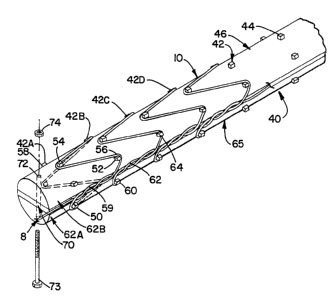
FIG. 4 shows the end view of an embodiment of a mandrel
used for forming the coil of a stent of FIG. 1. The
mandrel 40, as shown in FIG. 4, has a diameter of about
25 0.4 cm and rows (or rings) 42 of spikes 44 disposed
longitudinally 0 . 5 cm apart on the outer surface of the
mandrel. E~ach ring 42 of spikes rrnt:~;nF: six spikes 44
arranged evenly as a ring- like conf iguration on the
outer surface 46 of the mandrel. The spikes 44 are
3 0 about 0 .1 cm high and about 0 . 7 mm x 0 . 7 mm in cross
section .
FIG. 5 shows how a wire 8 made of nitinol is
wound on the mandrel around (or upon) the spikes to form
a stent with the conf iguration of FIG . 1. The wire 8 is
35 wound on the spikes 44 of two different rings to form a
generally sinusoidal winding 10 with curves . A f irst
end (not shown) of the wire can be anchored or secured -
.
W0 95~30385 2 ~ 8 9 3 ~ JOII ~
on the mandrel 4 0 so that the wire 8 can be wound upon
the spikes 44 tautly. The wire extends paGt the last
spike 50 o the first ring 42A of spikes to the second
ring 42B of spikes and winds upon the first spike ~2 of
5 the second ring of bpikes, then extends back to wind
upon a spike (the second spike 54) on the first ring 42A
of spikes, thereby forming the first valley 56 and peak
58 of the first winding (the order of the rings and
spikes are arbitrarily assigned for convenience of
10 reference). The wire is then wound upon the spikes in
the second and first rings of spikes alternately in a
wavy fashion to form the wavy loop of the first winding.
The wire wound upon the last spike 50 of the first ring
42A of spikes extends to the third ring 42C of the
15 spikes while intertwining with the portion 59 of the
wire ~lct~n~;n~ from the first end of the wire between
the first ring 42A of spikes and the first spike 52 of
the second ring 42B of spikes. Similarly, the wire
wound upon the last spike 60 of the second ring 42B
20 extends to the fourth ring 42D of the spikes while
intertwining with the portion 62 of the wire f~tPnf~i n~
between the second ring 42B of spikes and the f irst
spike 64 of the third ring 42C of spikes. This portion
61 of wire can be considered the interconnecting portion
25 interr~ nn~rtlng the first winding 42A and the second
winding 42B. The portion of wire 59 extending from the
first ring 4ZA of spikes to the first spike 52 of the
second ring 42B of ~ spikes i5 an end portion of the first
winding lOA (see FIG. 1) and the portion of wire 62
30 connected with the interC~nn~ tin~ portion 61 and
extending to the first spike 64 of the third ring 42C of
spikes is an end portion of the second winding lOB. The
process for forming a winding can be repeated to wind
the wire alternately upon the spikes of the second and
35 third rings of spikes to form the second winding.
Depending on the length of the wire in the
intercoImecting portion, the number of intertwining
WOgs/30385 2f 8g3.s~ P~
twists (or turns) therein can vary, typically from about
0 . 5 to about 4 . 5, preferably 1. 5 intertwining twists
between adj acent windings . An intertwining twist (or ==
turns) herein refers to a 360 turn of the double helix
5 formed by the intertwining of two wires. This process
for forming successive winding9 can be ~r)nt;nll~tl until a
stent of desired length is formed on the mandrel.
Af ter the wire i8 wound on the mandrel, the
other end (not shown) can be secured to the mandrel as
10 does the first end. The wire ends can be secured to the
mandrel by methods such as tying a knot on a structure
such as a spike or being gripped by a clamp. The wire
ends can be secured in such a way that they are turned --
radially inward on the mandrel 80 that when the f;n;Rh~
15 stent is deployed the wire ends will not protrude into
the body tissue. This can be done, for example, by
providing a depression on the surf ace of the mandrel on
which the wire end is clamped.
Subsequently, the mandrel with the wire
20 mounted thereon can be annealed at about 500C for about
3 o minutes and then cooled to room temperature . Methods
of i~nnf~l ;n~ nitinol material is widely known in the art ~_
and can be used for pL~LUyL ; n~ the stent of the
present invention to attain a desired configuration or --
25 shape. The stent can then be removed from the mandrel.
The mandrel can be made with a material that
can m~;nt~;n mechanical integrity for the wire to be
wound thereon and undergo temperature changes for : =
~nnP~l ;n~. An example of material effective for
30 construction of the mandrel is a metal such as aluminum,
titanium, or nickel; or an alloy such as carbon steel,
stainless steel, or MONEI-. As previously stated, the
mandrel of the present invention is preferably
disassemblable to facilitate the removal of a formed,
35 wound stent therefrom. The mandrel can have spikes or
projections disposed on the outer surface thereof upon
which the wire can be wound to form the stent. The
Wo 95/3038s ~ P~
2t89~S~ 18
spikes can be cylindrical, has a square cross section,
or other similar shapes as long as a wire can be firmed
wound upon them. Other than spikes, means on which the
wire can be wound, such as hooks, or 61its cut on the
5 mandrel, can be used to f orm the cu~ves of the stent .
Preferably, the mandrel can be di5assembled 80
that the stent can be released therefrom without being
distorted e~ther radially or longit--~l;n~lly. As used
herein, a stent is "distorted~ if after removal from the
10 mandrel, .~lrtl~rn;~l ~force is needed to shape the stent
back to the form prior to removal. Referring to FIGS. 4-
5, which shows the detail structures of an r-~c~;r^nt of
the mandrel of the present invention, the mandrel 40 has
spikes 44 and a cylindrical body 65 which includes three
15 layers. The two ~Yt~rn'll (or end) layers 68A, 68B of the
cylindrical body each has a lateral (or transverse)
cross section that is shaped generally like a segment of
a circle defined by an arc and a chord. All of the
spikes 44 arç disposed on such l~rt.orn;ll layers 68~, 68B .
20 The third layer 70 of the cylindrical body is an
intermediate layer disposed between and separating the
P~rt~rn~l layerg 68A, 68B. Such intermediate layer 70 is
a strip or A spacer having a lateral (or transverse)
cross section that is generally rectangular. Bach of
25 the three layers fan have holes 72 proximate its ends
through which screws 73 can extend. The three layers of
the mandrel can thus be secured together at the two ends
with screws 73 and bolts 74.
To remove a formed (or wound) stent from the
30 mandrel 8hown in FIGS. 4-5, the bolts 74 and screws 73
are removed from the ends of the mandrel 40 so that the
three layers of the mandrel are no longer secured
together. ~he intermediate layer 70 can then be
withdrawn ~or removed) from the mandrel 40, enabling the
35 two ,o~rt~-rn~l layers 68A, 68B to be brought together. ~ow
referring to FIG. ~ 6 (FIGs. 5-6 are not drawn to scale,
the spikes are shown larger proportionally compared to
.... . . . _ . _ _ _
WO95/30385 2~g354 . r~ .,
19
the overall size to show structure), the two external
layers 68A, 68B, after being brought together, results in
a structure that is still generally cylindrical but has
a smaller transverse, cross-s~t;~n~l area than that of
5 the three-layered mandrel. The thickness of the
intermediate layer 70 i8 selected such that the
transverse cross section of such two-layered structure
has a perimeter that is small enough to enable the
windings of the stent to be lifted off the spikes, thus
10 releasing the stent from the mandrel. At least one
layer (68A or 68B) of the two-layered structure can then
be slid out of the stent at a time. In this way, the
stent can be removed f rom the mandrel without
distortion .
In another embodiment, the mandrel can have
spikes that are movably affixed thereon. For example,
the spikes can be made of screws that can be removably
screwed into the cylindrical body of the mandrel. To
separate a formed stent from the mandrel without
20 distortion, the screws can first be removed by
unscrewing from the cylindrical body.
In yet another embodiment, as shown in FIG. 7,
the spikes can be slidably affixed in holes on the
cylindrical body. The spikes 144 can extend out o~ the
25 cylindrical outer surface 146 of the cylindrical body
(of the mandrel) for the wire to be wound thereupon.
When the stent has been formed, the spikes 144 can be
forced (for example, by pushing with a finger) proximate ~=
or below the outer cylindrical surface 146 of the
30 mandrel 140. This results in a mandrel 140 wherein the
spikes 144 do not extend out of the surface 146 of the
cylindrical body. The stent can then be slid off the
mandrel. The mandrel can have a annular outer shell 17~
through the spikes extend. A means, such as a core (not
35 shown) that fits inside the shell 17~, can be used to
push the spikes 144 out of the outer cylindrical surface
146 of the mandrel 140 for a wire to be wound thereon.
W095/30385 21893~ s~ - I~1/.J~ SI1~ 1 ~
~ r ~ 20
Such a mandrel is part; r~ rl y well adapted to be used
in mass-producing the stent. For example, to further
facilitate removal of a formed stent, a means can be
u3ed to extend the~ spikes as a group in and out of the
5 cylindrical surface 146 of the mandrel such that the
spikes do not have to be pushed individually.
A stent formed with a superelastic material by
the method described hereinabove can be deformed,
compressed or flexed to various shapes and will
10 immediately return to its preprogrammed, annealed shape
on release oii the externally applied force. Since the
stent can be formed to have any desired length, it can
be formed to have the exact length needed for a
particular application. Alternatively, a stent of a
15 desired length can be formed by trimming the ends o~f a
longer stent. A long stent can also be divided into
shorter stents.
The self -P~ri~n~; ng stent of the present
invention can be used to provide scaf f olding support to
20 hollow orga~;s or passageways or rh~nnPl ~ which are
narrowed. ~he stent can also act as a skeleton to
f ashion vascular graf ts f or the treatment of aneurysms
o~ vascular structures. The stent is preferably
delivered to, placed or deployed in a desired or
25 selected location by using a catheter-like instrument.
FIG. 8 and FIG. 9 ~show such an embodiment. The
instrument has an elongated tubular outer sheath (or
sleeve) 78 having a first or proximal end 79A and a
second or distal end 79B. The sheath can be a
30 conventional catheter. The instrument also includes an
elongated core or pusher device 80 having a first or
proximal end and a second or distal end movably or
slidably disposed within the sheath. The core 80 can
have a generally blunt, conical or h~ rh~rical tip,
35 nose, or terminal piece 81 proximate or at its distal
end. The sheath and core can be made with conventional
material efiective for making catheters. The conical
Wo g~/3038s ~ 8 g ~ S ~ P.~
, .
21
tip 81 of the core 80, being on the distal end 79B of -
the instrument, f acilitates the introduction of the
instrument through a narrow body channel into the
desired location of the body cavity. The conical tip 81
5 has a diameter at the base of t~e cone approximately
equal to the outside ~; i t~r of the eheath 78 . The
core 80 also has an annular recees 82 defined by the
conical tip 81 and a plug 83 some distance away from the
conical tip toward the proximal end of the core. The
10 core 80 can be drawn such that the conical tip is
proximate to the distal end of the sheath 78 so as to
conf ine a stent within the sheath . A stent 101 of the
present invention loaded (or seated) in the recess 82 of ~ -
the core 80 is compressed and secured (or restrained)
15 from ,o~i~nll; ng radially by the sheath 78 . Because the
stent of the present invention is highly compressible,
it can be rnnf; n~d in the recess of the core by the
sheath and the core. --
The instrument can have a channel running
20through the center of the core longitudinally. A guide- -
wire 84 can be provided to extend from one end of the
core 80 through the channel iqnd out the other end of the
core 80. The catheter-like sheath 78 with a stent
restrained therein is positioned across the area to be
25stented by passing over the guide-wire 84. Methods of
positioning catheters, such as one with the aid of a
guide-wire, are widely known in the art. Such standard
methods can be utilized to position the instrument with
the loaded stent in a desired location. After the
30instrument has been positioned in a desired location,
the sheath 78 is gently drawn or pulled back while the
core 80 is ~-;nti~irf~fl stationary. As the sheath 78 is
pulled backwards in the proximal direction, the now
unconstrained stent 101 springs open or expands radially
35due to its flexibility or memory for its predetermined
or preprogrammed state, as shown in FIG. lo. The
windings of the stent rest and press on the wall 86 of
WO 95~30385 2 1 8 ~ 3 ~ ~ i .; } , p~
22
the body cavity 87.~ The 6heath 78, the core 80, and
guide-wire 8~ are then withdrawn from the body cavlty
87. Once ~n~l~rl, the open cylindrical lumen of the
stent has a diameter larger than both the conical tip 81
and the sheath 78, therefore the instrument 76 can
easily be withdrawn without snagging or catching the
stent 101.
Due: to the wavy shape oi the windings, the
btent provides s~ffn1~1;ng support to m~;ntA;n patency
of the body cavity without the body ti6sue col l Are; ng or
protruding into the cylindrical lumen of the stent
around the wires, as shown in FIG. 11. The epine (not
shown in FIGS. 8-11 for reason of simplicity) prevent6
the stent 101 from ~ stretchirlg during the deployment of
the stent ae well as over time as the stent rests in the
body cavity af ter deployment . The high longitudinal
f lexibility of the: stent reduces trauma to the body
tissue .
In a further: ~ ~rl; t of the invention, the
Gtent can be made of a thermoela6tic shape-memory
material that can be rendered soft and 6hrunken at a
temperature below normal body temperature, for example,
a material that haa a transition temperature of about
30-60~C. A stent made of such a material can be
deployed in the bo~dy cavity by an instrument and method
as disclosed in U.S. Patent No. 5,037,427 (Harada et
al . ), which description relating to the instrument and
method of deployment of the 6tent i6 incorporated by
ref erence herein .
3 o The number of ~vindings, the longitudinal
di6tance between winding6 in the stent, and the number
of curvea in a winding can vary depending on f actors
such a6 the . dimensions of the body cavity in which the
6tent is to be positioned, the physiological cnntl;t;nn
of the tissue in the body cavity, the material selected
f or making the stent, and the wire thickness : interloop
gap ratio 6elected. FIG. 12 i6 another embodiment of
WO g5130385 21 8 9 3 5 4 I ~
23 ~ ~
the stent of the present invention. ThiE stent 201 has
two curves or waves 222A, 222B oriented in the same
direction in a single winding 210. Such a stent is made
by winding on a mandrel 240 having an end view as shown
5 in FIG. 13 and an isometric view of a portion thereof as
shown in FIG. 14. The mandrel 240 has rings 242 of ~ _
spikes 244 disposed on the outer surface 2~6 thereof.
Each ring 242 of spikes 244 contains four spikes evenly
spaced around the circumference of a circle
10 corresponding to the cross section of the mandrel 240.
As can be seen in FIG. 13 and FIG. 14, the corresponding
spikes 244 in different rings 242 are aligned (or
arranged) in longitudinal straight lines (or columns)
such that viewing f rom the end of the mandrel, only the
15 four spikes proximal to a viewer are visible. However,
the spikes in different rings can also be arranged in a
regular but nonstraight f ashion instead of in straight
columns, for example, as in twisted, parallel lines.
2 0 EXA~PLES
Stents were made according to the procedure
described hereinabove each using a gingle c~-n~; nll~U8
piece of superelastic nitinol wire and a mandrel shown
in FIG . 4 and FIG . 6 such that each wire was f ashioned
into a cylindrical stent about 3 cm long and 0 . 4 cm in
diameter in the fully ~ n~d state. A superelastic
nitinol wire 8 of 0.1 mm diameter was wound on a
generally cylindrical mandrel 40 of about 0 . 4 cm ~=
diameter with rings 42 of spikes 44 disposed about 0 . 5
cm longitudinally apart on the mandrel. Each ring 42 of
spikes c~mt~;n~d 6 spikes 44 evenly spaced apart on the
circumference (or perimeter) of the ring. Each spike 44
was 1 mm high and had a transverse cross section of
0 . 7mm x 0 . 7mm . Af ter a stent 1 was f ormed on the
mandrel and annealed at 500C for 30 minutes, it was
placed, compressed and confined (or secured) in the ==
recess space 82 of a catheter-like instrument 76
WO 9S/30385 A .
21893S~ ~2~
described hereinabove for pl ;l~r-^nt or deployment into a
coronary artery of a dog. Seven dogs, weighing between
22 Kg and 30 Kg, were used in the study. The stents
were deployed in the coronary arteries of the dogs by
5 llt;l;7;n~ a 5 French delivery instrument or catheter
(i.e., the sheath has an outside ~; t~r of 1.67 mm) as
illustrated in Figs. 8-10. Of the seven dogs, one died
during the procedure of placing the stent, six were
successfully stented, each with a stent deployed in the
10 coronary artery. The dogs were sacrificed after two to
eight months to investigate the patency of the coronary
arteries where the stents were deployed. All of the
stented arteries we~e found to be patent with minimal
intimal hyperplasia (or narrowing). The canine studies
15 were performed according to a protocol approved by the
Animal Care Committee (Protocol No. 9011022) of the
University of Minnesota.
Referring to FIG. 15, a 3tent 301 of another
embodiment has individual windings (or loops) 310
20 wherein adjacent loops are interconnected by a strip (or
portion) 362 of wire, which intertwines with a portion
of wire 309 that runs along the length of the stent to
maintain the geometry of the stent and prevents the
longitudinal stretching of the stent. ~ spine is formed
25 by the intertwining of interconnecting portions of wire
with the lengthwise-running portion of wire. One or
more of the windings (or loops) 310 in the stent can
have one or more eyelets 390 ~; Rh;t~n~d in it. The
eyelets are little~loops formed on the perimeter of the
3 0 individual windings which are interconnected by the
spine 320. A8 shown in FIG. 16, each winding 310 is
generally circular ~in outline. An eyelet 390 is formed
as a tight turn of ~the wire, forming a small loop on a
spike when the stent is wound on a mandrel (not shown).
35 Four eyelets 390 can be formed on a winaing 310, one on
the spine 320 and three spaced from the spine.
W0 9sl3o38s ~ 5 ~ F~
Such a stent can be f orm by running a
generally straight wire 309 along the length of the
mandrel (not shown) and then bendi~g the wire back to
run in the opposite direction at the end of the mandrel
5 before forming the first winding 310A. After a winding
(e.g. 310A) is formed, the portion of wire (e.g. 362A)
interconnecting the previously formed winding (e.g.
310A) and the next winding (e.g. 310B) is intertwined
with the generally straight portion 309 (i.e., the
10 portion that runs lengthwise on the mandrel) of the
wire. Then the next winding (e.g. 310B) is formed.
The eyelets can function as torsion springs
and permit the winding to be collapsed by f lexion at the =
eyelets when the loop or winding is compressed radially.
15 For example, when a winding 310 is compressed by
applying force on the eyelet 390A on the spine 320 and
the eyelet 390C opposite the spine, the winding will
collapse (or flex) about the other eyelets 390B, 390D.
In other words, the generally circular winding will be
20 transformed into two semicircular segments that are
hinged to flex in a book-like fashion about the eyelets
390B, 390D adjacent to but spaced from the spine 320 .
Likewise, when the eyelets adjacent to but spaced from
the spine 320 are compressed, the winding collapses
25 about the spine and about the eyelet 390C opposite the
spine . By compressing the winding to f lex about all the
eyelets simultaneously, the winding can be collapsed to
a geometry (or size) to fit inside a deployment
instrument ( such as the one shown in FIG . 8 ) that
30 constrains the radial dimensions of the stent. A stent,
having its windings thus collapsed, can be deployed in a
selected site in the body. Upon deployment, the stent
can spring back to its unconstrained dimension (i.e.
preprogrammed shape) to m-;nt;~1n the patency of the body
35 cavity or passageway. The eyelets, other than
functioning as torsion springs, also help to provide
more contact surface area for scaffold-supporting the
WO 95/3038~ 2 1 g ~ 3 ~ ~ P~
26
wall of the body cavity. Such an embodiment would be
preferred ior~arge~ stent3 with diameters of l0mm to
30mm .
Variou3 modif ication can be made on the above
5 embodiments. For example, the loop on an eyelet can be
wound on a spike clockwisely or counterclockwisely and
the number o~~~eyelets on a winding can vary. The eyelet
on the spine can be eliminated by, at the beginning and
end of a windlng, turning the wire g0 at the spike
10 aligned with the spine instead of forming a loop-shape
eyelet thereon. Such a stent can still be collapsed by
radial compre3sion. The elimination of this eyelet may
also enhance~he longitudinal integrity against
stretching. On the other hand, more eyelets can also be
15 made on a winding to provide more pointG upon which the
winding can f lex . In ~ 1 t; nn, in closing a winding on
the mandrel, a knot~ can be tied after the winding has
been wound on the mandrel, securing the two ends of the
winding together on the spine. The knot can be but is
20 preferably not tied onto a spike so that the wire on
which the knot is tied can be gripped more tightly by
the knot to further prevent the longitudinal stretching
of the stent. Such a stent with knots for closing
windings can even be made without the generally straight
25 wire running ~long the length of the stent for
intertwining with the interconnecting portions.
The above embodiment can further be modified,
as shown in FIG. 17 by using crimpable (or pinchable)
sections 492 of tubing to encircle and hold the
30 interconnecting portions 462 oi the wire onto the
generally straight portion 409 of the wire. After a
winding (e.g. 420A) is formed, the free ends of the wire
(i.e., the free end of the generally straight portion
409 of the wire and the free end of the wire ~tc~n~l;ng
35 from the newly formed winding which will become an
interconnecting portion 462 of the wire) are threaded
through a section of a crimpable tubing 492 made of a
WO 95/30385 , } ~ "
27
deformable material without shape-memory (e.g.,
stainless steel, titanium). These free end portions of
the wire are then threaded through the section 492 of
tubing until the section of tubing is proximate to the
5 newly formed winding (e.g. 420A). Then a tool, such as
a pair of crimping pliers (not shown), is used to crimp
or pinch the section of tubing 492 onto the portions of
wire 80 that they are held f ast by the crimped section
of tubing. Likewise,= before a winding (for example, the
10 fir8t winding formed on a stent) is formed, the portions
of the wire can be held fast in a similar manner. The
stents of the above Pmhn~;r^nts can be made with
superelastic nitinol with the ;InnP;~l ;nj method and
disassemblable mandrel described hereinabove.
In yet another embodiment, the stent can be
made, rather than from a single wire, by fashioning out
of larger pieces of material, such a8 punching the
desired stent conf iguration out of a single integral
piece of material (e.g. a nitinol tube), cutting the
20 desired stent configuration out of metallic structures
using dies, or chemically etching out of a tubular
structure. Methods of punching, die-cutting, chemical
etching, and the like, are known in the art and can be
used for making such stents. Stents can be fashioned
25 with such methods to have a structure substantially
similar to the single-wired stent described hereinabove.
Such a stent can have a cylindrical body which; n~ PA
a plurality of wavy clo5ed windings (or loops) and
strips interconnecting the windings 8uch that the stent
30 is prevented from stretching longitudinally. The strips
can be interconnected to form an aligned longitllrlin;~lly
oriented spine. Such a stent can be made of the
material described hereinabove and can be self-
P~r~n~l~hle from a first radially-constrained, lln~rîAn~lpd
35 diameter to a second, radially-unconstrained, `^~r?3n'l~'1
diameter .
WO 95/30385 ~ ~9~ 1 1
218935~ ~ --
2'8 ~ :
The~ methods for making the stent of the
present invention can also be automated to mass-produce
the stent.
It is to be understood that even though
5 numerous characteristics and advantages of the present
invention have been set forth in the foregoing
description, together with detail~ of the structure and
function of the invention, the disclosure is
illustrative only, and changes may be made in detail,
lC especially in manners of shape, size and arrangement of
parts without departing from the spirit and scope of the
invention .