Note: Descriptions are shown in the official language in which they were submitted.
P3360
1
CATHETER MAPPING SYSTEM AND METHOD
BACKGROUND OF THE INVENTION
This invention relates to systems and methods for mapping catheter
electrode position within a patient's body and, more particularly, a system
and method
which enables automatic real time three-dimensional measurement of catheter
electrode position with an accuracy well less than 1 cm.
As is known, accurate position information is necessary for mapping, or
localizing an accessory atrioventricular pathway. In this case, one can
localize the
ventricular or atrial insertion site of the pathway during antegrade or
retrograde
conduction through the pathway.
The need for accurate positioning information is further illustrated by
the standard method of cardiac mapping and subsequent ablation of the site of
a
ventricular tachycardia in a patient. The catheter is introduced into the
atrium or
ventricle, and the tip is positioned at an endocardial site. A tachycardia is
induced, and
the tip is moved to different positions, where the timing of sensed
intracardiac signals
is compared with ECG signals. Each position and the local activation moment
must be
accurately determined and recorded, so that an accurate map can be made from
which
the tachycardia focus can be determined. Following the mapping, the ablation
tip must
be accurately re-positioned with respect to the focus. This re-positioning
places a
great importance on being able to obtain accurate tip position information
when and as
the tip is moved to a position. Further, it is well known that frequently
multiple
ablations are often necessary in a relatively small area within the heart, in
order to
eliminate arrhythmogenic foci. Accordingly, the catheter is sequentially
positioned at
slightly different positions close to the focus, for producing lesions in the
heart wall.
These lesions are produced at different locations in order to ensure
elimination of the
foci. At present, it is difficult to obtain accurate and reliable information
concerning
the distances between successive ablation sites.
During hemodynamic and electrophysiologic cardiac catheterization
procedures, cardiologists generally employ a monoplane and sometimes biplane
fluoroscopic imaging to estimate the position of the catheter within the
heart.
However, with fluoroscopy, it is not yet possible to obtain automatic and
objective
P3360 ;. ,
_.. _ ....z. , . ,... ... _:.,,~,~~~. ~ ck
2 2189399
three-dimensional information about the catheter position without laborious
three-
dimensional reconstruction from fluoroscopic images. As is readily understood,
automatic measurement of the catheter position would be extremely useful
during
many interventional catheterization procedures.
Systems for obtaining three-dimensional catheter position data are
known, but have serious limitations. For example, a magnetic system employs a
special
element in the catheter tip, the size and configuration of which make it
useful for only
certain catheter types. Given the many different types of catheters in use for
different
applications, a system and technique that would be able to accurately locate
the
position of any type catheter would constitute a significant advance.
The patent art contains a great many devices and systems directed
toward catheter location. These systems embrace a number of different
approaches,
such as securing an inductor coil adjacent to the catheter tip with leads
extending from
the coil along the catheter for connection to external indicating equipment;
positioning
a varying magnetic field responsive component on the catheter or implement to
be
positioned, and using a movable external magnetic field source; use of a probe
which
generates a small magnetic field which is disturbed by a magnetically
permeable metal
in the device to be positioned; and the construction of various types of
cardiac
mapping probes and electrode configurations. However, these approaches have
not
proven commercially successful for one reason or another, and there remains a
substantial need in the art for an improved technique of catheter mapping,
particularly
as applicable to cardiac catheterization and ablation procedures.
SUMMARY OF THE INVENTION
In accordance with the above need in the art, there is provided a
catheter mapping system and method for mapping locations within a patient,
which
provides an improved accuracy of location, the accuracy being on the order of
a few
mm. The invention involves applying three orthogonal (x, y, z) current signals
through
the patient, directed substantially toward the area to be explored, such as
the patient's
heart, each of the signals having a respective characteristic which renders it
distinguishable from the other two orthogonal signals. The catheter, which has
been
introduced into the body area to be explored, has a tip electrode or another
mapping
CA 02189399 2006-05-29
66742-580
3
electrode positioned in the vicinity of the distal tip,
which is connected through to three sensing channels. The
sensing channels sense the signals induced at the electrode
location by the three respective applied signals, which
sensed signals are used to calculate the location of the
electrode. A simple calibration procedure uses two
electrodes at a known interelectrode distance on the
catheter, and three quick measurements for determining the
correlation of the respective sensed x, y and z signals with
the tip position.
In the preferred embodiment, the external signals
are orthogonal, but can be slightly off orthogonal. The
externally applied signals are suitably constant current
pulse currents at frequencies in the range of
about 25-50 kHz, with the constant current pulses having a
current in a range centered around about 0.1 mA. While
these parameters have been found to be useful for avoiding
interference with ECG pickups, other parameters can be used.
The sensed x, y and z signals are separated by digital
filters, or other suitable narrow pass filters, with the
resulting signals passed through a low pass filter which has
a cutoff designed to eliminate variations due to cardiac
contraction and patient respiration.
The ready availability of accurate three
dimensional position data will allow for numerous
improvements in visualization of catheter position. While
the X-ray state of the art only presents two separate images
in two usually perpendicular directions, three dimensional
information allows for a three dimensional presentation of
catheter tip position to cardiologists. This will make
catheterization a lot easier and quicker, and meet a
substantial long-existing need.
CA 02189399 2007-06-01
66742-580
3a
According to a broad aspect the invention provides
a system for use in carrying out catheter mapping of a body
location within a patient, comprising: external signal
means for applying three substantially orthogonal
alternating current signals across the patient via
respective electrodes adapted to be located on the patient,
such that in use said current signals are transmitted
through the body of the patient; each of said current
signals having a respective characteristic which renders it
distinguishable from the other two orthogonal signals; a
catheter adapted to be inserted into said patient's body and
manipulated to a plurality of locations, said catheter
having at least a mapping electrode; location means
connected to said mapping electrode for sensing a respective
voltage resulting from each of said current signals passing
through the body and processing said voltage to obtain
location signals indicative of the location of said mapping
electrode when positioned at respective different body
locations; means for calculating from said location signals
positions for each location where said orthogonal
alternating current signals were sensed; and output means
for outputting data corresponding to said positions.
BRIEF DESCRIPTION OF THE DRAWINGS
Fig. 1 is a block diagram illustrating the major
components of the system of this invention as used for
catheter mapping and related procedures.
Fig. 2(a) shows a normal electrocardiogram above
and a signal representative of respiration below,
illustrating the relative frequencies; Fig. 2(b) illustrates
a sensed location signal, and shows variations due to
cardiac contraction and patient respiration; Fig. 2(c)
illustrates the sensed location signal with the high
CA 02189399 2007-06-01
66742-580
3b
frequency filtered out, but still containing variations due
to contraction and respiration (Vc), and the signal after
the low frequencies have also been filtered out (V).
CA 02189399 2007-06-01
66742-580
4
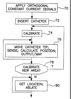
Fig. 3 is a flow diagram setting forth the primary steps of catheter
mapping and ablation in accordance with this invention.
Fig. 4 is a diagram representing a plot of three-dimensional location
data obtained in accordance with this invention.
DESCRIPTION OF THE PREFERRED EMBODIMENT
This invention is based upon using the patient, and in a specific
embodiment example, the patient's heart, as a potentiometer in three
orthogonal
directions. Orthogonal high-frequency current sources are utilized to transmit
a
relatively low current in each of three respective orthogonal directions
through the
patient, directed toward or near the body area of interest. As seen in Fig. 1,
respective
current sources 40, 41 and 42 are used to generate signals applied to
electrodes which
are shown diagrammatically as electrode pairs x, x_; y, y_; and z, z. A
catheter 46 is
introduced into the patient, and for purposes of the ongoina discussion it
will be
assumed that the distal end of the catheter is introduced into the patient's
heart. The
catheter has at least two electrodes, illustrated at 47, 48. Electrode 47 is
at about the
tip end of the catheter, and can be positioned at or adjacent to the heart
wall area of
interest. As used herein, the tip electrode may be actually at the tip, or
displaced
somewhat proximally from the tip but in the distal tip region of the catheter.
The
second electrode 48 is positioned a predetermined distance D from the
electrode 47.
Although just two such electrodes are shown, the catheter may contain three,
four or
more electrodes, so long as it contains at least a position-detecting
electrode,
preferably at or near the tip, and provides a pair of electrodes separated by
a
predetermined distance.D, for calibration purposes as set forth below. Note
that a
Percutaneous Transluminal Coronary Angioplasty (PTCA) catheter can have two
electrodes near its tip,
or on opposite sides of the balloon, with suitable connections for use in the
practice of this invention.
In a first embodiment, the three electrical signals applied to the patient
are high-frequency constant current pulse signals, of the form illustrated in
Fig. 2A,
each at a slightly different frequency. For example, the current source which
drives the
x, x electrodes, designated CS, may operate at 30 kHz, with a current of about
0.1
mA; CSy operates at 31 kHz; and CSZ operates at 32 kHz. In the alternative,
all three
sources can operate at about the same frequency, but are time multiplexed so
that they
P3360
. ...: a _ >.._. _
2 1 -.,~~,, ,,
~ 4
can be distinguished upon subsequent pick-up of sensed signals. The important
distinction is that some characteristic such as frequency, phase or time is
adjusted for
each of the three applied signals, so that three signals can be sensed in the
patient and
then separated out as respective x, y and z signals.
5 It is to be noted that the range of 25-50 kHz is advantageous for
practice of this invention, because it is well above the usual upper cut off
frequency of
bandpass endocardial electrogram amplifiers. Lower frequencies may also be
used, but
in such case specially trimmed filters are required for all electrogram
amplifiers to
eliminate the external signals. If, however, the invention is practiced with
procedures
where no endocardial electrograms are recorded, e.g. PTCA, then the external
source
frequencies may be much lower. Likewise, the orthogonal signals may have any
current
level suitable for avoiding noise pickup in other signals. And while current
pulses are
preferred because they eliminate the influence of varying skin contact
impedance, the
signals may be voltage pulses in some applications. Thus, the optimum
frequency, as
well as the signal level, will depend upon the application.
Continuing with the illustration of the invention, and assuming three
different frequency external signals, the mathematical basis for determining a
position
of the catheter tip is now explained. Still referring to Fig. 1, in the method
of
intracardiac mapping of this invention, the tip or mapping electrode 47 is
connected
through to the three detection filters 50, 54, 57, each of which is adjusted
to be
sensitive to a respective one of the three current source frequencies. At any
given
location, for each orthogonal current a voltage is sensed between electrode 47
and
reference electrode R, suitably a surface electrode on the patient's skin.
Presuming that
the body behaves linearly, the three different measured voltages give unique
x,y and z
values for the location of the tip electrode 47 within the patient's body, as
follows:
VX=ax
V,,=by
VZ = cz
The constants, or sensitivities, a, b and c, are unknowns which must be
determined, and are expressed in mV/mm. In order to automatically calibrate,
i.e.,
determine the three constants, in the preferred embodiment of this invention a
catheter
CA 02189399 2007-06-01
66742-580
6
is employed that has two electrodes with a known interelectrode distance (D,
in mm).
One of the two electrodes may be the tip electrode, or the two electrodes may
be two
separate electrodes, such as two electrodes of a quadripolar catheter. This
calibration
arrangement requires two sets of equally sensitive detection amplifiers and
signal
processing paths for each direction as indicated diagrammatically in Fig. 1.
Since each
of the two electrodes picks up a voltage for each of the x, y and z currents,
the
following equations are applicable:
V,1 = axl, Vy1= byi, and V1= czl
V,-; = ax2, Vy2 = byz, and V1.2 = czZ
To calculate the unknowns a, b and c, it is necessary to use the
measured value AV, = VXZ - V,;I along with the unknown Ox = X2 - xl; OVy = Vy2
- Vyl
together with Dy = y2 - y, aiid OVZ = VZ2 - Vzl together with Az = z2 - zl.
Then, knowing
that OVX = aAx, OVy = bAy, and AVZ = cAz, and AxZ + A y' + Oz2 = DZ, one
obtains:
(Av)2 T'Ab vJr +()2
With AV,, OVy, and OVZ as measured, and D a known, abz, and c'' can be
calculated. To simplify, let 1/a'' = A, 1/b2 = B, and 1/C2 = C, and OVX'' = X,
OVY 2 = Y and AVZ' _
Z. This produces the following simplified equation:
AX+BY+CZ+D2,
where X, Y and Z are measured and D is the known interelectrode distance. It
is now
required only to obtain measurements for three such equations, by placing the
catheter
in three different orientations, in the same heart chamber or other body area.
This does
not require any extra procedure, because the catheter in any event is being
continuously manipulated within the heart during catheterization. Note that it
is not
necessary to obtain these three orientations separately at the beginning of
the
procedure. Indeed, earlier position data can be corrected with later obtained
calibrations, When the three sets of orientation data are obtained, the three
simultaneous equations can be solved for A, B and C, the calibration values of
a, b and
P3360 ~ ;. .
7
c can then be calculated. While theoretically there are always two solutions
for a, b
and c from A, B and C, only the positive solution is the correct one.
In practice, the system may not be ideally homogeneous, meaning that
any given set of obtained measurements is not absolutely correct. This is not
a basic
problem to obtaining accurate measurements, since the calculations can be
continuously performed automatically during catheterization, and the results
can be
averaged. Thus, as long as the catheter is being manipulated, the calibration
measurements and calculations can be repeated any number of times, and a
resulting
average obtained that provides a very real and accurate position
determination. Note
also that it is easy with this invention to calculate the calibration
constants, or
sensitivities, for different areas of the heart chamber. This could be useful
since the
measurements may not be precisely linear. By recalculating the calibration
constants
for different areas of the heart chamber, calculated relative positions can be
reliably
obtained for clinical use in mapping and ablation purposes.
Even without any calibration, a catheterization can be performed by
assuming a "ballpark" sensitivity based, for example, on the weight or thorax
dimensions of the patient. Note also that it is not usually necessary to map
the whole
heart chamber. Mapping and subsequent ablation is usually only performed in a
certain
part or area of the chamber where the arrhythmia originates. Linearity is much
better
when the mapping is confined to a limited area of that heart chamber.
In another embodiment of the invention, calibration can be achieved
without using two electrodes in the heart, by assuming certain cardiac
dimensions
while only measuring VX, V,, and VZ on the mapping electrode. For example,
before
entering the left ventricle, the catheter has to be manipulated through the
aorta
descendens, the aortic arc, and the aortic valve. With the patient on his or
her back,
the depth from the aorta descendens to the aortic valve is approximately 5 cm.
The
distance from the aortic valve to the left ventricular apex is known to be
close to
10 cm. Using such approximate distance figures, together with the measured
voltages
at those sites, it is possible to generate a sensitivity calibration when the
system is
"told" where, in an anatomical sense, the catheter electrodes are positioned.
This
P3360
.:_, _. .. ,, r_..d,~';y.~ ,_, . . . r... .. .;:.;.; . .. .. ;_a tr. *:.. . . -
. , . . - . . - . . 2 1. 8 / ~~ 9--
8
results in catheter positions inside a normalized left ventricle. The same
technique can
be used in other heart chambers for obtaining reliable position data.
Referring again to Fig. 1, catheter 46 is shown having a tip electrode
47, which is manipulated into some position within the heart chamber. A
reference
electrode R, on the surface of the patient's body, is connected to a lead to
provide a
reference potential. For making position measurements, the sensed voltage
between
tip electrode 47 and electrode R is connected through a switch matrix 49 to
each of the
three filters 50, 54 and 57, which are digital filters or other suitable
narrow bandpass
filters designed to pick up the respective signals generated at 40, 41 and 42
respectively. The three current sources are driven by respective clocks
indicated at 44,
which generate the basic timing signals at f, fy and fZ. These clock signals
drive the
current source generators, and also are connected to the respective x, y and z
filters,
for time sampling of the received signals as illustrated in Fig. 2(a) at
points Vl and V2.
The output of each of the filters 50, 54 and 57 is coupled through a
corresponding
amplifier 51, 55, 58, and then through a low pass filter 52, 56, 59. The low
pass filters
have a cutoff of about 0.1 Hz, to filter out any more quickly moving
variations in the
signal from each amplifier. The purpose of this is to avoid problems arising
from heart
contraction and patient respiration. Accordingly, the low pass filters
suitably have a
long time constant in the range of 5-10 seconds, so as to filter out the
cardiac and
respiratory movements. However, it is to be noted that in some applications
the
respiration and cardiac movement information may be useful, such that use of
the low
pass filters is an option.
Figs. 2(a), 2(b) and 2(c) illustrate the effect of contraction and
respiration on the sensed x, y and z signals, and how these effects can be
filtered out.
Fig. 2(a) shows an electrocardiogram and a respiration signal; Fig. 2(b) shows
the
sensed signal which has been sampled at the plus and minus peaks, to develop a
signal
corresponding to the difference between the plus and minus portions of the
sensed 30
kHz pulses. The amplitude of the sensed signal is illustrated as varying due
to
respiration and contraction, giving rise to a signal V., as illustrated in
Fig. 2(c). Such
signal changes are removed by the low pass filter, resulting in an accurate
position
signal of voltage V, illustrated in Fig. 2(c).
CA 02189399 2007-06-01
66742-580
9
The x, y and z outputs from the three channels shown in Fig. 1 are
connected to a computer 65, or equivalent apparatus, for calculation of each
three-
dimensional location. The outputs are connected to a suitable output, or
display 66,
for vertical real time display. As discussed further below, position data can
be stored
for re-display later.
Referring back to Fig. 1, during the calibration steps each of the
electrodes of the electrode pair on catheter 46 that is used for calibration
is connected
to a pair of z processina channels, a pair of y processing channels, and a
pair of x
processing channels. Thus, the two sijnals are inputted to z filters 50, 50 y
filters
54, 54_; and x filters 5 7, 57_. These filters are accurately matched, in
order to provide
the OV,;, AVY and OVZ signals. As with the position measurements, the clock
signals
from block 44 are connected through to each of the six filters, to provide
digital
filtering of the respective pairs of x, y and z sianals. These six signals are
amplified
through amplifiers 51, 51_; 55, 55_; and 58, 58_; and then filtered through
low pass
filters 52, 52_; 56, 56_; and 59, 59_. The three pairs of x, y and z signals
are then
passed to computer 65 for carrying out the calculations set forth above, and
determination of the a, b and c constants. Instead of using 2 sets of channel
amplifiers
and filters, only one set can be used, with each electrode alternatingly
connected to the
same channel input. Using the same channel for processing the signals ensures
identical
amplification, and thus areater accuracy.
Referring now to Fig. 3, some of the salient steps taken in practicing
this invention are set forth. At block 70, the orthogonal constant current
signals are
applied across the patient, as represented in Fig. 1. As indicated previously,
these
respective signals are suitably about 30, 31 and 32 kHz, each with a current
of about
0. 1 mA. While somewhat lower frequencies can be used, it is noted that lower
frequencies, as well as higher currents, have the disadvantaje that they are
more likely
to be picked up in ECG tracings. While higher frequencies are suitable and
clearly
within the scope of the invention they require more exacting electronics. At
72, the
step of inserting the catheter so that the tip is in the region to be mapped
is indicated.
This step may, of course, be performed before applying the orthogonal signals.
At 74,
the step of calibrating the system for determining the a, b and c constants is
indicated.
CA 02189399 2007-06-01
66742-580
As set forth above, this step can be done simultaneously-while taking location
data. At
76, the catheter tip is moved into a position of interest. For a
catheterization
procedure which is to lead to ablation, sensing is performed to gather data
relating to
the heart, such as the location of an arrhythmia focus. Such data gathering
techniques
5 are well known in the art. The location information is determined, by the
calculations
set forth above, and the sensed information and location are stored and/or
mapped.
The flow diaaram indicates that the steps of block 76 may be repeated any
number of
times, at the discretion of the physician. Thus, the catheter tip may be moved
to any
number of locations, all of which can be identified and automatically mapped
in
10 accordance with this procedure. Next, at block 78, it is determined whether
there is a
need or a reason to calibrate again, because of locating in a new area. As
discussed
above, it may be desirable to recalibrate if the catheter tip has been moved
substantially, and, if so, the procedure goes back to 74. Again, note that the
calibration can be undertaken together with the steps of moving the catheter
tip,
sensing, getting location, and mapping. When the mapping has been finished,
the
procedure goes to 80, for ablation. Here, the previously obtained mapping
information
is utilized to position the catheter tip, i.e., the catheter tip is moved and
located, and
when it is at the desired mapped position, ablation is performed for removing
a source
of the arrhythmia. As is known, ablation is suitably performed by applying a
pulse of
radio frequency energy to the heart tissue for a period of time, e.g., several
minutes.
The typical ablation procedure makes a lesion of about 1 cm in diameter. The
ablation
can be repeated at different locations in the vicinity of arrhythmia focus,
using the
previous mapping data, and determining the exact location, or position of the
catheter
tip in accordance with this invention.
Referring now to Fig. 4, there is shown an example of electrical
localization, or position measurements, taken during a cardiac
catheterization. A
10 kHz current at .1 mA pulse height, was delivered in three orthogonal
directions
through the patient chest. Referring to the legend of Fig. 4, X was left to
right; Y was
head to legs; and Z was frontal chest to dorsal. Actual catheter tip positions
were
measured by means of calibrated Roentgen images (centimeters, horizontal axis)
and
plotted versus measured electrical potentials amplified five times (mV,
vertical axis),
P3360
11
for each of the X, Y and Z directions. In this patient, the catheter tip was
positioned at
four different places, namely in the high right atrial appendage; on the
bundle of HIS in
the left ventricle near the mitral annulus; and inside the coronary sinus. One
of the four
positions is represented as the reference, at the intersection of the
horizontal and
vertical axes. Note that linearity for each of the x, y and z directions is
very good.
Heretofore, it has been very difficult to obtain a locational accuracy within
several cm.
With this invention, the accuracy is within mm, depending upon the degree of
filtering
of variations induced by respiration and heart movement. This accuracy leads
to a
significant improvement in ablation procedures, since previously after the
initial
mapping, the physician essentially had to again do fine tuning or remapping
when
returning to ablate. Using the technique of this invention, the physician can
return very
quickly to the primary ablation position, and reposition the ablation
electrode for
producing lesions at precisely defined positions so as to effectively cure and
control the
arrhythmia.
As discussed above, in the preferred embodiment, the reference
electrode is placed somewhere on the skin, which has the advantage that it is
unlikely
to be displaced during the procedure. However, a disadvantage of this
arrangement is
that the cardiac contraction and respiration induced signal amplitude
variations are
relatively high. Another option is to use one electrode of a stable catheter
within the
body area, eg, heart, as the reference electrode. Positioning the reference
electrode in
another chamber of the heart has the advantage that displacement is relatively
unlikely;
and respiration and contraction influences are reduced because for a given
position of
the mapping electrode, both electrodes move simultaneously with respiration
and
contraction. However, even here the reference catheter electrode may
occasionally
displace, which renders previous measurements substantially useless.
. In another embodiment of the invention, and which addresses the need
of a reliable reference electrode which has minimal sensitivity to
contractions and
respiration, the source electrodes are used also as reference electrodes, and
the source
signals are time multi-plexed, as disclosed above. For example, a 90 kHz
signal source
is used, with respective successive pulses being connected across first the x-
x_
electrodes, next the y-y_ electrodes, and then the z-z_ electrodes, so that
each pair of
. _ ,.
P3360
~.-~ . .. , . ..; . . <~ _ ._
=-.,~ , .
12
electrodes transmits a respective 30 kHz signal. In this case, separation of
the sensed
voltages is achieved by timing as contrasted to frequency, in a known manner.
When
one signal is being detected, the two other pairs of electrodes are available,
and can be
used as reference electrodes. For example, when either the x or y measurement
is being
made, both the z and z_ electrodes are connected together as the reference
electrode;
when the z measurement is being made, the y and y_ electrodes are connected as
the
reference electrode. The advantage of this arrangement is that the effective
electrode is
located roughly in the middle of the patient, close to the heart, and is not
likely to
dislocate because it involves skin electrodes.
It is to be noted that, with some of the source electrodes positioned
around the heart, there may be some mapping locations which are rather close
to a pair
of skin electrodes. Due to the curvature of the equipotential lines of the
resulting
electric field, a slight error could be introduced when the mapping position
is not
roughly midway between the electrode pair. However, such an error can be
compensated for by estimating the position of the mapping electrode between
each
such electrode pair, and making an appropriate mathematical adjustment. The
approximate position of the mapping electrode can be checked by comparing the
y
voltage on the z-z_ electrodes with the y voltage on the mapping electrode;
comparing
the z voltage on the y-y_ electrodes with the z voltage on the mapping
electrode; and
comparing the x voltage on the y-y_ and/or the z-z_ electrodes with the x
voltage on
the mapping electrode.
The system and method of this invention are applicable to a number of
important medical techniques. A primary application, as indicated above,
involves
identification of the focus, or exit-site of tachycardia, e.g., ventricular
tachycardia
(VT). As is known, in the catheterization process, surface ECGs are obtained
during
ventricular VT and compared with intracardiac ECGs obtained through the tip
electrode at various places within the ventricle. By known techniques, the
exit-site of
the VT can be identified. By using the three-dimensional localization of this
invention,
the pacing sites and the corresponding correlations between paced and VT ECGs
can
be used to determine the optimal site at which the best correlation between
both can be
expected.
P3360
2189399
13
By way of further illustration of the scope of the invention, the system
and method of this invention are also applicable for three-dimensional imaging
of
coronary stenosis. This can be done by combining echo tip data with three-
dimensional data. A single catheter can be equipped with an echo tip at the
end, as
well as two distally located electrodes for obtaining dimensional information.
By
combining echo tip and three-dimensional data, an accurate three-dimensional
map can
be made for identifying coronary stenosis.
Yet another application is stent placement. By obtaining three-
dimensional information in accordance with this invention, previously explored
catheter positions can be accurately re-located so that a stent can be placed
at exactly
the same site where, for example, a PCTA had been applied, or where an
intraluminal
echo-image has been obtained. It is also to be noted that the technique can
likewise be
used for obtaining two-dimensional, or even one-dimensional position data, in
applications not involving three-dimensional positioning.
While the invention has been illustrated by the use of orthogonal
signals, they need not be absolutely orthogonal, although preferably
substantially
orthogonal. The angles may vary from strictly orthogonal and still be three-
dimensional
for purposes of practicing this invention; as long as the actual angles are
known, a
mathematical correction can be applied to compensate for any difference from
truly
orthogonal.