Note: Descriptions are shown in the official language in which they were submitted.
218946~
DEVICE WITH A THREE-DIMENSIONAL COMPENSATING FILTER FOR A
GANTRY MOUNTING RADIOGRAPHY APPARATUS.
BACKGROUND OF THE INVENTION
The spatial variations in the thickness and the
composition of the patient through which the X-ray passes
allows an image of the internal structure of the patient to be
formed. When making arteriography about the thorax with digital
subtraction (DSA), hyper radiation, due to air inside and/or
out~ide the portion of the patient through which the X-ray
passes, seriously degrades the image by saturing the image
device. Compensating the hyper transparency of an organ such
as the lungs inside the human body is quite delicate.
Some attempts were made for correcting the problem by
using metal compensating plates, curved or wedged, set directly
against the patient or near the image device. Although
sufficient for simple radiography, they are not always adequate
for all incidences required in volume arteriography. For
example, US patent No. 4,472,828 describes an X-ray filter for
chest X-rays which operation principle is similar to the one
of the metal compensating plates. This device may improve the
image of radiography about the thorax, but it is stationary and
as a result, it limits radiography to one view of the organ at
a time and has to be reset for a different view.
Another prior art attempt in an effort to improve
radiography images is the X-ray compensating masks, such as in
US patent No. 4,497,062, which are used to attenuate the X-ray
fluency passing through both mask and the patient.
2189~68
SUMMARY OF THE INVENTION
It is an object of the present invention to provide a
device that is designed to be used with a gantry mounting
radiography apparatus to allow a suitable internal or external
compensation of the hyper transparency area independently of
the positioning of the two opposite arms of the apparatus.
More particularly, the object of the present invention is
to provide a device for compensating hyper transparency area
due to air for use with a gantry mounting radiography
apparatus, the apparatus having a first and second opposite
arms rotatable around a rotation axis, the first arm supporting
an X-ray source and the second arm supporting an image
intensifier in registered position with the X-ray source, the
apparatus being used for radiographing an organ of the patient
or an object, the organ or object defining a reference plane
in which the rotation axis is substantially lying, the device
compr1slng:
a three-dimensional compensating filter defining a filter
plane; and
a supporting assembly to connect the filter to the first
arm of the gantry mounting apparatus, in front of
the X-ray source and in registered position with the
X-ray source and the hyper transparency area while
keeping the filter plane parallel to the reference
plane during rotation of the arms around the axis.
A non restrictive description of preferred embodiments
will now be given with reference to the appended drawings.
2189~ 68
BRIEF DESCRIPTION OF THE DRAWINGS
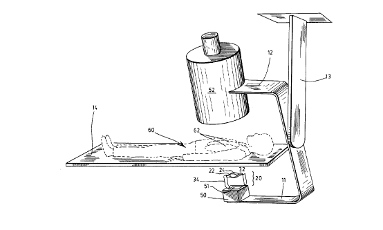
FIG. 1 is a schematic perspective view of the device
according to a first possible embodiment of the present
invention, showing an example of an internal compensation.
FIG. 2 is an enlarged schematic view of the device of FIG.
1, showing the filter in three different positions.
FIG. 3 is a side view of the device according to a second
possible embodiment of the present lnvention, showing an
example of an external compensation.
FIG.4 is an enlarged schematic view of the device of FIG.
1, showing the device in three different positions.
IDENTIFICATION OF THE COMPONENTS
The following is a list of the reference numerals, along
with the names of the corresponding components, that are used
in the appended drawings and in the description.
10 gantry mounting radiography apparatus
11 first arm
12 second arm
13 vertical arm
14 table
20 compensating device
21 filter plane
22 left part of the filter (inside compensation)
24 right part of the filter (inside compensation)
26 left part of the filter (outside compensation)
28 right part of the filter (outside compensation)
30 supporting assembly
2189~68
31 line
32 supporting plate (inside compensation)
34 arms (of the supporting assembly)
36 threaded hooks
50 X-ray source
51 collimator
52 X-ray tube amplifier
60 patient
62 lungs (of the patient)
64 head (of the patient)
66 reference plane
70 first plate
72 second plate
DETAILED DESCRIPTION OF PREFERRED EMBODIMENTS
Gantry mounting radioyraphy apparatus
FIG. 1 is a general representation of the rotating gantry
mounting radiography apparatus (10). The apparatus (10)
comprises two opposite arms (11,12). The opposite arms (11,12)
are connected to each other, forming a fork and are attached
to a vertical arm (13) of the apparatus (10) at substantially
the center of the fork. The connection point of the two
opposite arms (11,12) to the apparatus (10) allows rotation of
an X-ray source (50), a corresponding collimator (51) and an
X-ray tube amplifier (52) around the patient or any object that
has to be analysed. The first arm (11) bears the X-ray source
(50) while the second arm (12) bears the amplifier (52).
The attaching point of the two opposite arms (11,12)
defines a rotation axis that usually lies in a horizontal
2189468
plane. The patient, or object, rests on a horizontal table (14)
and the height of the table (14) is set in order that the
rotation axis of the apparatus (10) be aligned as close as
possible to the centroid of the organ or object in an imaginary
vertical plane. This setting maintains the X-ray source (50),
the collimator (51), the organ or object and the X-ray tube
amplifier (52) constantly aligned with each other independently
of the rotation of the arms (11,12). This is referred to as a
registered position.
In use, the X-ray source (50) generates an X-ray beam
aimed at the X-ray tube amplifier (52). The organ of the
patient that lies in the path of the beam is scanned by the
apparatus (10) at various angular positions and the data
collected by the amplifier (52) is sent to a computer (not
shown) for reconstructing the image or images for analysis. Of
course, the same principle applies to the part of an object
other than an organ of a patient. However, to simplify the
hereinafter description, reference will be made only to an
organ of a patient.
Inside compensation
FIG. 2 shows an example of an inside compensation of an
organ, essentially consisting of the lungs, using a
compensating device (20) according to a possible embodiment of
the present invention. In that case, the three-dimensional
compensating filter comprises two parts, namely, a left par~
(22) and a right part (24). Each part (22,24) is in registered
position with a corresponding lung (62) of the patient (60).
Each part (22,24) of the filter is a three-dimensional
scaled filter of the corresponding human lung (62), preferably
218946~
of an average size. The volume of the filter is reduced in the
inverse ratio of the distance to the focal spot of the X-rays
and the distance of the lungs (62) of the patient (60) to the
focal spot. Commonly, if the filter is 50% closer than the
lungs (62) of the patient (60), its volume is 50% of the size
thereof. A filter of 33~ of the size of the lungs (62) would
be located at 1/3 of the distance. It preferably has a volume
between 1/2 and 1/3 of the volume of such average human lungs,
depending on their relative position with reference to the
patient (60) and the density of the material. In general, it
is desirable that the image density of the compensated lungs
(62) attain the density of the tissues located between them,
which is that of water or muscles. Therefore, the compensation
required from the filter depends on its size. As an example,
if the filter is twice as small as the lungs (62) of the
patient (60), its absorption will be about twice the one of
water. One possible material is polyurethane. The penumbra of
the filter is usually sufficient for a gradual demarcation of
the shadow, but a thin coating of a slightly less absorbing
material (not shown) would diminish such demarcation.
The parts (22,24) of the filter are supported by a
supporting assembly (30) which allows them to be held in
position on the arm (11) and in front of the X-ray source (50).
In accordance with the present invention, the parts (22,24)
define a plane, called the filter plane (21), which remains
substantially parallel to a virtual reference plane (66)
defined by the organ of the patient. In the embodiment shown
in FIGS. 1 and 2, both planes are horizontal.
In use, the parts (22,24) of the filter remain in
registered position with the X-ray source (50), the lungs (62)
of the patient and the amplifier (52), and simultaneously the
2189468
filter plane (21) remain parallel to the reference plane (66),
as shown in FIG. 2, such as to create penumbra over the desired
portion of the lungs (62) to be X-rayed in function of the
angle of the arms (11,12). FIG. 2 shows examples of three
different positions for the compensating device (20) with
reference to the lungs (62) of the patient (60).
Preferably, the filter plane (21) remains parallel to the
reference plane (66) by gravity. To do so, the supporting
assembly (30) may comprise a line (31) transparent to X-rays
so to allow X-rays to be absorbed uniformly by the filter,
preventing, hence, disruption of its regular pathway. An
example of such material is nylon. The line (31) extends
between two opposite arms (34). The arms (34) are attached to
the collimator (51) and are adjustable in height.
The supporting assembly (30) further comprises a plate
(32) on which the parts (22,24) of the filter are placed and
are in equilibrium with gravity by providing the center of
gravity right below the line (31). The plate (32) is held on
the line (31) by means of threaded hooks (36) which allow
sliding of the plate (32) longitudinally for positioning and
to change the distance between the parts (22,24) of the filter
and the X-ray source (50).
Of course, one may choose to provide a supporting assembly
(30) with a motorized actuator (not shown) for keeping the
filter plane (21) parallel to the reference plane (66).
Outside compensation
The outside compensating device (20) for outside
compensation is similar to the inside compensating device (20),
21894 6~
except that the parts (72,73) of the filter are not directly
connected and are aligned differently.
FIGS. 3 and 4 show an example of a compensating device
(20) well adapted for a radiography of the head (64) of the
patient (60) using a gantry mounted radiography apparatus (10).
In that case, the parts (72,73) of the filter, which is still
mounted in front of the X-ray source (50), are each supported
by a corresponding pair of arms (34) between which extends a
line (31) similar to the one for the inside compensation. Each
line (31) passes through the centroid or slightly over the
centroid of the corresponding part (26,28) to maintain proper
alignment under the effect of gravity.
As aforesaid, the filter for outside compensation
comprises two parts (26,28), namely a left part (26) and a
right part (28). The exact shape of these parts (26,28) depends
on the range of angles. For instance, if the radiography, as
is FIG. 4, is only in a range of about 90~, it is possible to
only provide a quarter of the complete shape. For a 180~ range,
it should be half of the complete shape. For a 360~ range, it
should be the complete shape. The complete shape for the head
(64) of the patient (60) is an ovoid cylinder.
In FIG. 4, each part (26,28) of the filter is a three-
dimensional isometric part having a shape substantially similar
to a quarter of an ovoid cylinder. The left filter (26) is in
a position to cover the left hyper transparent area of the head
(64) while the right filter (28) is in position to cover the
right hyper transparent area, hence allowing the X-rays to pass
between them. The parts (26,28) are diametrically opposite and
have their curved surfaces facing each other. In that
particular case, the filter plane (21) is horizontal and
._ . 2189~ 68
substantially coincide with the centroid of both parts (26,28)
taken together.
Preferably, the arms (34) are mounted on a first plate
(70) in sliding relationship with a second plate (72) connected
in front of the X-ray source (50) by means of Velcro~ patches.
This allows the compensating device (20) to be moved
longitudinally for initial adjustment with reference to the
patient (60).
In use, and once properly aligned, the parts (26,28) of
the filter will create a window with gradually peripheral
compensation. The positioning is made by gravity and may of
course be replaced by a motorized actuator (not shown).
Although preferred embodiments of the invention have been
described in detail herein and illustrated in the accompanying
drawings, it is to be understood that the invention is not
limited to these precise embodiments and that various changes
and modifications may be effected therein without departing
from the scope or spirit of the invention.