Note: Descriptions are shown in the official language in which they were submitted.
. .~. .,
218~~41
Docket No. P-3555
FIELD OF THE INVENTION
The present invention relates to methods for detecting nucleic acid target
sequences
and in particular to detecting such target sequences by changes in
fluorescence polarization.
S
BACKGROUND OF THE INVENTION
Fluoresence Polarization (FP) is a measure of the time-average rotational
motion of
fluorescent molecules. It has been known since the 1920's and has been used in
both research
and clinical applications for sensitive determination of molecular volume and
microviscosity.
The FP technique relies upon changes in the rotational properties of molecules
in solution.
That is, molecules in solution tend to "tumble" about their various axes.
Larger molecules
(e.g., those with greater volume or molecular weight) tumble more slowly and
along fewer
axes than smaller molecules. There is therefore less movement between
excitation and
emission, causing the emitted light to exhibit a relatively higher degree of
polarization.
Conversely, fluorescence emissions from smaller fluorescent molecules, which
exhibit more
tumbling between excitation and emission, are more multiplanar (less
polarized). When a
smaller fluorescent molecule takes a larger or more rigid conformation its
tumbling decreases
and the emitted fluorescence becomes relatively more polarized. This change in
the degree of
polarization of emitted fluorescence can be measured and used as an indicator
of increased size
andlor rigidity of the fluorescent molecule.
In fluorescence polarization techniques, the fluorescent molecule is first
excited by
polarized light. The polarization of the emission is measured by measuring the
relative
intensities of emission (i) parallel to the plane of polarized excitation
light and (ii)
perpendicular to the plane of polarized excitation light. A change in the rate
of tumbling due
to a change in size and/or rigidity is accompanied by a change in the
relationship between the
plane of excitation light and the plane of emitted fluorescence, i.e., a
change in fluorescence
polarization. Such changes can occur, for example, when a single stranded
oligonucleotide
probe becomes double stranded or when a nucleic acid binding protein binds to
an
oligonucleotide. Fluorescence anisotropy is closely related to FP. This
technique also
measures changes in the tumbling rates of molecules but is calculated using a
different
equation. It is to be understood that polarization and anisotropy are
interchangeable
techniques for use in the present invention. The term fluorescence
polarization is generally
used herein but should be understood to include fluorescence anisotropy
methods. In steady
state measurements of polarization and anisotropy, these values are calculated
according to the
following equations:
2
.. .,
Docket No. P-3555 ~ ~ 8 9 9 41
P (polarization) - Ipa - Ipe
Ipa + Ipe
r (anisotropy) - Ipa - Ipe
Ipa + 2Ipe
where Ipa is the intensity of fluorescence emission parallel to the plane of
polarized excitation
light and Ipe is the intensity of fluorescence emission perpendicular to the
plane of polarized
excitation light.
As FP is homogenous, this technique is ideal for studying molecular
interactions in
solution without interference by physical manipulation. Fluorescence
polarization is therefore
a convenient method for monitoring conversion of single-stranded fluorescently
labelled DNA
to double-stranded form by hybridization (Murakami, et al. 1991. Nucl. Acids
ReS. 19, 4097-
4102). The ability of FP to differentiate between single and double-stranded
nucleic acid
conformations without physical separation of the two forms has made this
technology an
attractive alternative for monitoring probe hybridization in diagnostic
formats. European
Patent Publication No. 0 382 433 describes fluorescence polarization detection
of amplified
target sequences by hybridization of a fluorescent probe to the amplicons or
by incorporation
of a fluorescent label into the amplification products by target-specific
extension of a
fluorescently-labeled amplification primer. PCT Patent Publication No. WO
92/18650
describes similar methods for detecting amplified RNA or DNA target sequences
by the
increase in fluorescence polarization associated with hybridization of a
fluorescent probe.
Fluorescence polarization may be monitored in any of three different states:
steady
state, transient state, or dynamic state. In transient state FP, the
excitation light source is
flashed on the sample and polarization of the emitted light is monitored by
turning on the
photomultiplier tube after the excitation light source is turned off. This
reduces interference
from light scatter, as fluorescence lasts longer than light scatter, but some
fluorescence
intensity is lost. In steady state FP, excitation light and emission
monitoring are continuous
(i.e., the excitation source and photomultiplier tube are on continuously).
This results in
measurement of an average tumbling time over the monitoring period and
includes the effects
of scattered light. Dynamic FP may be monitored in either the time- or
frequency-domain.
Dynamic fluorescence techniques involve determining the lifetime of the
fluorescent molecule
in nanoseconds. The theory of dynamic fluorescence monitoring is described in
"Principles of
Fluorescence Spectroscopy" (Lakowicz, Plenum Press, 1983). Whereas steady
state FP
J
Docket No. P-3555 ~ 2 ~ 8 9 ~9 41
provides an average or "snapshot" of the fluorescence phenomena, dynamic FP
allows one to
observe the individual contributions of the fluorescent components in the
system being studied.
Use of these three fluorescence techniques is described by Kumke, et al.
(1995. Anal. Chem.
67, 3945-3951), Devlin, et al. (1993. Clin. Chem. 39, 1939-1943), and G.T.
Walker, et al.
S (1996, Clin. Chem. 42, 9-13).
Analysis of nucleic acids, and in particular detection of specific nucleic
acid target
sequences provides an extremely sensitive tool for diagnosis and
identification of biological
materials. Typically, nucleic acid target sequences are detected by specific
hybridization to a
labeled oligonucleotide probe. Several probe hybridization methods for
detecting nucleic acid
target sequences are known in the art (e.g., dot blots, Southern blots,
Northern blots), but
these are somewhat insensitive and are generally only applicable to samples
containing
relatively large amounts of the target sequence to be detected. Nucleic acid
amplification
techniques have greatly improved the sensitivity of target sequence detection
by providing
methods for specifically increasing the amount of target sequence prior to
detection. Nucleic
acid amplification methods can be grouped according to the temperature
requirements of the
procedure. The polymerase chain reaction (PCR; R. K. Saiki, et al. 1985.
Science 230,
1350-1354) , ligase chain reaction (LCR; D. Y. Wu, et al. 1989. Genomics 4,
560-569; K.
Barnnger, et al. 1990. Gene 89, 117-122; F. Barany. 1991. Proc. Natl. Acad
Sci. USA 88,
189-193) and transcription-based amplification (D. Y. Kwoh, et al. 1989. Proc.
Natl. Acad
Sci. USA 86, 1173-1177) require temperature cycling. In contrast, methods such
as Strand
Displacement Amplification (SDA; G. T. Walker, et al. 1992. Proc. Natl. Acad
Sci. USA 89,
392-396 and G. T. Walker, et al. 1992. Nr~c. Acids. Res. 20, 1691-1696, and
U.S. Patent No.
5,455,166), self sustained sequence replication (3 SR; J. C. Guatelli, et al.
1990. Proc. Natl.
Acad Sci. USA 87, 1874-1878), Nucleic Acid Sequence Based Amplification (U.S.
Patent
No. 5,409,818), restriction amplification (U.S. Patent No. 5,102,784) and the
Q~i replicase
system (P. M. Lizardi, et al. 1988. BioTechnology 6, 1197-1202) are isothermal
reactions.
Isothermal amplifications are conducted at essentially constant temperature,
in contrast to the
cycling between high and low temperatures characteristic of amplification
reactions such as the
PCR: The SDA reaction originally reported in the publications cited above
("conventional
SDA") is typically conducted at a temperature between about 35°C and
42°C, and is capable of
108-fold amplification of a target sequence in about 2 hours. Recently, SDA
has been adapted
for higher reaction temperatures (about 45-65°C - "thermophilic SDA" or
"tSDA"). tSDA is
capable of producing 109-101 fold amplification in about 15-30 min. at about
50-60°C. In
addition to increased reaction speed, there is a significant reduction in non-
specific background
amplification in tSDA as compared to conventional SDA.
4
21 X994 ~
Docket No. P-3555
Either unamplified or amplified target sequences may be detected by
hybridization of a
labeled oligonucleotide probe. This often requires separation of free and
hybridized probe
before the signal can be measured. However, monitoring changes in FP allows
differentiation
of free and hybridized probe without physical separation, thereby reducing
operating steps and
procedural complexity. As an alternative to probe hybridization, target
amplification may be
detected by generating double-stranded secondary amplification products from a
single-
stranded signal primer in a target amplification-dependent manner during the
amplification
reaction. Generation of secondary amplification products during target
amplification is
described and illustrated in published European Patent Application Nos. 0 678
582 and 0 678
581. In the process, a single-stranded oligonucleotide signal primer
comprising a detectable
label is converted to double-stranded form in a target amplification-dependent
manner.
Conversion of the signal primer occurs concurrently with the amplification
reaction and may be
detected as a change in FP when the label is fluorescent. The increase in FP
associated with
conversion of the signal primer to double-stranded form as a result of target
amplification is
approximately 20 mP using fluorescein or La Jolla Blue as the fluorescent
label. When
amplification is conducted at lower temperatures (e.g., about 35-45°C),
the change in FP can
be enhanced (e.g., to about 133-185 mP) by binding a double-stranded DNA
binding protein to
its specific binding sequence incorporated into the signal primer. In this
system, enhancement
is amplification-specific because protein binding can occur only when the
binding sequence in
the signal primer becomes double-stranded as a result of target amplification.
At temperatures
less than about 45°C, where the duplex is entirely double-stranded,
enhancement of
polarization is probably primarily the result of the DNA binding protein
fiarther slowing the
tumbling time of the molecule.
The specificity of probe hybridization and/or amplification is increased at
higher
temperatures (e.g., 45-75°C). It is therefore desirable to combine the
advantages of FP For
detecting nucleic acid target sequences with elevated reaction temperatures.
However,
increased temperature was expected to be incompatible with FP detection. Many
fluorescent
labels are not stable at higher temperatures. In addition, higher temperatures
promote
"breathing" of the duplex and "fraying" of the ends, leading to increased
single-strandedness.
This increased single-strandedness near the fluorescent label, particularly at
the end of the
duplex, could significantly decreases the magnitude of the change in FP for
the double-
stranded form and potentially eliminate it at temperatures which are optimized
for hybridization
specificity. These concerns were supported by preliminary experiments
evaluating the change
in FP upon hybridization at 55°C. At this temperature there was no
difference in polarization
between the single-stranded and double-stranded forms of oligonucleotides.
Further, FP is
sensitive to sample viscosity, which is altered at higher temperatures. The
effects of altered
S
21 ~9~41
- Docket No. P-3555
sample viscosity on the ability to use changes in FP for detection of nucleic
acid target
sequences at increased reaction temperatures were therefore uncertain.
SUMMARY OF THE INVENTION
In one embodiment, the present invention provides methods for detecting
amplified or
unamplified nucleic acid target sequences by changes in fluorescence
polarization upon
hybridization of a labeled probe at increased temperatures. In a second
embodiment, the
inventive methods are used for detection of target sequence amplification at
increased
temperatures. Amplification may be detected using methods in which double-
stranded,
fluorescent secondary amplification products are detected by an increase in
FP. Preliminary
experiments indicated that, at higher temperatures, increased single-
strandedness of nucleic
acid duplexes would severely reduce or eliminate the associated change in FP.
However, it has
been found that at increased temperatures double-stranded DNA binding proteins
restore, and
often enhance, the magnitude of the change in polarization associated with
double-
strandedness. It is believed that when hybridization or amplification is
conducted at higher
temperatures for improved specificity (e.g., about 45-75°C), binding of
double-stranded DNA
binding proteins to the double-stranded product may stabilize the double-
stranded form and
reduce the increased single-strandedness which contributes to the temperature-
associated
decrease in polarization.
DESCRIPTION OF THE DRAWINGS
Fig. 1 illustrates real-time detection of target sequence amplification at
53.5°C.
DETAILED DESCRIPTION OF THE INVENTION
When a nucleic acid target sequence is present in sufficient amounts, it may
be detected
by hybridization of an oligonucleotide probe comprising a detectable label.
Many methods for
direct detection by hybridization are known in the art. They include, for
example, methods in
which a single-stranded oligonucleotide probe is simply hybridized to the
target sequence and
detected by means of the detectable label as well as methods in which the
hybridized probe is
extended by polymerase to a diagnostic length prior to detection. To improve
the specificity of
hybridization and therefore the specificity of detection of the target
sequence, it is desirable to
hybridize the probe to the target at a temperature which is at or near the
highest temperature at
which efficient hybridization will occur. This temperature is partially
dependent on the
6
~ ~ ~9 ~ 41
Docket No. P-3555
particular sequences of the probe and the target, but may easily be determined
experimentally
or by calculation for any desired target sequence and probelprimer.
Hybridizing at higher
temperatures increases stringency, minimizing non-specific cross hybridization
of the probe to
similar sequences and promoting hybridization predominantly to the target
sequence of
interest. Either amplified or unamplified target sequences may be detected by
hybridization of
a labeled probe. When hybridization of a probe comprising a fluorescent label
is conducted at
higher temperatures to improve specificity, the double-stranded structure may
be stabilized by
a sequence-specific or sequence-non-specific double-stranded DNA binding
protein to maintain
the change in FP associated with conversion of the single-stranded
oligonucleotide to double-
stranded form.
The improved specificity of primer hybridization at higher temperatures also
makes it
desirable to perform nucleic acid amplification reactions at temperatures
close to the maximum
for efficient amplification primer hybridization. This minimizes mispriming
and as a result
reduces the amount of non-specific background amplification. As discussed
above,
hybridization of a labeled probe to the amplified target sequence may be used
for detection.
However, recently developed methods for detection of target sequence in
amplification
reactions employ at least one signal primer (also referred to as a detector
probe, EP 0 678 582
and EP 0 678 581 ). The signal primer is included in the amplification
reaction to facilitate
detection or monitoring of target amplification. During target amplification
the single-stranded
oligonucleotide (the signal primer) hybridizes to the target sequence and is
extended by
polymerise. The single stranded signal primer is rendered double-stranded as a
result of target
amplification to produce a secondary amplification product. Conversion of the
single-stranded
signal primer to double-stranded form in the secondary amplification product
is an indication of
target amplification, as secondary amplification products are not produced in
the absence of
target amplification. The decrease in the local mobility of the fluorophore
which accompanies
the change in probe conformation (primarily strandedness) results in a
detectable change in
correlation time (tumbling time) for the fluorescent label. Single- to double-
stranded
conversion of a signal primer comprising a fluorescent label may therefore be
monitored by
measuring changes in fluorescence polarization or fluorescence anisotropy.
At typical temperatures for isothermal nucleic acid amplification (e.g., about
35-45°C),
conversion of a 5' fluorescein-labeled signal primer from single-stranded to
double-stranded
form produces a readily detectable increase in FP of about 20 mP. As described
in EP 0 678
581 and EP 0 678 582, this increase may be enhanced by addition of a sequence-
specific
double-stranded DNA binding protein such as a restriction endonuclease,
repressor protein,
receptor binding protein, etc. By incorporating the appropriate recognition
site for the double-
stranded DNA binding protein into the signal primer, the recognition site
becomes double-
7
Docket No. P-3555
stranded as a result of target amplification, ensuring specific binding of the
protein to
secondary amplification products with enhancement of the amplification-
specific change in FP.
At lower temperatures, specific protein binding sequences are necessary to
ensure that the
protein binds only to secondary amplification products. This is believed to be
due to the
S relatively high levels of mispriming by amplification primers at lower
temperatures. In the
absence of specific recognition sequences in the secondary amplification
products, the double-
stranded DNA binding protein binds to these non-specific amplification
products in sufficient
amounts to prevent detection of any amplification-specific enhancement in FP.
In contrast, in
the present invention either a sequence-specific or a sequence non-specific
double-stranded
DNA binding protein may be used to maintain the change in FP at higher, more
stringent,
amplification temperatures.
The changes in FP observed when the single-stranded probe or primer becomes
double-
stranded may be monitored on a variety of fluorometers appropriate for
detection of the
selected fluorescent label, including transient-state fluorometers (e.g., from
Diatron), steady
state fluorometers (e.g., from Jolley Instruments), or frequency-domain
fluorometers (e.g.,
from SLM-Milton-Roy). Fluorescence polarization measurements may be taken post-
hybridization or post-amplification (endpoint measurement). Alternatively,
fluorescence
polarization measurements may be taken during, or concurrently with, the
hybridization or
amplification reaction (real-time measurement). Real-time monitoring of
fluorescence has
significant advantages in that it provides an essentially immediate result, is
quantitative,
improves sensitivity (analysis of a change in slope is more accurate than a
single endpoint), and
the sample acts as its own internal standard. This last advantage is
particularly important for
analysis of clinical specimens, as sample viscosity may significantly affect
endpoint readings.
Preliminary experiments suggested that the magnitude of the change in FP
associated
with conversion of nucleic acids from single- to double-stranded form would
decrease with
increasing temperature for end-labeled oligonucleotides. In nucleic acid
hybridization studies,
the change in FP (OmP) was substantially unai~ected at temperatures below
about 45°C.
However, ~mP began to decrease at about 45°C, and was essentially
absent as hybridization
temperatures approached about 60°C. However, it was unexpectedly found
that the change in
FP upon hybridization could be maintained, and even enhanced, at temperatures
of 45°C or
above when a double-stranded DNA binding protein was present. Further, probe
hybridization
studies indicated that the increase in FP associated with conversion of single-
stranded signal
primer to double-stranded secondary amplification product would be
substantially eliminated at
the reaction temperatures typical for thermophilic amplification reactions
such as tSDA and
PCR. However, it was unexpectedly found that increases in FP were maintained
when
generation of secondary amplification products was monitored in amplification
reactions at
8
v
~l X994 i
Docket No. P-3 5 5 5
about 45-75°C. As the polymerases used to amplify nucleic acid targets
are double-stranded
nucleic acid binding proteins, Applicants believe that this phenomenon is due
to sequence-
nonspecific binding of the amplification polymerase to the secondary
amplification products,
producing an effect similar to that observed upon addition of a double-
stranded DNA binding
protein in the probe hybridization studies.
It also appears that the lower levels of mispriming associated with
amplification at
higher temperatures unexpectedly permit the use of sequence non-specific
double-stranded
DNA binding proteins to maintain or enhance changes in FP. FP detection of
amplification in
such amplification systems is therefore significantly simplified, as there is
no need to engineer
specific binding sequences into the signal primer, and the additional reaction
component (a
separate double-stranded DNA binding protein) is rendered optional. That is,
in contrast to
amplification at lower temperature, the enzymes already present for target
amplification at
higher temperatures (e.g., polymerase) also serve to maintain or enhance the
target
amplification-specific increase in FP. Of course, if desired, binding
sequences for sequence-
specific double-stranded DNA binding proteins as taught in EP 0 678 582 and EP
0 678 581
may be used in the present invention for monitoring changes in FP at increased
temperatures in
either target amplification or probe hybridization assays.
Binding of the double-stranded DNA binding protein may counteract the tendency
of
the duplex toward increased single-strandedness at higher temperatures,
thereby stabilizing the
double-stranded form. That is, binding of the protein may reduce end-fraying
and breathing in
the duplex. The stabilizing effect of these proteins has often been found
sufficient to fully
restore the increase in FP to at least the levels typical of lower assay
temperatures. That is,
double-stranded DNA binding proteins generally maintain at least the FP
increase observed at
about 37°C when the amplification temperature or hybridization
temperature is between about
45°C and 75°C. As discussed below, the presence of the double-
stranded DNA binding
proteins in higher temperature assays may also enhance the magnitude of the
change in FP.
The present disclosure uses SDA as an example of the methods of the invention
in
target amplification reactions, however, the invention may also be applied to
any amplification
method in which a target amplification-specific double-stranded secondary
amplification
product can be produced from a single-stranded probe or primer. This may be
accomplished
by using the amplification polymerase to displace a downstream signal primer.
The inventive
methods may therefore be used in isothermal amplification reactions other than
SDA, e.g.,
3SR, as the detection method is independent of whether the target sequence is
RNA or DNA.
In 3SR, target-dependent generation of double-stranded secondary amplification
products
occurs generally as it does for SDA. The T7 RNA polymerase used in 3SR lacks
5'-3'
exonuclease activity and the degradative activity of reverse transcriptase is
an RNAse H
9
2189941
Docket No. P-3555
activity which is active only on RNA hybridized to DNA. Therefore, in the 3SR
amplification
scheme of Guatelli, et al. (1990. Proc. Natl. Acid. Sci. 87, 1874-1878), the
signal primer may hybridize to
the RNA target sequence and be displaced by extension of the 3' amplification
primer ("A" in Fig. 1 of
Guatelli, et al. supra): Alternatively, the signal primer may hybridize to the
cDNA generated by
reverse transcription at a position downstream from the 5' amplification
primer ("B" in Fig. 1
of Guatelli, et al.). In either case, the extended signal primer is displaced
by the polymerise
when the upstream 3' ("A") or 5' ("B") amplification primer is extended. The
opposite
amplification primer then binds to the signal primer extension product and is
extended,
converting the labeled signal primer to double-stranded form. Signal primer
extension
products which include the T7 RNA polymerise promoter sequence are amplifiable
by 3 SR
and provide a source of additional copies of the signal primer. The
Transcription Mediated
Amplification (TMA) and NASBA reactions are essentially the same as 3 SR and
would
perform similarly to produce double-stranded target amplification-specific
secondary
amplification products with addition of a signal primer. Although 3 SR and
related
amplification methods are currently conducted at temperatures below the
thermophilic
temperature range (i.e., at less than about 45-75°C), substitution of
thermostable enzymes as
necessary should allow fluorescence polarization detection of amplification
under thermophilic
conditions according to the present invention, as all of these amplification
reactions include a
sequence non-specific double-stranded DNA binding protein which would
stabilize duplexes
and maintain FP changes at the higher temperatures.
The inventive methods may also be applied to detecting amplification by the
PCR,
although fluorescence polarization measurements must be taken during the low
temperature
periods of the amplification cycle for "real time" monitoring of
amplification. In PCR, the
primer hybridization and extension step is typically conducted at about 60-
75°C. Using a 5'-3'
exonuclease deficient polymerise (e.g., exo- Vent, exo- Pfu or the Stof~el
fragment of Taq),
extension of a PCR amplification primer hybridized to the target sequence
displaces the
extended downstream signal primer. The opposite PCR amplification primer
hybridizes to the
extension product of the signal primer and is extended, resulting in
conversion of the single-
stranded signal primer to double-stranded form. The double-stranded signal
primer is
amplifiable by hybridization and extension of one amplification primer and one
signal primer in
subsequent cycles, providing an additional source of double-stranded signal
primer. The
increase in fluorescence polarization or fluorescence anisotropy may then be
detected after
conclusion of the PCR under conditions in which amplification products remain
double-
stranded. Alternatively, secondary amplification products may be detected
during PCR at the
low temperature points of the cycling protocol (about 60-75°C), with
the amplification
A
'"' Docket No. P-3SSS
polymerase serving to stabilize the secondary amplification product and
maintain a detectable
change in FP.
As an alternative to using a signal primer, the amplification primers of any
of the
foregoing amplification methods may be fluorescently labeled. This generates
double-stranded
fluorescently-labeled amplification products from the single-stranded
amplification primers
with an associated change in FP. Because background will be higher in this
embodiment,
sensitivity may be reduced as compared to use of a signal primer.
Any fluorescent molecule known in the art for labeling nucleic acids may be
used in the
methods of the invention, for example, fluorescein and fluorescein derivatives
such as S-(4,6
dichlorotriazin-2-yl) amino fluorescein (S-DTAF); eosin; rhodamines such as
Texas Red, 6
carboxy-X-rhodamine (ROX) and tetramethylrhodamine; cyanine dyes such as
thiazole orange,
oxazole yellow and related dyes described in U.S. Patent Nos. 4,957,870 and
4,888,867;
pyrene; porphyrin dyes such as La Jolla Blue. The fluorescent label should be
selected such
that its fluorescent lifetime is comparable in magnitude to the correlation
time being measured,
1 S taking into account that temperature, viscosity, and the size of the
oligonucleotide to which the
fluorescent dye is conjugated all affect tumbling time. For example,
fluorescein (lifetime
approximately 4 nanosec.) and LaJolla Blue (lifetime approximately 2 nanosec.)
are both useful
for correlation times of about 0.1 - 100 nanosec. If a nucleic acid binding
protein is used in
conjunction with the fluorescent label, the correlation time is generally
increased. For example,
correlation time for a free fluorescein label is about 0.2 nanosec. The
correlation time
increases to about 0.4 nanosec. when the fluorescein label is conjugated to a
single stranded
oligonucleotide and increases further to about 2 nanosec. when conjugated to a
double-
stranded oligonucleotide. When FP is enhanced by binding the fluorescein-
labeled double-
stranded oligonucleotide with a double-stranded DNA binding protein the
correlation time
increases again to about 20 nanosec. At temperatures less than about
4S°C there is essentially
no end-fraying or breathing of the duplex nucleic acid. The increased
correlation time in the
presence of a DNA binding protein at these temperatures is therefore a
reflection of the effect
of the protein to further slow the tumbling time of the double-stranded
molecule. La Jolla Blue
(Devlin, et al. 1993. ('lin. (_'hen~. 39, 1939-1943) is particularly useful
for labeling primers and
probes for detection of nucleic acid target sequences in biological samples,
as this dye absorbs
and emits light in the near-infra red spectrum, a region of relatively low
background
fluorescence with clinical specimens (peak maxima at about 68S nm and 70S nm,
respectively).
It has also been found that S-DTAF is superior to fluorescein for FP analysis
when used as a
label on nucleic acids. This label provides a significantly increased dynamic
range as compared
to fluorescein or La Jolla Blue and therefore improves the sensitivity of the
FP assay.
v
"~' Docket No. P-3555 ~~~ 1
The fluorescent label is covalently linked or conjugated to the probe or
primer so as not
to interfere with either emission of fluorescence from the label or
hybridization of the probe or
primer to the target sequence. As FP changes occur when the label is near or
involved in a
conformational change, the linkage should be in proximity to the site where
the conformational
change is expected. This may be, for example, at an internal site in the probe
or primer, at the
5' end of the primer, or at either end of the probe. In general, the label is
not linked to the 3'
end of a primer, as the 3' end must be available for extension by polymerase.
The fluorescent
label is covalently coupled to the probe or primer via a linker or "tether"
suitable for use in
conjugating labels to oligonucleotides, e.g., amino-ethyl, amino-hexyl and
amino-propyl linking
arms (Applied Biosystems, Clontech, Glen Research, Devlin, et al.,
sa~pf°a.). Other amino
linkers are described in WO 92/18650. The label may also be conjugated to the
oligonucleotide at CS of pyrimidines or C8 of purines, as generally described
by Goodchild,
1990. Biocorlj. (..'hem. 1, 165. Fluorescein may be linked internally by
synthesis of an
oligonucleotide containing a phosphorothioate, and subsequent reaction with
iodoacetamidofluorescein. Methods for linking 5-DTAF to oligonucleotides
typically involve
reaction of an amino-modified oligonucleotide with 5-DTAF in a NaHC03/Na2C03
buffer.
The labeled oligonucleotide is purified from unreacted excess dye by column
chromatography
and unlabeled oligonucleotide is removed to produce the final product. A more
rigid tether,
such as one containing double bonds, slows the tumbling time of the
fluorescent label and
allows measurement of longer correlation times.
It should be noted that when a change in FP is used for detection of nucleic
acid
hybridization or amplification in real-time (concurrently with conversion of
the single-stranded
oligonucleotide to double-stranded form during amplification or hybridization
rather than after
its completion), it is not necessary to "zero" the sample to compensate for
background
fluorescence as is required for endpoint measurements. This is because in FP
detection of a
change in polarization or a rate of change in polarization (not the magnitude
of a change)
indicates a positive result. Lower concentrations of fluorescently labeled
signal primer or
probe improve detection sensitivity by ensuring that a greater percentage of
single-stranded
signal primer or single-stranded probe is converted to double-stranded form
for a given
concentration of target. However, low signal primer or probe concentrations
may result in
saturation over a broad range of target levels when endpoint measurements are
taken. End-
point measurements of FP, taken after completion of the amplification or
hybridization
reaction, may therefore not be strictly quantitative with regard to the
initial target levels.
Monitoring FP in real-time overcomes the problem of saturation because samples
containing
higher target levels exhibit more rapid increases in FP values than those
containing less target.
Of course. the correlation between the rate of FP increase and initial target
levels is valid only
12
'""'' Docket No. P-3555
when comparing samples in which the rate of amplification or hybridization is
essentially
identical. For clinical specimens, which contain varying levels of inhibitors,
the assay may not
be strictly quantitative. For example, it may be diffcult to differentiate a
sample which
contains a high amount of initial target and undergoes inefficient
amplification from a sample
which contains a low amount of initial target but undergoes amplification at a
high rate.
Nevertheless, real-time monitoring of FP values provides at least a semi-
quantitative estimate
of initial target levels. Quantitation may be improved by including an
additional target
sequence at a known initial concentration as an internal positive control
(Walker, et al. 1994.
Nucl. Acic~r Re.s. 22, 2670-2677), or assaying a parallel sample containing
the positive control.
The internal positive control not only provides an indication of general
amplification or
hybridization performance for a sample lie., a control for false negatives),
it also provides a
standard for quantitating the amount of target in the sample.
EaAMPLE 1
A primary amine-labeled oligonucleotide was synthesized using AMINO-MODIFIER
C6-TFA (Glen Research) on an ABI DNA Synthesizer Model 380B employing standard
synthetic protocols (TAGAGTCTTCAAATATCAGAGCTTTACCTAACAA, SEQ ID NO:1 ).
The complementary oligonucleotide was also synthesized. The oligonucleotides
were
deprotected by heating with concentrated ammonium hydroxide at 55°C for
15 hours and
purified by standard PAGE techniques. SEQ ID NO:1 (56 ~L of a 150 ~M solution)
was
mixed with 60 p,L of NaHCO~/Na2C03 buffer (25 mM, pH 9). To this solution was
added 10
p,L of 40 mM S-DTAF in DMF. The reaction was allowed to incubate at
37°C for 72 hrs. in
the dark. The labeled oligonucleotide was first purified from excess unreacted
dye by column
chromatography on a NAP-5 column (Pharmacia) equilibrated with 25 mM
NaHC03/Na2C03
buffer. Several 0.5 mL fractions were collected and the labeled
oligonucleotide was found in
Fraction 2. Fraction 2 was then further purified to separate labeled from
unlabeled
oligonucleotide using an Oligonucleotide Purification Cartridge (OPN, ABI) and
conventional
protocols. The final fraction was assayed for spectral purity on an HP 89532A
spectrophotometer, scanning 240-600 nm. Optical densities were: A2~~t~
0.11273, A494
0.0215, A2ool2so 1.62, A2~,UI49a 5.25.
Three 1 mL samples were prepared in disposable borosilicate glass tubes (
12x75 mm,
Fisher) for analysis in the FPM-1 fluorometer. The first sample was a buffer
blank (55 mM
NaCI, 111 mM TRIS-HCI (pH 7.5), 0.7 mM K2HP04 (pH 7.4), 1.1 mM EDTA, 0.7 mM (3-
mercaptoethanol, 0.27 pg/mL BSA, 0.02% TRITON X-100, 7% (v/v) glycerol), the
second
contained the single-stranded 5-DTAF labeled oligonucleotide, and the third
contained the 5-
13
Docket No. P-3555
DTAF labeled oligonucleotide hybridized to its complement. The FPM-1
maintained a
temperature of 37°C during FP measurements. At this temperature, the
single-stranded 5-
DTAF labeled oligonucleotide produced an FP reading of 121 mP and the double-
stranded 5-
DTAF labeled oligonucleotide produced an FP reading of 233 mP. This represents
a change in
polarization (~mP) of 1 12 mP upon conversion of the oligonucleotide from
single- to double-
stranded form.
The effects of temperature on OmP with conversion of SEQ ID NO:1 to double-
stranded form by hybridization were studied in the same buffer system. Four 2
mL samples
were prepared: the buffer blank, 10 nM 5-DTAF single-stranded oligonucleotide,
10 nM 5-
DTAF double-stranded oligonucleotide, and 10 nM 5-DTAF double-stranded
oligonucleotide
with a thermophilic double-stranded DNA binding protein (a thermophilic DNA
polymerase).
The complementary oligonucleotide was present in 50% excess in the double-
stranded
samples. The samples were incubated at 37°C for 30 min. and initial FP
readings were taken
on the FPM-1 fluorometer. The samples were then transferred to 10 mm quartz
fluorescence
cuvettes (Spectrosil Far UV Quartz, Starna) and the temperature study was
carried out on the
SLM 8100 spectrofluorometer. The temperature (37°C, 50°C and
55°C) was controlled by a
water bath (Lauda M-20) through the sample turret. The four samples were
incubated in the
turret for at least 1 hr. and then read with the excitation monochromator
slits set at 8/4 mm,
wavelength 494 nm and the emission monochromator slits set at 10/10 mm,
wavelength 520
nm. For consistency, all polarization results were reported in the FPM-1
format (mP). The
experiment was repeated at each temperature after addition of 275 units of the
DNA
polymerase in a volume of 5 ~L.
The FPM-1 results for hybridization at 37°C, with (+) and without (-)
the double-
stranded DNA binding protein, are shown in Table 1
Table 1
DS DNA Bindi~ Protein - +
Single-stranded 5-DTAF labeled Oligonucleotide 163 mP 163 mP
Double-stranded S-DTAF labeled Oligonucleotide 231 mP 353 mP
~mP ~ 68 ~ 190
Even at low hybridization temperature (37°C), the double-stranded DNA
binding
protein provided nearly a three-fold enhancement in the magnitude of ~mP with
conversion of
the single-stranded oligonucleotide to double-stranded form. Presence or
absence of the
binding protein had no effect on polarization of the single stranded
oligonucleotide. However,
14
Docket No. P-3555
FP of the double-stranded form was 122 mP higher in the presence of the DNA
binding protein
than in its absence, illustrating the specificity of the enhancement for
double-stranded nucleic
acids. At this hybridization temperature the hybridized duplex is entirely
double-stranded, and
the enhancing effect is therefore believed to be due to effect of the bound
protein on the
tumbling time of the molecule, adding to the increase in correlation time
contributed by the
single- to double-stranded conversion.
The SLM 8100 results for hybridization without double-stranded DNA binding
protein
at 37°C, 50°C and 55°C are shown in Table 2:
Table 2
Temperature 37C 50C 55C
Single-stranded 5-DTAF Oligonucleotide178 mP 158 139 mP
mP
Double-stranded 5-DTAF Oligonucleotide259 mP 193 144 mP
mP
OmP 81 55 5
As the hybridization temperature increased, the polarization values of both
the single-
stranded and double-stranded oligonucleotides decreased. This may be partly
due to the
decrease in sample viscosity as temperatures increase. The decrease in FP was
much more
pronounced for the double-stranded oligonucleotide (115 mP vs. 39 mP between
37°C and
55°C), however, and this probably reflects increased single-
strandedness due to the breathing
and end-fraying which would not occur in the single-stranded oligonucleotide.
The AmP
therefore also decreased with increasing temperature and was only 5 mP at
55°C. This is not a
significant change in polarization and is not a reliable indication of
conversion to double-
strandedness.
The SLM 8100 results for hybridization at 37°C, 50°C and
55°C with addition of DNA
polymerase are shown in Table 3:
Table 3
Temperature 37C 50C 55C
Single-stranded 5-DTAF Oligonucleotide138 mP 121 mP 102 mP
Double-stranded 5-DTAF Oligonucleotide400 mP 388 mP 355 mP
4mP 262 26l 253
At all hybridization temperatures, addition of the double-stranded DNA binding
protein
significantly enhanced the change in polarization associated with single- to
double-stranded
""""° Docket No. P-3555
conversion of the oligonucleotide. At 37°C, 4mP was more than three-
fold larger in
magnitude in the presence of the binding protein than in its absence (compare
Table 2 and
Table 3). At higher hybridization temperatures (50°C and 55°C),
not only was the previously
observed loss of polarization overcome by the presence of the DNA binding
protein, the
magnitude of 4mP was further enhanced to a level similar to that observed at
37°C with
protein enhancement. These results suggest that the DNA binding protein
maintains or
stabilizes double-strandedness to overcome the loss of polarization at higher
temperatures.
The DNA binding protein is also capable of enhancing ~mP by slowing the
tumbling time of
the stabilized double-stranded form.
EXAMPLE 2
Additional experiments were conducted to confirm that the maintenance of FP at
higher
hybridization temperatures was a generalized effect of double-stranded DNA
binding proteins
and that the effect was also observed for other fluorescent labels. A 33-mer
oligonucleotide
containing a recognition site for the restriction endonuclease ApoI (GAATTC)
was
synthesized and labeled at the 5' end with 6-FAM. The complement of the 33-mer
was also
synthesized. Four 100 pL samples containing 100 nN1 of the single-stranded 6-
FAM
oligonucleotide were prepared in 4 mM TAE, 50 mM NaCI pH 7.8. The
complementary
oligonucleotide (300 nM) was added to two of the samples. All samples were
incubated at
37°C for 30 min., after which they were diluted into 900 pL of 55 mM
NaCI, 1 11 mM TRIS-
HC1 pH 7.5, 0.7 mM K2HP0,~ pH 7.4, 1.1 mM EDTA, 0.7 mM j3-mercaptoethanol, 27
p,g/mL
BSA, 0.02% TRITON X-100 and 7% glycerol. At this point in the procedure the
concentration of the fluorescent oligonucleotide was 10 nM. The samples were
then diluted
1:10 in the same buffer to give a final concentration of 1 nM fluorescent
oligonucleotide in
both the single- and double-stranded samples. The restriction endonuclease
ApoI (2100 units,
New England BioLabs) was added to one single-stranded sample and one double-
stranded
sample. All samples were initially incubated at 37°C for 1 hr. and FP
was measured on the
FPM-1 fluorometer. They were then incubated at 56°C for 1 hr. and re-
read on the FPM-1.
The results are shown in Table 4:
16
Z~~~941
Docket No. P-3555
Table 4
mP at 56C
mP dmP mP at 56C 4mP +ApoI OmP
at
37C
SS 43 33 33
DS 59 16 42 9 59 26
ApoI requires Mg2+ to restrict double-stranded DNA. The absence of magnesium
in
this experiment allowed the enzyme to bind to its double-stranded recognition
site in the
hybridized oligonucleotides but prevented cleavage. The results demonstrate
that sequence-
specific double-stranded DNA binding proteins also stabilize and maintain ~mP
at higher
hybridization temperatures. In this case, binding of the restriction
endonuclease at 56°C not
only restored the change in FP associated with conversion of the
oligonucleotide to double-
stranded form but also enhanced it. The 4mP at 56°C was increased more
than 50% as
compared to the OmP observed for hybridization at 37°C.
Similarly, a 41-mer oligonucleotide containing a recognition site for the
restriction
endonuclease BsmFI (GTCCC) was synthesized and labeled at the 5' end with 6-
FAM. The
complementary oligonucleotide was also synthesized. Six 100 p,L samples were
prepared as in
the ApoI experiment, adding the complementary oligonucleotide to three of the
samples.
BsmFI (10 units, New England BioLabs) was added to one single-stranded sample
and one
double-stranded sample. In addition, 40 units of BsmFI were added to another
of the single-
stranded and double-stranded samples. The results are shown in Table 5:
Table 5
mP at mP at 3
3 7C 7C
+BsmFI +BsmFI
mP ~mP ( 10 units)~mP (40 units)~mP
at
3
7C
SS 44.0 124.0 245.2
DS 63.2 19.2 158.8 34.8 303.9 55.7
mP at mP at
56C 56C
+BsmFI +BsmFI
mP OmP (10 units)~mP (40 units)OmP
at
56C
SS 35.2 43.7 142.7
DS 43.3 8.1 75.5 25.8 197.7 55.0
17
Docket No. P-3555
BsmFl is a Class Its restriction endonuclease, which binds to its recognition
site in
double-stranded DNA but cleaves at an adjacent site. In this experiment, the
BsmFI
recognition site was sufficiently close to the end of the oligonucleotide to
eliminate the
cleavage site from the double-stranded molecule. This structure, and the
absence of
magnesium, allowed the enzyme to bind but prevented cleavage of the hybridized
oligonucleotides. BsmFI was even more efFective than ApoI for restoring and
enhancing 4mP
at higher hybridization temperatures. At 56°C, in the presence of 40
units of BsmFI, ~mP was
almost three-fold larger in magnitude than at 37°C in the absence of
BsmFI. The degree of
enhancement of 4mP may also be related to the concentration of the double-
stranded DNA
binding protein.
A 27-mer oligonucleotide was synthesized and labeled at the 5' end with 6-ROX.
The
complement of the 27-mer was also synthesized. Four 100 pL samples were
prepared as
before and the complementary oligonucleotide was added to two of the samples.
Bst
polymerise (125 units, New England BioLabs) was added to one single-stranded
sample and
to one double-stranded sample. Fluorescence polarization was measured on the
SLM 8100
fluorometer (Ex/Em 584/604). The results are shown in Table 6:
Table 6
mP 4mP mP at 56C OmP
at
37C
SS 138.4 82.6
DS 160.4 22.0 86.8 4.2
mP mP at 47C mP at 56C
at
37C
+Bst 4mP +Bst 4mP +Bst dmp
SS 197.0 184.7 148.1
DS L 3 I 1.0 I 114.0 282.6 97.9 216.8 I 68.7
I
Bst DNA polymerise also restored and enhanced OmP at hybridization
temperatures
above 37°C. In this experiment, Bst increased the magnitude of ~mP at
56°C more than three-
fold as compared to the change in polarization observed at 37°C in the
absence of polymerise.
An approximately five-fold enhancement of ~mP in the presence of the double-
stranded DNA
binding protein was observed even at 37°C. In addition, the magnitude
of the change in
polarization upon hybridization in the presence of the binding protein was
more than 30 mP
greater at 47°C than at 56°C. Decreasing OmP with increasing
temperature may reflect
reduced viscosity of the medium and/or increased flexibility of the double-
stranded molecule at
higher temperatures. However, the tendency of the duplex toward increased
single-strandness
18
z ~ ~9~~ ~
"'~" Docket No. P-3555
at higher temperatures may also begin to overcome the ability of the double-
stranded DNA
binding protein to stabilize the duplex and maintain the increase in
polarization. Based on
these and similar studies, Applicants predict that double-stranded DNA binding
proteins will be
effective to maintain useful changes in polarization for detection of
hybridization and
amplification up to about 75°C.
EXAMPLE 3
An IS6110 target sequence of Mycobacterium tuberculosis was amplified by tSDA,
with inclusion of a signal primer for detection of amplification by generation
of secondary
amplification products. All oligodeoxynucleotides were synthesized using
standard techniques
and purified by gel electrophoresis. The 5'-fluorescein labeled signal primer
was prepared
using standard procedures and 6-FAM AMIDITE (Applied Biosystems, Inc.). The
signal
primer hybridized to nucleotide positions 985-1010 of the IS6110 element (D.
Thierry, et al.
1 S 1990. Nucl. Acids Res. 18,188) and had the following sequence:
5'-ATCCGTATGGTGGATAACGTCTTTCA (SEQ ID N0:2)
The amplification and bumper primers were as follows, with the BsoBI
recognition
sequence shown in bold italics and the IS6110 target binding sequence
underlined:
5'-CGATTCCGCTCCAGACTTCTCGGGI'CTACTGAGATCCCCT (SEQ ID N0:3, S1)
2O 5'-ACCGCATCGAATGCATCTCTCGGGrAAGGCGTACTCGACC (SEQ ID N0:4, S2)
5'-CGCTGAACCGGAT (SEQ ID NO:5, B1)
5'-TCCACCCGCCAAC (SEQ ID N0:6, B2)
The samples were placed in disposable borosilicate glass test tubes (12 X 75
mM) and
maintained at 37°C during polarization measurement on the FPM-1
fluorometer. tSDA was
25 performed in 100 p,L samples with the final concentrations of reagents as
follows: 35 mM
K2HPO4 (pH 7.5), 3 mM TRIS-HCl (pH 7.9), 15 mM NaCI, 0.3 mM DTT, 10.5 mM
MgCl2,
1.4 mM each dGTP, dATP, TTP and dCTPaS, 0.1 mg/mL bovine serum albumin, 500 ng
human placental DNA, 15 nM primer S~, 6 nM primer S2, 5 nM each primers B1 and
B2, 320
units BsoBI (New England Biolabs), 8 units Bca (Panvera), 5 nM 5'-fluorescein
labeled signal
30 primer and the amounts of M. tuberculosis DNA indicated in Table I. The
samples were
initially prepared in 70 pL of 50 mM K2HP04 (pH 7.5), 10.7 mM MgCl2, 2 mM each
dGTP,
dATP, TTP and dCTPa.S, 0.14 mg/mL bovine serum albumin, 21.4 nM primer S 1,
85.7 nM
primer S2, 7.1 nM each primers B 1 and B2, and 7.1 nM 5'-fluorescein labeled
signal primer.
Varying amounts of target were then added to each sample in a 10 p,L aliquot
of I 0 mM TRIS-
35 HCL pH 7.9, 10 mM MgCl2, 50 mM NaCI, I mM DTT with 500 ng of human
placental DNA.
These 80 p.L samples were denatured by heating for 2 min. in a boiling water
bath and
19
'~ Docket No. P-3555
equilibrated for 3 min. at 60°C for primer annealing. BsoBI and exo-
Bca polymerise were
diluted together to 16 units/pL and 0.4 units/pL, respectively, in 10 mM TRIS-
HCl pH 7.9, 10
mM MgCl2, SO mM NaCI, 1 mM DTT and added in a 20 p,L aliquot to each 80 pL SDA
sample equilibrated at 60°C. After mixing, SDA was allowed to proceed
for 15 min. at 60°C
S and was then terminated by addition of 6 ~tL of 0.5 M EDTA. The samples were
diluted with
0.9 mL of 55 mM NaCI, 1 1 I mM TRIS-HCl (pH 7.5), 0.7 mM KZHP04 (pH 7.4), 1.1
mM
EDTA, 0.7 mM (3-mercaptoethanol, 27 ~tg/mL bovine serum albumin, 0.02% TRITON
X-100,
7% (v/v) glycerol. Fluorescence polarization was measured after equilibration
at 37°C. A
preparation of an exonuclease deficient Klenow fragment of E. coli polymerise
I (United
States Biochemical) was then added (5 pL of a 5 units/p.L stock solution) and
fluorescence
polarization was recorded a second time at 37°C.
The signal primer exhibited a target amplification-dependent increase in
fluorescence
polarization as shown in Table 7 (mP):
Table 7
Number ofM. tuberculosis genomes
1000 100 10 1 0
114 (154) 108 (136) 79 (95) 62 (68) 57 (60)
* values in parentheses are post polymerise addition
Samples containing higher input target exhibited higher polarization values,
while the
negative control (0 input target) exhibited a polarization value comparable to
that of the single-
stranded signal primer. Amplification of ten M. tuberculosis genomes was
clearly detectable
over the negative control and amplification of one genome was slightly
increased above
background.
Upon addition of the polymerise and remeasurement of fluorescence
polarization, FP
values were considerably enhanced for the amplified samples which contained M.
tuberculosis
DNA, resulting in increased assay sensitivity. As FP was measured at
37°C, this is probably
primarily because binding of the polymerise to the double-stranded secondary
amplification
product further slows the tumbling time of the fluorescent label on the signal
primer. There
was essentially no increase in FP in the sample which did not contain the
target. Enhancement
of OmP in tSDA with addition of a polymerise was unexpected, as conventional
SDA requires
a sequence-specific binding protein in order for enhanced FP to be observed.
This may be due
to the higher incidence of mispriming at the lower operating temperature of
conventional SDA.
In contrast, the higher operating temperature of tSDA appears to reduce
background
Docket No. P-3555
amplification to a level which eliminates the need for sequence-specificity in
the DNA binding
protein. That is, at the completion of tSDA the double-stranded DNA present is
predominantly target-specific. A double-stranded DNA binding protein which
does not
specifically bind to the secondary amplification products can be used because
conditions are
such that background amplification is essentially absent. Any double-stranded
DNA specific
binding protein should therefore be effective to enhance the change in FP
under the conditions
of tSDA.
Evidence of enhancement of the change in FP was evident even before addition
of the
exo- Klenow polymerase. Similar effects were observed in mock SDA reactions
where the
signal primer was hybridized to a complementary oligodeoxynucleotide. In the
absence of
BsoBI and Bca, there was an increase in FP from 55 mP to 70 mP upon
hybridization.
Addition of BsoBI and Bca resulted in a hybridization-associated increase in
FP to about 125
mP. These results were unexpected because conventional SDA, with polarization
similarly
measured at 37°C, did not show any enhancement in the absence of added
DNA binding
protein. The results of the mock SDA reactions, and the observation of mP
values greater than
70 mP prior to addition of polymerase in the high target samples in Table 7,
suggest that
double-stranded DNA binding proteins already present in the amplification
reaction also serve
to enhance the change in FP. Further, FP begins to decrease if tSDA is
extended beyond the
time of maximum target amplification (generally about 15 min.). This is also
the point of the
amplification reaction at which non-specific background products begin to
increase.
EXAMPLE 4
Thermophilic SDA reactions to amplify a target sequence in the Chlamydia
trachomatis elementary body (EB) were performed in a 1 mL volume containing 5
mM MgCl2
(Sigma), 0.2 mM each dGTP, dATP, TTP (Pharmacia, 1.4 mM dCTPa,S (United States
Biochemicals), 20 p,g/mL non-acetylated bovine serum albumin (New England
BioLabs), 1 ng/
pL human placental DNA (Sigma), 40 mM K2HP04 pH 7.6, 5% (v/v) glycerol, 3%
(v/v)
DMSO, 0.75 pM primer S1, 0.1875 ~M primer S2, 10 nM S-DTAF labeled signal
primer,
0.075 ~M primers B1 and B2, 3.2 units/~L BsoBI (New England BioLabs), 0.25
units/pL
exonuclease deficient Bst DNA polymerase (Molecular Biology Resources) and 0
or 10~
Chlamydia elementary bodies (EB's). The reaction containing no target also
contained 10 ~L
0.5 M EDTA to ensure that no amplification could occur.
Prior to the addition of BsoBI, Bst polymerase, BSA and MgCl2 the reactions
were
heated at 95°C for 5 min. to denature the target DNA. After denaturing
the target, the samples
(800 ~tL) were transferred to a cuvette in an SLM 8100 fluorometer and allowed
to equilibrate
21
'""' Docket No. P-3555 ~ ~ ~9 9 ~ 1
at 53.5°C for 10 min. Amplification was initiated by adding 200 pL of
enzyme mix (100 pL 50
mM MgCl2, 24 l~L 1 mg/mL BSA. 20 yL 25 units/PL Bst polymerase, 20 pL 160
units/pL
BsoBI and 40 pL 1X NEB2 (New England BioLabs). FP was monitored every 2 min.
using
L-optics through a monochromator. The excitation wavelength was 494 nm and the
emission
wavelength was 520 nm, which are optimal for fluorescein and 5-DTAF.
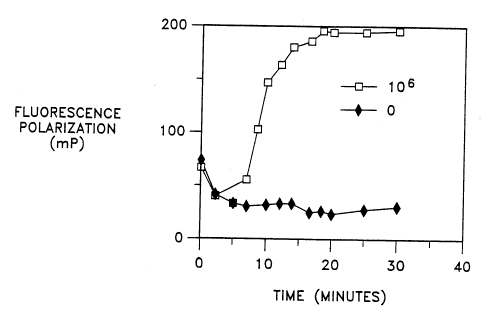
The results are shown in Fig. 1, which illustrates an increase in FP with time
in the
reaction containing target ( 10~ EB's). The maximum OmP was about 161.4 in
this reaction
and the target is detectable in about 6-8 min. at this initial concentration.
The reaction
containing no target and EDTA shows no increase in FP. The ~mP was similar
when the
IO reactions were monitored on the FPM-1 fluorometer, although the
polarization values were
different.
Based on the results of probe hybridization studies conducted at comparable
temperatures, it was unexpected that significant increases in polarization
would be detectable
in thermophilic amplification reactions such as tSDA. Applicants believe that
under these
I5 reaction conditions the polymerase used for amplification also functions as
a stabilizer of the
double-stranded secondary amplification product, thus reducing or preventing
the increase in
single-strandedness typical of elevated amplification temperatures.
Stabilization of the double-
stranded structure appears to maintain, and in some cases to even enhance, the
amplification-
dependent increase in polarization at higher temperatures.
22
Docket No. P-3 S S 5
SEQUENCE LISTING
(1) GENERAL INFORMATION:
S
(i) APPLICANT: Linn, Carl P.
Walker, George T.
Spears, Patricia A.
IO (ii) TITLE OF INVENTION: FLUORESCENCE POLARIZATION DETECTION
OF
NUCLEIC ACIDS
(iii) NUMBER OF SEQUENCES: 6
IS (iv) CORRESPONDENCE ADDRESS:
(A) ADDRESSEE: Richard J. Rodrick, Becton Dickinson
and
Company
(B) STREET: 1 Becton Drive
(C) CITY: Franklin Lakes
2O (D) STATE: NJ
(E) COUNTRY: US
(F) ZIP: 07417
(v) COMPUTER READABLE FORM:
2S (A) MEDIUM TYPE: Floppy disk
(B) COMPUTER: IBM PC compatible
(C) OPERATING SYSTEM: PC-DOS/MS-DOS
(D) SOFTWARE: PatentIn Release #1.0, Version #1.25
3O (vi) CURRENT APPLICATION DATA:
(A) APPLICATION NUMBER:
(B) FILING DATE:
(C) CLASSIFICATION:
3S (viii) ATTORNEY/AGENT INFORMATION:
(A) NAME: Fugit, Donna R.
(B) REGISTRATION NUMBER: 32,135
(C) REFERENCE/DOCKET NUMBER: P-3555
40
(2)
INFORMATION
FOR
SEQ
ID
NO:1:
(i) SEQUENCE CHARACTERISTICS:
(A) LENGTH: 34 base pairs
4S (B) TYPE: nucleic acid
(C) STRANDEDNESS: single
(D) TOPOLOGY: linear
$0
(ii) MOLECULE TYPE: DNA (genomic)
(xi) SEQUENCE DESCRIPTION: SEQ ID NO:1:
SS TAGAGTCTTC AAATATCAGA GCTTTACCTA ACAA 34
(2) INFORMATION FOR SEQ ID N0:2:
(i) SEQUENCE CHARACTERISTICS:
f)0 (A) LENGTH: 26 base pairs
(B) TYPE: nucleic acid
(C) STRANDEDNESS: single
(D) TOPOLOGY: linear
23
-~ Docket No. P-3555 ~ ~ ~'~ 9 41
(ii) MOLECULE TYPE. DNA (genomic)
S (xi) SEQUENCE DESCRIPTION: SEQ ID N0:2:
ATCCGTATGG TGGATAACGT CTTTCA 26
(2) INFORMATION FOR SEQ ID N0:3:
(i) SEQUENCE CHARACTERISTICS:
(A) LENGTH: 40 base pairs
(B) TYPE: nucleic acid
(C) STRANDEDNESS: single
1$ (D) TOPOLOGY: linear
(ii) MOLECULE TYPE: DNA (genomic)
(xi) SEQUENCE DESCRIPTION: SEQ ID N0:3:
CGATTCCGCT CCAGACTTCT CGGGTCTACT GAGATCCCCT 40
2S (2) INFORMATION FOR SEQ ID N0:4:
(i) SEQUENCE CHARACTERISTICS:
(A) LENGTH: 40 base pairs
(B) TYPE: nucleic acid
(C) STRANDEDNESS: single
(D) TOPOLOGY: linear
(ii) MOLECULE TYPE: DNA (genomic)
(xi) SEQUENCE DESCRIPTION: SEQ ID N0:4:
ACCGCATCGA ATGCATCTCT CGGGTAAGGC GTACTCGACC 40
(2) INFORMATION FOR SEQ ID N0:5:
(i) SEQUENCE CHARACTERISTICS:
(A) LENGTH: 13 base pairs
4$ (B) TYPE: nucleic acid
(C) STRANDEDNESS: single
(D) TOPOLOGY: linear
(ii) MOLECULE TYPE: DNA (genomic)
S0
(xi) SEQUENCE DESCRIPTION: SEQ ID NO: S:
SS CGCTGAACCG GAT 13
(2) INFORMATION FOR SEQ ID N0:6:
(i) SEQUENCE CHARACTERISTICS:
60 (A) LENGTH: 13 base pairs
(B) TYPE. nucleic acid
(C) STRANDEDNESS: single
(D) TOPOLOGY: linear
24
~ t a~9~ ~
' ° Docket No. P-3555
(ii) MOLECULE TYPE: DNA (genomic)
S (xi) SEQUENCE DESCRIPTION: SEQ ID N0:6:
TCCACCCGCC AAC 13