Note: Descriptions are shown in the official language in which they were submitted.
CA 02189958 2004-02-20
-1
METHODS OF CAPTURING SPECIES FROM LIQUIDS
AND ASSAY PROCEDURES
The present invention relates to methods of capturing
species from liquids and to assay procedures involving said
species.
Whilst the present invention is of broad and general
applicability, it has particular relevance to the problem of
monitoring organisms in water and will be described with
particular reference to that context.
Current methods for assaying the content of organisms
in water such as cryptosyporidium and giardia are time
consuming and labour intensive.
A major problem in such assay procedures is that the
organisms may be present in very low numbers in substantial
volumes of water and must first be concentrated into a
sample of substantially reduced volume. Conventionally,
this is done by passing large volumes of water through a
cellulosic filter material which is then broken up and
placed in a smaller volume of liquid in which it is agitated
over a prolonged period with a view to releasing the
captured organisms from the filter material. The
proportions of organisms present in the liquid samples which
are captured by this way and success-fully released from the
filter material is relatively poor and the operation is
prolonged taking typically about twentyfour hours to
perform. The product of this procedure is a sample in which
the organisms are still very dilute.
T_n WO-A-93/16383, an assay for cryptosyporidium oocysts
by an electrorotation assay techniques as described, which
requires direct visualisation of the oocysts under a micro-
scope. To run such an assay on water samples of the kind
normally encountered requires further concentration of the
organisms beyond the stage reached following the filtration
procedure described above. In unrelated assay procedures,
it is known that
CA 02189958 2004-02-20
-2-
magnetically attractable particles may be coated with
selective reagents such as antibodies or oligonucleotides.
Such coated magnetically attracted particles are used in
assay procedures by mixing the particles in suspension with
a species to be captured so as to form a complex in which
the species is captured to the particles. The magnetic
particles are then collected by magnetic attraction so as to
concentrate and localise the captured species for further
operations.
The present now provides a method of capturing a
species from a liquid comprising attracting magnetically
attractable particles to a solid support by magnetic forces,
which particles have an affinity for said species,
contacting said particles on said support with said liquid
to capture said species on to said particles, and r'leasing
said particles from said support by reduction of said
magnetic forces.
Because the particles are held on the solid support
during the time in which trey are being contacted with the
liquid containing the species to be captured, it is possible
for the volume of liquid co-a a_ning the species to be much
greater than the volume occupied by the partzcl~s during
this operation. Large volu:;~es of the liquid may be washed
through or over the solid support bearing the macnetically
attracted particles, so that the particles may capture said
species in sufficient quantity for further operations to be
carried out, even if the species is present at great dilu-
tions in the liquid. For ir_stG~:ce, the volume of the liquid
contacted with the particle. mayr be greater than the volume
occupied by the solid support by a factor of at least 5,
preferably at Least 10, still more preferably at lease 50,
and 100 or more.
The liquid may be passed repeatedly over she solid
support, e.g. by continuous recirculation, so as to i;-:Drove
the capture of said species.
The particles may be assa_~ed for the cap~ured species
whilst retained on the solid su~nort. It will =en~_~ali~~
~i8995~
WO 95131726 PCT/GB95I01056
-3-
however be more appropriate to release the particles with the
captured species. This may be done simply by vigorous washing
' or even air blasting whilst maintaining the magnetic
attraction but is preferably accomplished by reducing the
' 5 magnetic attraction.
When the particles are released from the solid support,
they may be collected in a much reduced volume of liquid, for
instance a volume similar to that occupied by the solid _-
support itself, or even less.
A very substantial concentration of the species to be
captured may therefore be achieved.
We have found that the same result cannot be achieved by
the alternative procedure of mixing the magnetically attract-
able particles with the sample to form complexes between the
particles and the species to be captured and then passing the
resulting dilute suspension of complexed particles through a
zone of magnetic attraction to capture them. The process of
magnetic attraction of the particles is too slow and too
easily disrupted by liquid currents and the formation of the
required complexes in the volume of a dilute sample would
require an excessive concentration of magnetically attractable
particles.
The solid support may be a superparamagnetic material or
ferromagnetic material. Superparamagnetism~~ is the magnetic
behaviour exhibited by materials which respond to a magnetic
field with an induced magnetic field without resultant per-
manent magnetisation.
There are many examples of materials which exhibit super-
paramagnetism or ferromagnetism which may be used in the
present invention. Particularly preferred materials are -
stainless steel, aluminium, chromium or platinum. Metallised
foams based on such metals may be used, e.g. aluminium coated
' polyester/polyether foams which are commercially available.
However, materials in which an induced magnetic field =.
results in a permanent residual field may also be used as
further described below.
CA 02189958 2004-02-20
-4-
A solid support material may be magnetised to attract
the magnetically attractable particles by placing the solid
support within a suitable container and applying an external
magnetic field from a permanent magnet or an electromagnet.
S The solid support, if of superparamagnetic material, may be
demagnetised simply by turning off the electromagnet or
physically removing the permanent magnet used to reduce the
field. The magnetic field applied may be a rapidly
reversing magnetic field obtained by passing an alternating
current through a coil.
Preferably, to prevent heat generated in the coil of an
electromagnet used for this purpose from reaching the solid
support, the solid support may be positioned in a pole gap
of a magnet core about which core a coil winding is
positioned remote from the solid support.
A solid support material which is not superparamagnetic
may be demagnetised by known methods such as gradual
reduction and periodic reversal of an externally applied
field.
Physically, the solid support may take ma-!y forms e.g.
mesh, wire, a wool, beads or one or more plates. The
material preferably has an open structure to assist easy
removal of the particles therefrom and easy passage on the
liquid containing the species to be captured. Structures
providing a sub-stantial surface area within a small volume
are preferred.
However, the solid support may simply be the walls of a
contai.~.er such as a glass tube to which the cartir_les are
attracted by an external magnetic field.
T~.e most preferred form of solid support is a stainless
steel mesh, e.g. of 40 x 40 wires per inch (16 x 16 wires
per cm', used as a flat strip of single or double thickness.
b'any forms of magnetically attractable particle are now
known and easily commercially available. Examples include
iron cxide particles as described in US-A-4,556,083 and US
A-3,91~,538, nickel oxide particles as described in Biotec.
and Bioengr. XIX: 101-124 (1977), Agarose-polyaldehyde beads
containing magnetic particles as in US-A-4,732,811. DYNRLT'~
CA 02189958 2004-02-20
-5-
beads (commercially available magnetic polystyrene coated
beads); MagnogelTU 44 (magnetic polyacrylamide-agarose
beads), ENZACRYT~ (poly-M-diaminobenzene/iron oxide) as
described in Clin. Chim. Acta. 69:387-396 (1976). Cellulose
containing ferric oxide particles are described in Clin.
Chem. 26:1281-1284 (1980) and albumin magnetic microspheres
as described in J.IMMUNOL. Methods 53:109-122 (1.982).
Magnetic porous glass particles are described in WO-A-
93/10162.
The particles may also be of superparamagnetic
material.
The particles may preferably have a specific binding
affinity for the species to be captured and for this purpose
they may bear antibody molecules, substances having an
epitope capable of reacting in a specific manner with an
antibody such as an antigenic protein or oligosaccharide,
biotin, avidin or streptavidin, or like materials. they may
bear a nucleic acid or nucleic acid analogue such as DNA,
RNA or a synthetic analogue thereof. Also, the particles
may have a chemical rather than a biochemical affinity for
the species to be captured. For instance, they may have
chelating activity for capturing ions from the liquid.
They may have affinity for a water borne organism such
as Legionella, cryptosyporidium or giardia. However, the
2S invention is of general applicability and may be used for
capturing a wide range of micro-organisms from a wide range
of sample sources including food products and body fluid
samples such as blood, serum, saliva, urine, cerebrospinal
fluid and so forth.
The invention includes assay methods comprising
capturing a species to be assayed or to be used in an assay
by a method of capture as described above, and conducting an
assay of or using said captured species. Optionally, the
captured species may be removed from the particles prior to
3S or during said assay procedure.
CA 02189958 2004-02-20
-6-
The assay procedures involved may take a wide
variety of forms including chemical assay procedures,
enzyme assay procedures such as RIA or ELISA or nucleic
acid procedures such as hybridisation assays.
Preferably however, the assay is an electro-.rotation
assay. WO-A-93/16383 describes apparatus in which
electro-rotation assays can be conducted. As described
there; particles such as plastics microbeads or the cells
of organisms like giardia and cryptosyporidium can be
made to rotate by the application of a rotating
electrical field. The field conditions under which
rotation is achieved, the direction of rotation and the
speed of rotation, all depend upon the dielectric
properties of the particle. Microorganism cells such as
cryptosyporidium oocysts can be concentrated by a capture
method as described above and can then be detected by
subjecting them to electro-rotation conditions and
observing their electro-rotation. The magnetically
attractable particles used in the concentration of the
oocysts need not be removed prior to electro-rotation and
indeed are an aid in observing the rotation, particularly
where automated image analysis systems are used to
perform the observation. The particle or particles bound
to the oocysts provide a useful visual marker which can
be seen rotating.
According to an aspect of the present invention,
there is provided a method of capturing a species from a
liquid, comprising attracting magnetically attractable
particles to a solid support by magnetic forces, which
particles have an affinity for said species, and
contacting said particles on said support with said
liquid to capture said species onto said particles on
CA 02189958 2004-07-05
-6a-
said solid support.
The invention includes apparatus for use in capturing a species from a
liquid comprising a reservoir for said liquid, a source of magnetic field, a
pump
for liquid circulation and means defining a flow path for liquid from said
reservoir via said pump and back to said reservoir, wherein said source of
magnetic field is outside said flow path, and said flow path contains a solid
support magnetisable by a magnetic field applied thereto by said source of
magnetic field.
The invention further includes apparatus for use in capturing a species
from a liquid, comprising a conduit for flow therethrough of said liquid, a
solid
support in said flow path in which a magnetic field can be induced, a source
of
magnetic field out-side said conduit for inducing a magnetic
CA 02189958 2004-07-05
field in said solid support, and magnetically attractable particles on said
solid
support which have an affinity for a said species.
The invention will be further described and illustrated with reference to
the accompanying drawing in which:
Figure 1 shows schematically apparatus for use in the invention.
Figure 2 shows a second form of apparatus for use in the invention;
and
Figure 3 is a plan view of the electromagnet in the apparatus shown in
Figure 2.
As shown in Figure 1, apparatus for use in the invention may comprise
a container such as a syringe body 10 containing a support matrix such as
expanded aluminium 12 surrounded by a helically wound copper wire coil 14
which may for instance comprise 4000 turns of enamelled 40 SWG (standard
wire gauge) wire to which is connected a suitable supply of alternating
electric
current e.g. a 50 volt 50 Hz supply, via suitable switch means. Generally,
frequencies of from 1 to 500 Hz may be employed at voltages of from 1 to 500
volts.
In a typical procedure according to the invention, antibody coated
magnetic beads in a suitable buffer (e.g. pbs) are exposed to the solid
support
and an external magnetic field is applied to induce a corresponding field in
the
solid support. Over a period of minutes, the particles are drawn on to the
solid
support. The attached particles may be washed by slowly running wash liquid
into the top of the syringe body 10 whilst letting liquid out at a
corresponding
rate so as to avoid the level of liquid falling to expose the solid support.
If this
were to happen, there would be a likelihood of surface tension forces pulling
the beads off.
CA 02189958 2004-02-20
-7a-
A sample containing organisms expressing surface anti-
bodies corresponding to the antibodies in the beads and
having a volume which may be of the order of 100 times the
volume o= the part of the syringe body 10 occupied by the
solid support 12 may then be slowly run through, optionally
followed by
218995
WO 95131726 PCT/GB95101056
-8-
further wash liquid, until the solid support is barely
covered.
The external magnetic field is then removed and the beads
are permitted to detach from the solid support, optionally
with agitation being used to disperse them. The beads may
then be run out of the syringe for analysis, bearing any
organisms which have bound thereto. An advantage of this
procedure is that there is no need to use any chemical treat-
ment to release the organisms from the solid support, which
could affect the viability or integrity of the organisms.
Chemical methods are, in contrast, normally needed in most
immuno-affinity capture and release methods.
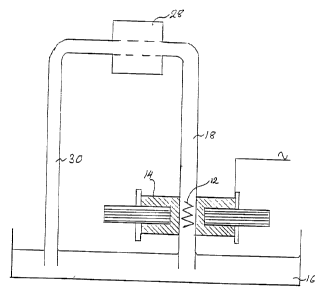
An alternative form of apparatus shown in Figure 2
comprises a reservoir 16 for liquid. A tube 18 dipping into
the reservoir 16 contains the solid support 12 and passes
through a pole gap 20 in a magnet core 22 which is C-shaped
in plan view having a long arm 24 remote from the pole gap 20
around which is positioned a coil 14 wound on a coil former
bobbin 26 and connected to an electrical supply as described
in connection with Figure 1. The tube 18 is connected via a
peristaltic pump 28 to a further tube 30 dipping back into the
reservoir 16.
In use, liquid to be treated in the system may be recir-
culated repeatedly using the peristaltic pump 28 to flow over
the solid support valve as described in more detail in Example
2 below.
The invention will also be further illustrated by the
following examples.
Exarnnle 1
Super-paramagnetic polystyrene beads containing magnetite
(average diameter 0.8 hem, 67% magnetic content - Sigma
Chemical Co.) were coated overnight at room temperature with
a mouse monoclonal antibody raised against cryptosporidium.
The resultant antibody coated beads were placed into an
WO 95!31726 L ~ ~ / ~ ~ ~ PCT/GB95101056
_g_
apparatus similar to that shown in Figure 1 and described in
the above text.
An A/C field (50 Hz, 50 volts) was applied to the coil
to generate a magnetic field, and the beads were incubated -
with the aluminium solid phase for 6 minutes. Following this
incubation, excess unbound beads were washed away with PBS
(phosphate buffered saline).
A 10 ml sample containing cryptosporidium oocysts
(obtained from Moredum Institute Animal Health) was added to
l0 the tube and incubated for 10 minutes (fn the presence of the
applied magnetic field).
After incubation, the solid phase was washed/rinsed with
ml of PBS whilst the magnetic field was present.
Following washing, the magnetic field was removed, i.e.
field generator was switched off; and the magnetic bead/crypto -
were flushed out in 1 ml of PBS.
The presence of cryptosporidium/bead complexes was
determined by immuno-fluorescence staining techniques using
an anti-cryptosporidium fluorescent FITC conjugate (Bradsure
Biochemicals Ltd.). Cryptosporidium complexes were detectable
using the procedure clearly indicating that specific capture
and the subsequent elution (from the solid phase) had been
achieved.
Exaam~e 2
Using the apparatus described above with reference to
Figures 2 and 3, 50 ml PBT (phosphate buffered saline + 0.05
Tween 20) was circulated over the solid support at a flow rate
of approximately 100 ml/30 sec. The solid support was a thin
strip of stainless steel formed into the zig-zag configuration
illustrated in Figure 2. A suspension of antibody coated
beads (200 ~1) was added to the reservoir and the electrical
power was turned on at 50 volts/50 Hz. Circulation was
continued for 45 minutes.
W O 95!31726 ~ ~ ~ ~ ~ ~ ~ PCT/GB95/01056
-10-
Most of the PBT was drained off from the reservoir and
100 ml of fresh PBT was added as a wash.
A spike of cryptosporidium oocysts in a volume of 50 ml
PBT was added_and allowed to circulate for 45 minutes. The
bulk of this was then drained off from the reservoir and 100
ml of fresh PBT was added as a further wash. This wash liquid
was drained off and combined with the remainder of the 50 m1
spike liquid fgr later determination of the cryptosporidium
remaining in the circulating liquid.
The solid support was further washed with 400 ml PBT.
Circulation was then halted. The power was turned off from
the magnet and the cryptosporidium was eluted from the solid
phase using 5 ml PBS. This eluate was.collected for
determination of the numbers of cryptosporidium oocysts
captured.
To determine the number of oocysts present, the liquid
was in each case pushed through a membrane filter which was
then stained and the numbers of oocysts determined by immuno-
fluorescence. The results are as shown in the table below:-
Run Oocysts Oocysts Percent
not Captured and Captured and
Captured Recovered Recovered
1 272 352 56
2 218 316 59
2S 3 374 559 60
CA 02189958 2004-02-20
-11-
Exam l a 3
Determination of Capture Efficiency of Cryptosporidium
oocysts.
S The apparatus described above with reference to Figures
2 and 3 was modified by substituting as the solid support a
1 cm x 3 cm strip of stainless steel mesh (40 x 40 wires per
inch (16 c 16/cm)) folded longitudinally in half to make a
double thickness strip 0.5 cm wide. PBT (Phosphate Buffered
Saline + O.OS% TweenT~ 20) (2S ml) was circulated over the
solid support at a flow rate of approximately 100 ml/30
seconds. A suspension of antibody coated beads specific to
Cryptosporidium (500 ~1) was added to the reservoir and the
electrical power was turned on at 60 volts/SO Hz.
1S Circulation was continued for 60 minutes and ~~urrent
adjusted to 7S mA. Following this, excess beads were run to
waste together with a 20 ml wash of PBT. The beads were as
described in Example 1.
A spike of Cryptosporidium oocysts of known number in
25 ml was added to the reservoir and allowed to circulate
for 60 minutes. At the end of the period, the solid support
was washed by running through S00 m1 of PBT to waste. The
flow was then halted and the Cryptosporidium bead complexes
eluted from the solid phase using 20 ml PBS. This eluate
was collected for determination of numbers of
Cryptosporidium oocysts captured. To determine the number
of oocysts present, the liquid in each case was pushed
throug:~: a membrane filter wish was then stained and the
numbers of oocysts determined by immunofluorescence. The
results are shown in the table below:-
WO 95131726 PCTIGB95101056
-12-
Run Oocysts Oocysts Captured Percent
and Recovered Captured and
Recovered
1 515 163 31.6%
2 390 162 41.5%
3 502 232 46.2%
Example 4
Detexmiaatioa of Capture Efficiency of Giardia oocysts.
Using the apparatus as used in Example 3, 25 ml PBT
(Phosphate Buffered Saline + 0.05% Tween 20) was circulated
over the solid support at a flow rate of approximately 100
ml/30 seconds. A suspension of antibody coated beads specific
to Giardia (500 ~.1) was added to the reservoir and the
electrical power was turned on at 60 volts/50 Hz. Circulation
was continued for 60 minutes and current adjusted to 75 mA.
Following this, excess beads were run to waste together with
a 20 ml wash to PBT. The beads, prior to coating, were as
described in Example 1.
A spike of Giardia cysts of known number in 25 ml was
added to the reservoir and allowed to circulate for 60
minutes. At the end of this period the solid support was
washed by running through 500 ml of PBT to waste. The flow
as then halted and the Giardia bead complexes eluted from the
solid phase using 20 ml PBS. This eluate was collected for
determination of numbers of oocysts captured. To determine
the number of cysts present, the liquid in each case was
pushed through a membrane filter which was then stained and
the numbers ofGiardia determined by immunofluorescence. The
results are shown in the table below:-
21~9~58
WO 95!31726 PCTIGB95101056
-i3-
Run Giardia Giardia Captured Percent
Spike and Recovered Captured and
Recovered
1 584 405 69.3
2 584 221 37.g
3 423 246 58.2
~xamnle 5
Datermi.aation of Capture Efficiency of Cryptosporidium
aad Giardia.
Using the apparatus described in Example 3 25 ml PBT
(Phosphate Buffered Saline + 0.05 Tween 20) was circulated
over the solid support at a flow rate of approximately 100
ml/30 seconds. A suspension of antibody coated beads specific
for Cryptosporidium and Giardia (500 ~1) was added to the
reservoir and the electrical power was turned on at 60
volts/50 Hz. Circulation was continued for 60 minutes and
current adjusted to 75 mA. Following this, excess beads were
run to waste together with a 20 ml wash of PBT. Prior to
antibody coating the beads were as described in Example 1.
A spike of combined Cryptosporidium and Giardia of known
number in 25 ml PBT was added to the reservoir and allowed to
circulate for 60 minutes. At the end of this period, the
solid support was washed by running through SOD ml of PBT to
waste. The flow as then halted and the cryptosporidium and
Giardia complexes eluted from the solid phase using 20 ml PBS.
This eluate was collected for determination of numbers of
oocysts/cysta captured. To determine the number of cysts
present, the liquid in each case was pushed through a membrane
filter which was then stained and the numbers of organisms
determined by immunofluorescence. The results are shown in
the table below:-
2189958
W0 95131726 PCTlGB95/01056
-I4-
Run Spike Spike Organisms Present
Captured Captured
and and
Recovered Recovered
C G C G C G
1 552 680 260 502 471 73.8
2 552 680 187 269 33.9 39.6
3 482 453 82 227 17.0 50.1
Example 6
Capture of Cryptosporidi"- oooysts is river sediment.
Using the apparatus described above in Example 3 25 ml
PBT (Phosphate Buffered Saline + 0.05% Tween 20) was
circulated over the solid support at a flow rate of
approximately 100 ml/30 seconds. A suspension of antibody
coated beads specific to Cryptosporidium (500 ~C1) was added
to the reservoir and the electrical power was turned on at 60
volts/50 Hz. Circulation was continued for 60 minutes and
current adjusted to 75 mA. Following this, excess beads were
run to waste together with a 20 ml wash of PBT. The beads
were as used in previous examples.
A spike of Cryptosporidium oocysts of known number in 25
ml PBT and river sediment (-100 NTU) was added to the
reservoir and allowed to circulate for 60 minutes. At the end
of this period, the solid support was washed by running
through 500 ml of PBT to waste. The flow as then halted and
the Cryptosporidium eluted from the solid phase using 20 ml
PBS. This eluate was collected for determination of numbers
of Cryptosporidium oocysts captured. To determine the number
of oocysts present, the liquid in each case was pushed through
a membrane filter which was then stained and the numbers of
oocysts determined by immunofluorescence. The results are
shown in the table below:-
W 0 95/31726 PCTIGB95101056
-15-
Run Oocysts Oocysts Captured Percent
and Recovered Captured and
Recovered
1 515 111 21.5%
2 515 104 29.2%
3 492 115 23.3%
Example 7
Capture and Concentration of Legionella paeumophila.
Using the apparatus as described in Example 3 25 ml PBT
(Phosphate Buffered Saline + 0.05% Tween 20) was circulated
over the solid support at a flow rate of approximately 100
ml/30 seconds. A suspension of antibody coated beads specific
to Legionella pneumophila (500 ~.1) was added to the reservoir
and the electrical power was turned on at 60 volts/50 Hz.
Circulation was continued for 60 minutes and current adjusted
to 75 mA. Following this, excess beads were run to waste
together with a 20 ml wash of PBT. The beads were as used
previously.
A spike of Legionella pneumophila of known number in 25
ml was added to the reservoir and allowed to circulated for
60 minutes. At the end of this period, the solid support was
washed by running through 500 ml of PBT to waste. The flow
as then halted and the Legionella bead complexes eluted from
the solid phase using 20 mls PBS. This eluate was collected
for determination of numbers of bacteria captured. To
determine the number of cells present, the liquid in each case
Was pushed through a membrane filter which was then stained
and the numbers of Legionella determined by
immunofluorescence. The results are shown in the table
below:
X189958
W 0 95131726 PCT/GB95101056
-16-
Run Legionella Legionella Percent
Spike Captured Captured and
and Recovered Recovered
I
1 1044 564 54%
2 11854 4444 37.5%
3 I 5611 3121 56.6%
Many modifications and variations of the invention as
illustrated and described above are possible within the broad
scope of the invention. In particular, the invention may be
applied to a wide range of analyte species. It will be of
particular benefit where the analyte species is dilute and/or
present in association with large amounts of particulate
material, e.g. in the food industry for detecting organisms
in foodstuffs such as cheese.