Note: Descriptions are shown in the official language in which they were submitted.
Pca885
2190237
-1-
APPARATUS AND METHOD FOR
TRANSURETHRAL FOCUSSED ULTRASOUND THERAPY
Background of the Invention
This invention relates to an apparatus and method for the treatment of benign
prostatic hyperplasia (BPH), prostate cancer, and other diseases by
application of
focussed ultrasonic energy from a probe placed near the site of the lesion.
BPH is a very common disease in men over 50 years of age,. in which swelling
of the prostate results in obstruction of the urethra and consequent inability
or difficulty
in urinating. In its early stages it causes discomfort and inconvenience.
Permitted to
progress, it can result in severe pain and serious consequences. It is
traditionally
treated by transurethral resection of the prostate (TURP), a surgical
procedure with
good effectiveness but an unfortunate level of pain, blood loss, morbidity,
complications, expense, lost time, and in some cases death. Other methods,
using
lasers or radio frequency or microwave energy, have not been proved to
approach
TURP in effectiveness. A method combining high effectiveness with fewer short-
term
bad effects than TURP is still urgently required.
Prostate cancer is the second leading cause of cancer-related death in men.
In its early stages it can be treated successfully by radical prostatectomy,
but this
procedure has all of the disadvantages of TURP and in addition often results
in
incontinence, impotence, or both. Prostate cancer can also be treated by
radiation
therapy, but similar serious side effects are common if a sufficient dose is
used to have
a good chance of a favorable result. A curative method with less initial
trauma is
needed. More advanced prostate cancer is also treated by radical prostatectomy
or
radiation therapy, but this procedure usually does not result in cure, though
it may
achieve palliation. Since less is accomplished in these cases, a less invasive
method
is even more necessary.
Ultrasound is well known to urologists for its ability to image a volume of
tissue,
creating pictorial slices without the need to cut. It can do this because
ultrasonic waves
are transmitted through tissue without being too strongly attenuated, yet,
because there
is significant absorption by tissue, intense ultrasound can produce very
substantial
heating in the interior of an organ. The goal in exploiting this effect is to
create a large
ultrasound intensity at the interior region to be treated while minimizing the
ultrasound
intensity in tissue that is to be spared. Prior attempts have been made to use
the
2190237
-2-
capabilities of focussed ultrasound for treatment of BPH and prostate cancer.
One
approach utilizes extracorporeal ultrasound focussed from outside the body;
another
uses a transrectal probe.
U.S. Patent No. 5,344,435, to Turner et al. describes the transurethral
application of unfocussed ultrasound energy for the treatment of prostatic
disease. The
disclosed apparatus, however, does not exploit the ability of ultrasound to
reach a focus
within the tissue, and thus to deliver a higher intensity. at an internal
point than is
present at the urethral wall. Accordingly, and despite the use of urethral
cooling, the
inventors do not recommend temperatures greater than 48°C. Use of these
temperatures diffusely in the prostate may be of some clinical value, but does
not
produce effects comparable to application of higher temperatures in a sharply
defined
volume of tissue, as taught in the present invention.
The apparatus of Turner et al. '435 operates in what is generally termed a
hyperthermal mode. Energy transfer utilizing the apparatus of Turner et al.
'435 is by
radiation, that is, energy is transmitted from a source within the apparatus
into a
treatment volume much larger than the source itself. As a consequence of the
hyperthermal irradiation temperature being limited to a maximum of
48°C, the diseased
prostatic tissue must be irradiated for relatively long periods of time, often
up to 60
minutes or more. This is disadvantageous in that it requires the patient to be
immobilized during such lengthy treatment sessions.
When a transrectal probe is used, the ultrasound must pass through 4 cm or
more of healthy tissue before reaching the tissue that is to be destroyed. If
the probe
is outside the body, the ultrasound must pass through an even greater depth of
healthy
tissue. In either case, the large distance between the probe and the tissue to
be
treated is disadvantageous because it increases the difficulty of targeting
the ultrasound
accurately, because healthy tissue is exposed to the potentially damaging
effects of
high intensity ultrasound, and because a higher initial power must be used to
make up
for attenuation in tissue between the probe and the target.
A further drawback to prior systems is that they focus the ultrasound at peak
intensity on each individual volume of tissue to be treated. This requires
extremely
accurate targeting, generally requiring an elaborate and costly targeting
system such
as diagnostic ultrasound. It further requires the provision of accurate
relative motion
between the probe and the patient. Because of the high power required to
compensate
71493-312 ca o2i9o23~ Zooo-os-io
3
for attenuation, and because of the accurate targeting
required, prior art systems are extremely expensive, costing
well over one hL~.ndred thousand dollars and in some cases many
times more.
Summary of the Invention
The broad principal objects of the present invention
are to provide a device capable of treating BPH, prostate
cancer, and other diseases by the application of high intensity
ultrasound.
The invention provides an apparatus for treatment of
diseases of the ;prostate in a mammalian body, said apparatus
comprising: (a) a generator of a radio frequency electrical
signal, having a frequency in the range of from about 1 MHz to
about 10 MHz, ca~~able of generating a constant power level and
~_5 capable of operav-ing at said constant power level for a period
of time of at le<~st 30 seconds; (b) ultrasound probe means
including a transducer housing containing a transducer with a
single transducer unit made of one or more piezoelectric
crystal elements and an output aperture having an area, and
~:0 corresponding coupling means and focussing means therefor, for
converting at least a portion of said electrical signal into a
beam of ultrasound energy, said beam having an area and
sufficient power to produce thermal effects in prostatic tissue
and to cause coac~ulative necrosis in selected portions of
25 diseased prostati.c tissue, and for coupling said ultrasound
energy into diseased prostatic tissue, and focussing said
ultrasound energy at a focal plane, such that said area of said
beam of ultrasound energy at said focal plane is less than said
area of said aperture; (c) delivery means for transurethrally
30 introducing said ultrasound probe means into the prostatic
urethra of a mammalian body; and (d) positioning means for
71493-312 ca o2i9o23~ 2ooo-os-io
3a
fixing said ultrasound probe means in a desired position in
said prostatic urethra; (e) at least one visualization means
for enabling the remoi:e observation of at least one of the
positioning of ;aid ultrasound probe means, and the treatment
of said diseased prosi=atic tissue, said visualization means
being selected from the group (i-ii) consisting of: (i)
endoscopic mean for viewing the position of said ultrasound
probe means within thE: urethra, and (ii) diagnostic ultrasound
means for generating an ultrasound imaging signal for producing
an ultrasound image of= at least a portion of the prostatic
tissue to be treated.
Treatment can be made minimally traumatic by avoiding
incision of any tissue and by entering only a single body
cavity; minimizing damage to any tissue other than that which
is to be treated; minimizing the required power output from the
device so as to avoid unnecessary heating of nearby tissue;
simplifying the ::monitoring procedure by using direct endoscopic
visualization as far as possible; minimizing the cost of the
treatment; permitting treatment without the requirement for
?0 anesthesia beyon~~ topical agents such as lidocaine, so that the
procedure is no more painful or acutely traumatic than
examination with a flexible cystoscope; permitting treatment in
which the urethra is neither pierced nor heated, and treatment
of the prostatic parenchyma is well controlled; and permitting
?5 treatment in whi~~h post procedure catheterization is
unnecessary, and such that patients without comorbidities can
be treated at a medical services-providing facility such as a
hospital, clinic,, or even at a doctor's office, on an out-
patient basis.
~~0 A compact, intraluminal device produces a focussed
beam of ultrasound energy, and utilizes a single ultrasound
71493-312 ca o2i9o23~ 2ooo-os-io
3b
transducer consisting of one or more piezoelectric elements.
The device is capable of causing greater than hyperthermal
therapeutic temperatures in selected regions of diseased tissue
of a body organ in a particular area of the body, without
causing the tem~~eratune in surrounding non-diseased tissue or
in adjacent anatomical_ areas to be raised to damaging levels,
thereby enabling the device to be much simplified by being able
to dispense with the need for means for cooling adjacent non-
diseased tissue and organs not being treated, to avoid thermal
damage thereto. The device is also capable of effecting a
course of therapy in a shorter period of time than is required
for a course of therapy utilizing unfocussed radiating
ultrasound energy in a. hyperthermal mode of operation having a
much lower maximum temperature limitation, as is necessary to
L5 avoid damage to surrounding non-diseased tissue and other
anatomical areas.
2190237
-4-
A still further specific object of the present invention is the provision of a
transurethral focussed ultrasound device for the treatment of BPH and other
diseases
of the prostate, having the above features, and which, because of the faster
course of
therapy, is simpler in design than a device for hyperthermal treatment, and
which is
able to dispense with the need for a urine drainage system because of the much
shorter period of time the device is required to be present in the prostatic
urethra of the
patient during administration of a course of therapy. - The present apparatus,
in fact,
when operationally positioned, does not need to extend beyond the prostatic
urethra,
either to the bladder neck or further into the bladder itself.
The novel apparatus and method of this invention are based on a therapeutic
modality which we have termed Transurethral Ultrasound Therapy (TUT). This
treatment modality utilizes the application of focussed ultrasound energy to
the prostate
from a probe in the prostatic urethra to effect hyperthermal or above
hyperthermal
heating of selected diseased prostatic tissue to be treated, thereby causing
coagulative
necrosis of the diseased tissue. The great advantage of the apparatus
according to the
present invention utilizing TUT is the superiority of the geometrical aspects
of the
treatment; the ultrasonic energy wave only has to travel about 1 cm through
tissue.
This is about one quarter as far as for transrectal application and represents
a still
greater advantage over extracorporeal application. The improved geometrical
factors
resulting from use of the apparatus of the present invention utilizing the TUT
therapeutic modality allows the ultrasound energy to be focussed into a
defined tissue
volume, with minimal intensity being directed at tissue further away from the
source.
There is far less attenuation in this short path length, so the probe need not
emit a
great excess of ultrasound energy to compensate for attenuation. At the same
time,
non-diseased tissue nearer to the energy source is spared, because the
ultrasound
energy intensity in those areas is low. Because the TUT probe resides in the
urethra,
direct cystoscopic observation is a great aid in locating the probe,
eliminating the need
for more expensive monitoring systems. Further, while some transurethral
devices can
be uncomfortable, the improved geometry of the apparatus of the present
invention
allows a small, non-traumatic probe to be used. The TUT apparatus therefore
combines high effectiveness with low invasiveness similar to flexible
cystoscopy, which
is commonly performed with only topical lidocaine jelly.
2190237
-5-
Certain further advantages arise out of operation of the present apparatus
utilizing a high intensity, focussed beam of ultrasound energy. One advantage
is that
higher therapeutic temperatures can be attained in more precisely defined
diseased
regions in the interior of the prostate than can be attained with conventional
hyperthermal treatment. Because these regions are removed from anatomical
regions
where higher temperatures can cause damage, there is no need for the device to
provide-for cooling these other anatomical regions. The present apparatus,
therefore,
also offers the advantage of enabling the achievement of high temperature
where it is
called for, while enabling the maintenance of safe, lower temperatures in
surrounding
areas.
Another advantage of the present apparatus is that because of the higher
therapeutic temperatures attainable with focussed ultrasound in the limited
area of the
diseased tissue, the duration of treatment is shortened considerably over the
time
required for the typical course of conventional hyperthermal treatment. A
further benefit
of the shortened treatment time utilizing the present apparatus is that a
urine drainage
system extending into the bladder is not required as part of the present
apparatus. In
devices utilizing conventional hyperthermal treatment, such a urine drainage
system is
necessary to remove the accumulation of urine forming in the patient's bladder
over a
lengthy treatment session.
The present apparatus, therefore, has the still further advantages of being
considerably simpler in construction and being easier to manufacture by not
requiring
means for cooling adjacent tissue or means for urine drainage, although in
certain
embodiments of the apparatus, one or both of these features may optionally be
present.
The utilization of a focussed beam of ultrasound energy in the present
apparatus, moreover, enables the apparatus to be constructed utilizing a
single
ultrasonic transducer consisting of one or more piezoelectric elements. This
is in
contrast to devices of the radiating ultrasonic applicator type which require
a plurality
of transducers to produce an ultrasound energy field capable of being
simultaneously
radiated in ~ many directions, usually omnidirectionally, into a volume
considerably
greater than the volume at the source.
All of the foregoing features and advantages of the present apparatus are
lacking in the various apparatuses of the known prior art. Accordingly, the
apparatus
2190237
-6-
of the present invention is deemed to satisfy a need in the art for such a
device and
to make a novel and innovative contribution to the art in this field.
Brief Description of the Drawings
Figure 1 shows an ultrasound emission pattern from a circular aperture of an
ultrasound probe according to the present invention.
Figure 2 shows a first preferred embodiment of the apparatus according to the
present invention in anatomical perspective.
Figure 3 shows an ultrasound transducer housing according to the present
invention.
Figure 4 shows a cross-sectional view through 4-4 of the housing of Figure 3.
Figure 5 shows a cross-sectional view through 5-5 of the housing of Figure 3.
Figure 6A shows a detailed view of one embodiment of the ultrasound probe of
the apparatus of Figure 2, including the transducer, focussing means and
coupling
means.
Figures 6B-6D show alternative embodiments of the transducer, focussing
means and/or coupling means of the ultrasound probe means of the apparatus of
the
present invention.
Figure 7 shows a focussed ultrasound beam emanating from the ultrasound
probe of Figure 6.
Figure 8 shows a second preferred embodiment of the apparatus according to
the present invention with a transurethrai imaging ultrasound probe.
Detailed Description of Prefen-ed Embodiments of the Invention
The heating effect of ultrasound depends on the intensity, or power per unit
area, of the ultrasound. When the ultrasound is focussed into a spot whose
area is
small, the intensity is con-espondingly high. If the-same total power is
spread over a
larger area, the intensity is correspondingly lower. The amount of heat
generated at
a point in tissue, and thus the temperature increase that results, is
generally
proportional to the ultrasound intensity at that point.
The TUT apparatus of the present invention uses a probe in the urethra quite
close to the tissue to be treated. One important advantage of being able to
treat the
diseased tissue from such close proximity is that a high relative aperture can
be
utilized. The relative aperture, n, is defined as the focal length divided by
the diameter
of the aperture through which ultrasound energy is emitted. If a small value
of n is
64680-928
219027
-7-
used, the ultrasound energy intensity at the focus is much higher than the
intensity
either nearer to the aperture or beyond the focus. Accordingly, tissue a
distance from
the focus is spared.
This is expressed quantitatively by the following formulae, wherein the
emission
of ultrasound from circular aperture 4 of ultrasound probe 6 in Figure 1 is
considered.
Aperture 4 has a diameter A and area (rr/4)A2, and is in contact with tissue
surFace 8.
The emitting probe is of a focussing configuration causing ultrasound energy
beam 10
to be focussed at focal plane 14 a distance f into the tissue. If the power
emitted from
the aperture is W watts, the initial intensity lo, or power per unit area, is
given by
to = W / (n/4)A2
At plane 18 a distance x from the aperture, or f-x from the focal plane, the
volume of tissue exposed to the ultrasound has a circular cross section of
area
[(nl4)Az] ~[(f-x)/f]z. The intensity at x is therefore given by
Ix = W / [(rd4)[A~(f-x)/f]2]
in the absence of attenuation. The ultrasound energy is, however, actually
attenuated as it passes through tissue, so that
IXa = W~exp[ Nx]/[(rr/4)[A(f-x)/f]Zl
where N is the attenuation per unit length, and has the approximate numerical
value 0.16v crri', if v is the frequency expressed in megahertz (MHz).
The area of the exposed tissue at focal plane 14 does not drop to zero, as
suggested by these equations. Figure 1 shows that the focus is not infinitely
sharp.
At the focus, the diameter of the exposed tissue is given by diffraction
theory as 1.2M,
where n is the relative aperture defined above, and .1 is the wavelength of
the
ultrasound; in tissue its approximate numerical value in mm is 1.SIv, if v is
expressed
in MHz. The focal intensity If is given by
If = W~exp (-~ / [(rr/4)[1.2M]2]
These equations show that the ratio of intensity at the focus to initially
emitted
intensity is given by
I~lo = exp ( ~ / [1.2M/A]2
Similarly, the ratio of the intensity at a distance (f-x) from the focus to
the
intensity at the focus is
IX/lf = exp[ N(x-f)]~[1.2Mf/A(f-x)]2
_g_
Thus, in order to minimize intensity in healthy tissue while delivering as
much
power as possible near the focus in the tissue to be treated, it is best to
use a small
relative aperture and a short focal length. The focal point should be in the
tissue to be
treated, so the focal length is about equal to the distance from the probe to
the tissue
to be treated. In other words, the probe should be as near as possible to the
target.
This configuration reduces the attenuation and therefore eliminates the need
for very
high power from the probe. The small relative aperture causes the intensity to
be
substantially less at a distance from the focal point. Locating the probe in
the urethra,
about four times nearer to the tissue to be treated than with a transrectal
procedure,
is the only way to meet these two requirements in the case of prostate
therapy. The
first preferred embodiment described below has a focal length of 12 mm and an
aperture of 8 mm, for a relative aperture of 1.5. When operated at a frequency
of 5
MHz and emitted ultrasound power of 10 watts, it delivers over 1600 watts/cm2
to the
focal point. In order to deliver this much power to the focus, a transducer
operating at
5 MHz, 40 mm from the focal point, would have to emit over 60 watts of
ultrasound
power. At this power level, it could not be operated continuously for a long
enough
time to produce extensive coagulation without a cooling system that would be
impractical for use within the body. For best results, the relative aperture
should be no
more than 1.7 and the focal length not more than 20 mm. A relative aperture,
n, of 1.7
is often denoted as f11.7 optics.
A further advantage of the invention is that it makes possible the use of a
higher
ultrasound frequency. The length of the lesion, in the direction parallel to
the direction
of propagation of the ultrasound, is proportional to the depth of focus. But
the depth
of focus, in turn, is inversely proportional to the ultrasound frequency. Use
of a low
frequency, therefore, tends to produce an elongated lesion, which is
disadvantageous
because tissue in critical regions may be heated, with the danger of harm to
the
patient. Anatomical regions placed at risk by a large depth of field, and thus
by a,low
ultrasound frequency, include the prostatic capsule, anterior rectal wall,
external
sphincter, and neurovascular bundle. For this reason it is desirable to use as
high a
frequency as possible. But since ultrasound attenuation increases with
frequency, the
range of the treatment is limited more severely at higher frequency. The
apparatus of
the current invention permits use of higher frequency because the ultrasound
does not
need to travel as far through tissue as is required with transrectal or
extracorporeal
2190237
_g_
ultrasound. Because a higher frequency can be used, prostate disease is
treated with
less risk of harm to critical structures of the patient's anatomy. For the
typical 1 cm
distance utilized by the apparatus of the current invention, 20% of the
ultrasound
energy would be transmitted to the focal point even at a frequency as high as
10 MHz,
while 87% would be transmitted at a frequency of 1 MHz. The typical 4 cm
distance
required for transrectal treatment requires a frequency no higher than 2.5 MHz
to be
used in order for 20% of the emitted ultrasound energy to reach the focal
point. While
lower transmission can be tolerated, it requires a more expensive high power
transducer, and results in deposition of substantial quantities of heat in non-
diseased
tissue that is not intended to be affected. Prior systems therefore compromise
by using
a lower ultrasound frequency than would be desired for maximum patient safety.
Two preferred embodiments of an apparatus and method for transurethral
ultrasound therapy, according to the present invention, will now be described.
The first
embodiment is used under visual control. Its advantages include efficacy, low
cost, and
lack of trauma. Its most preferred use is for treatment of BPH, although it is
also useful
for treatment of prostate cancer. The second embodiment combines therapeutic
and
imaging ultrasound. It allows the position of the focal spot within the tissue
to be
controlled to within less than 1 mm, and permits the evolving effect to be
monitored in
real time. In addition to BPH, this embodiment is particularly useful in the
treatment
of prostate cancer.
First Preferred Embodiment
The first preferred embodiment of an apparatus according to the present
invention is a therapeutic ultrasound system powered by a simple, inexpensive
generator. This system relies on visual control and is used without
simultaneous
ultrasonic imaging. A reusable flexible imaging/illumination bundle is
included within
the catheter.
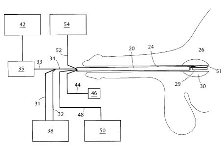
Referring to Figure 2, catheter 20 is inserted via the patient's urethra 24
until
it reaches prostatic urethra 26 within prostate 30. Ultrasound probe 29
extends beyond
catheter 20. Probe lines 31, 32, and 33 are combined into bundle 34 which
passes
through the interior of catheter 20. Probe lines 31 and 32 carry cooling water
between
supply 38 and ultrasound probe 29. Probe line 33 carries radio frequency
electricity
between power unit 42 and ultrasound probe 29. Matching network 35 minimizes
inefficiencies in coupling of the radio frequency electrical power into the
transducer.
219237
Flexible endoscope 44, which terminates in eyepiece 46, also
passes through the interior of catheter 20 to provide a view
of prostatic urethra 26 and probe 29. Cable 48 carries
illumination from light source 50 to endoscope 44. Positioning
balloon 5i extends beyond catheter 20 and is inflated with
fluid from reservoir 54 carried through tube 52.
Figure 3 shows transducer housing 55 of ultrasound
probe 29 extending from proximal end 56 of catheter 20. Lines
31-33 are attached to the transducer housing. Distal end 58
of flexible endoscope 44 extends slightly out of the catheter.
Positioning balloon 51 is adjacent to the transducer housing.
When balloon 51 is inflated, front face 60 of transducer
housing 55 is pressed firmly against wall 62 of prostatic
urethra 26 assuring good acoustic coupling of ultrasound
energy into prostatis tissue.
Figure 4 shows section 4-4 of Figure 3. Wall 66 of
catheter 20 defines lumen 68, which accommodates bundle 34,
endoscope 44, and tube 52, which is used to inflate
positioning balloon 51. Figure 5 shows section 5-5 of Figure
3. Probe line 32 carries a cooling liquid from supply 38,
which preferably includes a chiller to lower the temperature
of the cooling liquid below room temperature, to transducer
housing 55. Probe line 31 carries return liquid from the
transducer housing to supply 38, allowing continuous flow of
cooling 7_iquid. Probe line 33 provides power to the
transducer and may carry other electrical signals.
Figure 6A shows ultrasound probe 29 in further
64680-928
2~9J237
- l0a -
detail. Transducer 72, which includes a single ultrasonic
transducer consisting of one or more piezoelectric elements
made of piezoelectric material, such as hard lead
zirconate/lead titanate piezoelectric ceramic, receives radio
frequency power from line 33, and vibrates in response to
create ultrasound energy. Because of the concavity of front
surface 73 of transducer 72, the ultrasound energy is focussed
as shown in Figure 7. The ultrasound energy is coupled by
quarter wave plate 74, minimizing reflection back to the
transducer, and passes through front face 60 of transducer
housing 55. The transducer and quarter wave plate are
supported by back plate 78 and periphery 80, defining gap 76,
which damps backward prapagation of ultrasound. The outer
housing comprises front face 60, back face 70, arid periphery
71 of transducer housing 55. If necessary, cooling liquid
from line 32 enters the housing at inlet 82, moves through
passage 86, and exits through outlet 84 to line 31, carrying
off heat generated within the transducer housing.
64680-928
2190237
-11-
This heat could otherwise damage the transducer and quarter wave plate, and
could
cause undesirable heating of the urethral wall.
Figures 6B-6D show alternative techniques for achieving focussing of the
ultrasonic energy. In Figure 6B the transducer 72 is flat rather than concave
as in
Figure 6A, and planoconcave lens 73a, of a suitable material that transmits
ultrasound,
provides the focussing. In Figure 6C transducer 72 is concave but quarter wave
plate
74 is flat, with gap 75 filled by a material that transmits ultrasound. . In
Figure 6D the
transducer comprises a plurality of flat, ring-shaped elements 77. Electrical
energy is
provided to each ring with a phase that is advanced with the respect to the
phase of
electrical energy supplied to the next inner ring, creating a phased array
that focusses
ultrasound energy.
In use, catheter 20 and probe 29 are advanced to the prostatic urethra.
Endoscope 44 is used to position the probe as desired. When the position is
correct,
positioning balloon 51 is inflated to fix the probe's position and to assure
good contact
between front face 60 and the urethral wall. Blood and other bodily fluids are
thus
excluded from the area between the transducer and the prostatic tissue. When
the
position has been fixed, power from power supply 42 is applied to transducer
72 via
line 33 and matching network 35.
As illustrated in Figure 7, ultrasound energy from transducer 72 passes
through
quarter wave plate 74, cooling liquid in passage 86, front wall 60 of the
transducer
housing, and urethral wall 62, then entering the prostatic parenchyma 81.
Ultrasound
energy is absorbed in the prostatic parenchyma, depositing energy as heat
generally
proportional to the ultrasound intensity. Because of the focussing effect,
outer rays 84
converge so that the intensity is greatest near focal plane 14. As a result,
substantial
heat is deposited in central region 88 while much less heat is deposited
elsewhere
within the prostate. In regions beyond the focal plane, attenuation and
spreading of the
ultrasound over a larger area combine to cause more rapid decrease in
intensity. This
has the desirable effect of tending to spare tissue beyond the focal point,
including
several critical structures. When the temperature of central region 88 has
increased,
a generally~spherical surrounding volume is heated by thermal conduction.
Isotherms
90 define spherical shells with temperature increasing toward the center. The
ultrasound power, frequency, and duration can be chosen so that an ultrasound
exposure of between 30 seconds and 10 minutes causes a volume of several cubic
2190237
-12-
centimeters to be heated to a temperature of at least 60°C. It is known
in the art that
prostate tissue heated to this temperature undergoes coagulative necrosis and
is
subsequently resorbed. The apparatus of the present invention, therefore,
causes
elimination of a clinically useful volume of tissue without frequent
retargeting of the
ultrasound, and without the need for a complex system to produce and monitor
motion
of the probe relative to the tissue.
According to a particularly preferred method, the concentration of generated
heat in tissue within central region 88 is increased still further. It is well
known that the
ultrasonic propagation properties of tissue are modified by changes in the
tissue such
as coagulative necrosis. N.L. Bush, I. Rivens, G.R. ter Haar, and J.C. Bamber
have
reported measurements of this effect in an article titled "Acoustic properties
of lesions
generated with an ultrasound therapy system" appearing in Ultrasound in
Medicine and
Biolo Volume 19, Number 9, pages 789-801. They find that attenuation of
acoustic
waves is increased when tissue has been sufficiently heated to undergo
coagulative
necrosis. The average increase in their measured values was over 98%. In the
particularly preferred method, ultrasound is applied at a relatively high
intensity for a
short time, so that tissue is denatured in central region 88. Other regions of
the tissue,
where the ultrasound intensity is lower, are not heated as much and are not
denatured.
The ultrasound power is then decreased, preferably in a short time of about 5
seconds
or less to prevent unnecessary heat loss, to a level at which tissue away from
the focus
is not significantly heated. In central region 88, because of the increased
attenuation
in the tissue that has been denatured, heat continues to be deposited at a
high rate.
This additional heat then moves by thermal conduction to tissue in the region
surrounding the focus. A large, targeted volume of tissue is thus treated
without
excessive heating of tissue that is not targeted. The ultrasound power can be
decreased by the operator after a predetermined time interval or according to
some
other criterion. This power change is preferably accomplished by an automatic
system
responsive to a timer or to sensing of some condition by means familiar to
those skilled
in the art. In one embodiment, the reflected ultrasound echo is detected by
the
ultrasound transducer. This is accomplished by means similar to the second
preferred
embodiment described below, but a simpler system can be used because there is
no
need to form an ultrasound image. Thus the reflection of some or all of the
ultrasound
used to heat the tissue can be measured. The changes induced in tissue near
the
2~9~237
-13-
focal point cause changes in the reflected ultrasound including changes in
reflectivity,
sound velocity, and others. In one embodiment, the change in reflected
ultrasound
intensity is detected. This change signals that tissue near the focal point
has been
denatured. The ultrasound power is then decreased, either automatically or by
operator intervention. It is also possible for the device to respond
automatically to
detection of a fault condition. For example, the temperature of the cooling
fluid exiting
the transducer housing can be measured by means such. as a thermocouple placed
in
outlet line 31. If this temperature is excessive, indicating that the
electrical energy
supplied to the transducer is not being efficiently converted to ultrasound
energy
coupled into the interior of the prostate, automatic circuitry responsive to
this
temperature can lower the electrical power level, avoiding damage to the
transducer.
Alternatively, as is known in the art, a conventional ultrasonic imaging probe
could be
placed in the rectum to monitor the placement of the transurethral device
and/or the
development of the lesion.
Because the probe is very small and is delivered by a flexible system, and
because the urethral wall is neither pierced nor excessively heated,
discomfort during
the procedure is no worse than in flexible cystoscopy, which is routinely
performed
without anesthesia other than topical lidocaine. The need for postprocedure
catheterization is limited by the absence of trauma to the urethral wall, so
that a patient
without complications or comorbidities can return home the same day he is
treated.
Second Preferred Embodiment
A second preferred embodiment of an apparatus according to the present
invention integrates the therapeutic ultrasound transducer with a
transurethral imaging
ultrasound probe, as shown in Figure 8. This system is generally known to
those
skilled in the art, but in the apparatus of this preferred embodiment of the
present
invention, is sized for use in a small conduit such as the urethra. Its
dimensions,
construction, and method for delivery and endoscopic visualization are similar
to those
of the apparatus of Figures 2-6. Power and control unit 92 provides electrical
energy
to excite transducer 106 for both imaging and therapeutic purposes. All of
these lines
are contained in cable 105. Ultrasonic image 95 is generated by mechanical
motion
or by an electric array, both of which techniques are known in the art, and is
displayed
on screen 94. For therapeutic use where a small numerical aperture is
preferred,
power is provided to all of transducer 106.
2190237
-14-
For imaging, which requires greater depth of field, power is provided only to
central portion 108 of transducer 106.
The combination system provides transurethral ultrasonograms in real time
before, during and after therapy. Therapy may be interrupted briefly to
acquire an
updated sonogram, allowing the progress of the treatment to be monitored.
Location
of and positioning of the apparatus in the parenchymal lesion within the
prostate is
precise, because heating tissue to a temperature above 60° C results in
a bright area
96 on the ultrasonogram. Internally generated symbol 97 in the ultrasound
image
specifies the focal point of the ultrasound therapy transducer to an accuracy
better than
one millimeter. Because of its proximity to the lesion, the transurethral
imaging
transducer shows the development of echogenic zone 96 with great clarity. When
probe 106 is so positioned that symbol 97 coincides with the image of tissue
to be
treated, the therapeutic ultrasound is known to be accurately targeted on that
tissue.
Round-trip attenuation of the diagnostic ultrasound and return echo is less
than 85
percent, for a frequency of 5 MHz and 12 mm distance from probe to focus. This
allows an excellent signal-to-noise ratio. Images of treated zones can be
stored in
memory and displayed even after the immediate echogenicity has faded. Multiple
lesions, precisely targeted and monitored for size, can be produced in minimal
time.
It is also possible to monitor therapeutic ultrasound therapy using a
conventional
transrectal ultrasound probe.
Beyond its use for difficult BPH lesions, this system may offer the first
effective
minimally invasive system for treating prostate cancer. Focal lesions can be
targeted
for obliteration. In addition, as much as necessary of the prostatic
parenchyma can be
heated to coagulation temperatures. In the event of recurrent or residual
tumor, a
repeat procedure causes minimal morbidity. This system should approach or
exceed
the effectiveness of radical prostatectomy while preserving continence and
sexual
function in most cases, because of its low trauma.
In an alternative related use, hyperthermia from heating of the prostate with
either embodiment of the device of this invention can be used in combination
with
ionizing radiation therapy. The combination of hyperthermia and ionizing
radiation is
known to be effective in treatment of malignant tumors. The tissue
temperatures used
in this application are lower than those required for coagulative necrosis,
and preferably
are less than 50° C.
2190237
-15-
While the invention has been described with particular reference to prostate
diseases such as BPH and prostate cancer, there are many other organs,
including but
not limited to the heart, liver, urinary bladder, gall bladder, and organs of
the circulatory
system, that can be treated by devices within the scope of the invention.
The foregoing two preferred embodiments of the apparatus of the present
invention are illustrative. Other embodiments of the apparatus, within the
scope of the
invention, which is established by the claims following hereinafter, will be
recognized
by those skilled in the art.