Note: Descriptions are shown in the official language in which they were submitted.
- 21904~9
Implant for susPension of the urinary bladder in cases of
incontinence of urine in women
The invention relates to an implant for suspension of the urinary
bladder in cases of incontinence of urine in women.
Treatment of incontinence of urine in women is distinguished by
a large number of different treatment concepts. While milder
forms of stress incontinence can still be treated successfully
by training of the pelvic floor and pharmacological treatment,
only surgical treatment remains for severe forms of incontinence
of urine. The aims of surgical treatment are to achieve
anatomically adequate and permanent displacement of the neck of
the bladder cranio-ventrally in the abdominal pressure region,
and in the event of descensus genitalis and prolapse to take
reconstructive measures to render the insufficient suspension and
support apparatus capable of carrying the load again.
-
About 200 operating methods, modifications and modifiedmodifications demonstrate the disagreement in the therapeutic
procedure. In view of the multifactorial development of stress
incontinence, the surgeon as a rule chooses the method which most
closely meets the requirements existing in the individual case.
Transvaginal and suprapubic accesses are combined, as are
gathering of tissue, fixations and suspending bridles of fascial
ribbon, lyodura or alloplastic material. The individually
different constellations of findings do not allow a general
preference to be given to a particular operating method. Rather,
219U~g
it is a matter of carefully choosing, considering and deciding
from the entire range of therapeutic possibilities available.
The object of the invention is to provide an implant for reliable
treatment of incontinence of urine in women, especially in cases
of extreme weakness of the pelvic floor with prolapsing
anatomical displacement of the organs of the lesser pelvis, and
in patients following several unsuccessful previous operations
using the usual techniques.
This object is achieved by an implant for suspension of the
urinary bladder in cases of incontinence of urine in women,
having the features of Claim 1. Advantageous embodiments result
from the sub-claims.
After the implant according to the invention has been inserted
in a surgical operation, as described below, the urinary bladder
lies with a wide surface area on the implant, which means that
an absolutely stable bilateral fixation of the urinary bladder
is achieved both in the bladder neck region and in the region of
the apex of the bladder. By hanging at four points by means of
the two first projections and the two second projections starting
from the base of the basic structure of the implant, the urinary
bladder.is supported elastically as on a hammock as a result of
the implant, regardless of the condition of the pelvic floor.
Renewed prolapse or descensus even under load can thus reliably
be avoided. With the implant according to the invention, not
only the bladder outlet but the entire urinary bladder is
incorporated in a broad stable support which supports the entire
base of the bladder. This results in no increase in discharge
resistance, but exclusively stress-proof relieving of the
sphincter vesicae externus. There is no risk of obstruction if
the implant is incorporated correctly.
In a preferred embodiment, the basic structure of the implant has
several layers. In this, a net of polypropylene, which is not
21gO4~9
absorbable, can be provided with a porous coating of an
absorbable composite material of polyglactin 910 (a copolymer of
glycolide and lactide in a ratio of 9:1) and polydioxanone on
both sides.
In the course of breakdown of the absorbable contents of the
implant in the body, replacement of these contents by connective
tissue with construction through the net, which remains
permanently, takes place. The net is therefore secured from
dislocation and the surrounding tissue is protected from
mechanical irritation or erosion. A stable, permanent suspension
of the bladder results.
The invention is described in more detail below with the aid of
an embodiment example. The drawing shows in
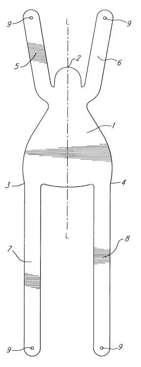
Figure 1: a plan view of an embodiment of the implant.
Figure 1 shows a plan view of an embodiment of an implant
according to the invention. The implant comprises a flat,
flexible basic structure.
A base 1 is triangle-like to elongated oval in shape and has a
longitudinal axis L-L. In the embodiment example, the base 1
rather-resembles a triangle with the corners 2, 3 and 4, the
corner 2 (through which the longitudinal axis L-L of the base 1
runs) being rounded. The area of the base is about 30-50 cm2.
From the base 1, close to the corner 2 but at a distance from
this, a first projection 5 which is bridle-like in construction
starts. The first projection 5 runs on the left-hand side of the
longitudinal axis L-L at a sharp angle with respect to the
longitudinal axis L-L, which is less than 20~ in the embodiment
example. Another first projection 6 which has the same form as
the first projection 5 is arranged in mirror symmetry to the
longitudinal axis L-L.
219~4~9
-- 4
From the corner 3 of the base i, a second projection 7 which runs
on the left-hand side of the longitudinal axis L-L and
essentially parallel to this starts. The second projection 7 is
thus generally directed in the opposite direction to the first
projection 5, i.e. while the first projection 5 in Figure 1
extends (at an angle) upwards, the second projection 7 runs
downwards. Like the first projection 5, the second projection
7 could also form an angle to the longitudinal axis L-L of the
base 1 which differs from 0~. From the corner 4 of the base 1,
another second projection 8 which runs in mirror image to the
second projection 7 in respect of the longitudinal axis L-L
starts. The two second projections 7 and 8 have the same
dimensions and are longer than the two first projections 5 and
6.
Between the two first projections 5 and 6, in the region of the
corner 2, the base forms a semicircular or oval extension about
2-3 cm2 in area. The purpose of this extension is suspension of
the bladder neck and of the proximal urethra, so that after the
implant has been inserted, complete support of the base of the
bladder, bladder neck and proximal urethra is achieved overall.
Holes 9 which can be of assistance during insertion and fixing
(suturing in place) of the implant can be provided close to the
ends of the first projections 5 and 6 and of the second
projections 7 and 8.
In the embodiment example, the implant has three layers. The
middle layer comprises a non-absorbable, flexible net of 0.7 mm
thickness made from monofilament polypropylene threads. This
material does not lose its physical properties in the body in the
long-term, and is insensitive to variations in pH. It is elastic
and unidirectionally extendable, to allow changes in shape, such
as exist, for example, during pressure on the abdomen, due to the
filling level of the bladder or during micturition.
21904~9
-- 5 --
A porous coating of an absorbable composite material comprising
polyglactin 910 and polydioxanone is applied to both sides of the
polypropylene net. In the embodiment example, these outer layers
are absorbed without residue within about 120 days after
implantation.
The basic idea of this material combination is to combine the
connective tissue-conductive properties of the layers of the
composite material, which are preferably constructed as nonwoven
layers (fleece), with the permanent stability of the
polypropylene net. Findings from animals experiments show that
a large quantity of fibrohistiocytic tissue is constructed
through the nonwoven layer on the surface within the first three
weeks. In particular, filaments of polyglactin 910 serve as
conductors for the formation of aligned collagenic connective
tissue fibres which, in contrast to scar tissue, show no tendency
to shrink at all. As absorption of the outer layers progresses,
loose collagenic connective tissue is constructed through the net
of polypropylene and surrounds it. These healing-in processes
secure the implant against dislocation and protect the
surrounding tissue from mechanical irritation or erosion, with
firm and permanent suspension of the urinary bladder. The good
tissue compatibility of the materials used is confirmed by wide
clinical use.
-
The bursting pressures of the implant measured in vitro are farabove the forces which occur physiologically under load in
humans. The implant is extendable unidirectionally by about one
third of its starting length.
The implant shown in Figure 1 has a base 1 of triangle-like
shape. Deviations from this are possible. As already mentioned,
the first projections 5 and 6 also do not have to run in exactly
the opposite direction to the second projections 7 and 8,
respectively. The precise shape of the implant can be adapted
to suit the anatomical circumstances of the patient.
2190449
-- 6
The build-up of the layers described for the implant and the
choice of material also serve only as an example. Other tissue-
compatible materials can similarly be used.
One possibility of how the implant according to the invention can
be inserted in a surgical operation is described in the
following.
The spatium retropubicum is exposed by a Pfannenstiel's incision.
After the apex of the bladder has been loosened from the
peritoneum down to the vagina, the bladder is removed from the
roof of the vagina proximally approximately a good two finger-
widths up to the bladder neck and there only approximately one
finger-width. The vessels running laterally and the ureter are
to be protected carefully. This preparative procedure can be
facilitated by preoperative injection under the vagina with
saline solution alone or by addition of suprarenin 1:200,000.
It is advisable to tampon the vagina. With an indwelling
catheter, preparation is effected with an Overholt and scissors
between the rear wall of the bladder and vagina distally and
laterally, so that the Overholt tip can be seen on both sides
paraurethrally in the spatium retropubicum.
From retrosymphyseally to both sides of the urethra/bladder neck
angle,-in each case a strong guide thread is drawn with the
Overholt between the bladder and vagina. On these guide threads
the two second projections 7, 8 or the front (proximal) bridles
of the implant can be drawn retrosymphyseally between the vagina
and bladder and positioned exactly. The wide base 1 of the
implant comes to rest between the bladder and vagina. It should
be ensured that the position of the alloplastic implant extends
sufficiently far below the bladder neck in order to eliminate an
existing insufficiency of the bladder neck or so that such an
insufficiency cannot develop. The two first projections 5, 6 or
rear (distal) retropubic bridles are passed by the urethra on
21904~9
both sides and fixed to the ligamentum pubicum superior behind
the two pubic rami.
The rear wall of the bladder from the bladder neck to the apex
of the bladder rests with a large surface area on the implant.
When the implant is introduced, it is to be ensured that the
often very thin wall of the bladder is not pushed towards the
urethra, but is tightened abdominally. If the bladder tissue
wrinkles in the region of the bladder neck, prolapse of this
excess portion of the wall of the bladder may later result here.
Nevertheless, to avoid dislocation a sufficient number of fine
monofilament fixation sutures (e.g. with thread thicknesses 4/0)
is advisable. It is appropriate to pin the vagina to the implant
on both sides with single-knot sutures and thus also to
incorporate the vagina into the suspension.
Before fixing the first projections 5, 6, the urinary bladder is
filled with about 300 ml saline solution, to ensure that
sufficient space remains for the urinary bladder to expand and
to maintain an adequate bladder capacity. The two second
projections 7, 8 of the implant are pulled right and left through
the musculus rectus and apposed crosswise over this.
Pulling too tightly on the second projections 7, 8 carries the
risk of severely limiting the retropubic space and therefore the
possibility of expansion of the urinary bladder.
To avoid infection, opening of the bladder or suprapubic draining
of urine should be refrained from as far as possible.
The use of the implant according to the invention is not to be
interpreted as a universal method, but is, in particular, a
reliable method in extreme situations of a descensus vesicae with
generalized weakness and rarification of the tissue texture in
the pelvic floor. Suspension of the urinary bladder with the
21~0449
implant is particularly suitable in the event of pronounced
recurrences, including after several previous operations. In
addition to rapid postoperative mobilization, rapid occupational
integration is also ensured if the floor of the pelvis is exposed
to high stress due to heavy lifting. In extremely obese patients
with vertical and rotatory descensus, it would be conceivable to
use the implant according to the invention as a primary
intervention. The essential advantage lies in the wide-area,
absolutely stable, bilateral fixation of the urinary bladder both
in the bladder neck region and in the region of the apex of the
bladder. The urinary bladder is supported elastically as on a
hammock as a result of the implant, regardless of the condition
of the pelvic floor. Renewed prolapse or descensus can reliably
be avoided even under load. Since with the implant not only the
bladder outlet but the entire urinary bladder is incorporated in
a broad fixed support which supports the entire base of the
bladder, by suspension of the implant it is not an increase in
the discharge resistance which is aimed for but exclusively
stress-proof relieving of the sphincter vesicae externus. There
is no risk of obstruction if the implant is introduced correctly.