Note: Descriptions are shown in the official language in which they were submitted.
2193306 EM4 ~'~ 1 ~ 8 81
4
r TECHNICAL FIELD
z This disclosure relates to surgical joining of bone bodies, and
3 more particularly to instant fixation and staged bone fusion of bone
bodies, such as spinal vertebrae.
si
BACKGROUND OF THE INVENTION
7 Although the immediate effort leading to this disclosure is directed
a toward the lumbar spine (anterior or posterior in approach), the
described vertebral implants for immediate fixation and staged
stabilization leading to arthrodesis (bone fusion) of bone bodies may be
m used in a bone fracture or osteotomy to fuse together resulting bone
~z bodies, and across one or more joints or articulations. Furthermore,
,3 the implants may be used in the lumbar, thoracic and cervical spine.
_ ~, To facilitate fusion and healing of fractured bones, it has long
~s been known to utilize fixation plates and screws to hold together
r6 disunited bone bodies. Typically, the separate bone bodies are formed
when a single bone fractures, requiring bone reunion. Plates are
rs secured across a fracture region with screws, joining together the bone
bodies. The plates hold the bone bodies together in proximate relation,
zn facilitating bone growth and fusion there between. In this manner, the
z~ bone bodies are supported in close proximity, or in direct contact which
z1 facilitates fusion there between. For cases where it is impossible to
z3 fixture together bone bodies internally of a patient's skin, external
z~ fixation is used. For external fixation, threaded pins are rigidly secured
BA9-OOS.P03 A279512)91110N 1 PAT-USIAP-00
2193306
1 into each bone body. The pins, which extend outwardly of a patient's
1 skin, are fixtured together with an external fixation device, placing the
bone bodies in adjacent proximate position to promote healing there
a between. However,' this is not practical for certain joints such as joints
s formed between spinal vertebrae.
s An early technique for achieving arthrodesis between adjacent bone
7 bodies across a joint or articulation is the well known Cloward
Technique for use in the human cervical spine. A solitary dowel of
s bone is tapped into place in a prepared circular bed that is smaller
to than the dowel of bone. The dowel acts as a wedge, distracting the
11 surrounding soft tissues of the joint, and separating the bone bodies or
11 vertebrae joined there along. The intervertebral disc substantially
m comprises the soft tissues of the joint. The dowel of bone is inserted,
I or wedged into place, prodding its own stability by putting an annulus
Is of the disc on stretch. Additionally, simple friction of the inserted
16 dowel between adjacent vertebral bodies stabilizes axial dislocation.
17 However, a second surgical procedure must be performed to extract or
18 harvest the dowel of bone, substantially adding trauma to the procedure,
19 increasing costs, as well as increasing the threat of infection to the
1o patient. Alteratively, bank bone from human donors can be used, but
11 bank bone is less osteogenic and may introduce infection, or even
a transmission of Acquired Immune Deficiency Syndrome (AIDS) or
13 hepatitis. Furthermore, bone morphogenic protein, hydroxyapatite, or
1a other bone stimulating material may be utilized. Additionally, there has
BA9-OOS.P03 ,4179511191110N 2 PAT-UStAP-00
2193306
been a need to ensure the implant remains axially secured which has
2 lead to further developments.
3 As a step forward from the Cloward Technique, the Bagby metal
dowel (U.S. Patent No. 4,501,269) utilizes the same principle. A
s perforated cylindrical hollow implant is inserted between prepared
surfaces across a vertebral joint. The inserted implant immediately
7 stabilizes the joint by spreading the bony surfaces apart in wedged
s opposition to surrounding tissue. This initial stabilization is more
9 substantial because a metal dowel, unlike a bone dowel, will not be
absorbed or fatigue in use. Over time, fusion occurs through and
m around the implant which is filled with bone fragments. Use of the
~2 metal dowel eliminates the need for a second operation to harvest a
r3 dowel of bone. Bone fragments to be inserted in the implant are
m retrieved during preparation of the circular beds in each vertebra.
~s Furthermore, such a metal implant avoids the disadvantage of having to
use bone bank to obtain donor bone. The Bagby implant described in
m U.S. Patent No. 4,501,269 has a smooth outer surface, interrupted only
r8 by numerous openings or fenestrations through which bone ingrowth and
through growth can occur. Bone morsels or bone grafts are typically
zo harvested when preparing the circular bed in each vertebra, after which
m they are placed into the fenestrated metal cylindrical implant. The
22 Bagby implant is then driven or tapped into place in a manner similar
23 to the placement of Cloward's Bone Dowel, which was solely directed
2~ for use in the cervical spine.
BA9-OOS.P03 A279512I9121ON 3 PAT-USIAP-00
2193306
Improvements have also been made to "Cloward's Technique"
z wherein two dowel bone grafts are posteriorly inserted (Wiltberger's
3 Technique) between adjacent lumbar vertebral bodies. Furthermore,
threaded surfaces have been added to such bone grafts in order to
s keep the grafts in place (Otero Vich German Application Number
3,505,567, published June 5, 1986). More recently, a number of U.S.
7 Patents have proposed combining the threaded features from threaded
s bone grafts with a metal implant, resulting in rigid threaded implant
9 structures for placement between adjacent spinal vertebrae.
One threaded metal fusion implant disclosed in Michelson (U.S.
m Patent No. 5,015,247) provides a cylindrical fusion implant having an
~z outer diameter sized larger than the space between adjacent vertebrae
13 to be fused. Threads provided on the exterior of the member engage
r.r the vertebrae to axially secure the implant there between. The implant
is has a plurality of openings configured along the cylindrical surface to
promote bone ingrowth. However, the threads per se of the implant
do not function as a fastener to fix together the adjacent vertebral
rs bodies. Instead, the implant functions as a wedge, imparting a
r9 distraction force across the disc which stabilizes the articulation formed
zo there between by stretching the annulus of the disc. In fact, the
a threaded implant relies solely on the annulus to provide stabilization
zz between the vertebrae, in direct responsive to wedge-induced distraction
z3 created there between. Distraction of the annulus stabilizes the two
z~ vertebrae, enabling ingrowth to later occur within the implant.
BA9-OOS.P03 A17951z191zlON 4 PAT-USlAP-00
2193306
r Therefore, through-growth and fusion (arthrodesis) occur between the
1 adjacent vertebrae subsequent thereto depending on the immobilizing
3 potential of an intact healthy annulus which may or may not be
.r present. Therefore, there is a need to provide an implant that
s produces immediate fixation per se between bone bodies following
s insertion and independent of the annulus. Particularly for cases where
7 the annulus structure is substantially or completely weakened or damaged
a at surgery of implantation, the wedge-effect of prior art threaded
implants will not produce any distraction forces across the annulus.
ro Also, when the implant is used to arthrodese and change angulation, a
rr healthy annulus cannot be totally corralled to be placed on stretch. As
r1 a result, there is no form of stabilization or fastening between bone
rs bodies sufficient to enable the occurrence of arthrodesis there between
r.r when the annulus is weakened or inadequate.
rs Another threaded implant disclosed in Ray (U.S. Patent No.
r6 5,005,104) provides a threaded fusion cage that is configured to be
r7 implanted in close adjoining pairs between adjacent vertebral bodies.
ra Threads of adjacent cages are configured in overlapping relation when
r9 they are implanted. However, the fusion cages function only as wedges,
Io imparting distraction forces across the annulus. The distraction forces
zr immediately stabilize the intervertebral articulation by stretching the
z1 annulus of the disc immediately after implantation. Over time, the
z3 adjacent vertebrae fuse together. However, where a stretched annulus
Ia does not provide sufficient stabilization, initial early bone growth is
BA9-OOS.P03 A179512r9I1rON S PAT-USIAP-DO
2193306
r seriously hindered, if not completely prevented. Furthermore, a
1 stretched annulus can still allow slight motion.
3 For bone fusion to occur with any of the above devices, the
a invasion of new delicate blood vessels from the adjacent healthy bone
s is necessary for the creation of new living interconnecting bone. Where
complete stabilization does not occur instantaneously upon implantation,
7 motion can disrupt the in growth of delicate blood vessels. Disruption
a of the vessels then restricts or even prevents bone healing there
9 between. The same problem occurs with any of the above mentioned
1o implant techniques, including the threaded techniques of Otero Vich and
11 Michelson. Even when the annulus is completely on stretch, the threads
r2 per se of these constructions do not function in the manner of
m conventional screws, extending through one object and into another.
m Namely, they do not function to fasten together adjacent bodies by
1s coaction of the thread with each body. Alternatively, they do not
!6 fasten together bodies by action of the thread with one body, and
17 action of a fastener head with the other body. Instead, the threads
1s I~ merely act as a series of ridges that engage with each adjacent bone
19 body, while the implant body functions as a wedge. The implant
1o distracts apart the vertebral bodies which stretches the annulus, and
a stabilizes the articulation as a consequence thereof, while the thread
1z functions solely to prevent axial dislodgement.
13 A further area of prior art relates to implants having surface
1a features that enable bony ingrowth to occur. For example, beads of
BA9-OOS.P03 A179511191110N 6 PAT UStAP-00
2193306
! titanium have been provided on the stems of hip implants to form such
1 features. Ingrowth by a bone bed with the structural features occurs
3 some time after implantation. Therefore, fixation is not immediately
Present as a result of the surface features, and some other fixation
s must be relied upon until ingrowth occurs. With the exception of the
Cloward Bone Dowel and Otero Vich, the above-mentioned vertebral
7 body implant devices incorporate fenestrations or openings that tend to
a facilitate bony ingrowth into the metal spinal implants.
Additionally, Lin et al. (U.S. Patent No. 4,778,469) teaches a
ro surface construction of a space occupier having a pattern for tissue
r! ingrowth in the surface of an implant. Tapered posts having undercuts
r1 are provided along a surface of a hip implant. Subsequent to
m implantation, physiological bone ingrowth occurs within the undercuts,
!.r helping to fix the implant within the bone. However, this construction
!s does not enable immediate fixation via the undercuts. Instead, it relies
!6 upon physiological bone ingrowth which takes time to occur. Therefore,
!7 other mechanisms must be relied upon to maintain implant fixation
rs within the bone prior to ingrowth. Such is also the case with the
!9 previously mentioned vertebral implants.
zn Therefore, there is a present need to provide implant devices that
1r fasten bone bodies together directly upon implantation. There is also
z1 a need to provide such a device that facilitates staged stabilization,
13 ultimately leading to bone fusion there between. The final stage of
m bone fusion through and around the implant substantially eliminates any
BA9-OOS.P03 A179511l9l110N 7 PAT-USIAP-00
2193306
I need for the implant to maintain the fusion, thus allowing the bone
z union to provide primary support there between, i.e. the implant can
3 be removed without reversing the arthrodesis in such cases as chronic
a infection. Furthermore, there is a need to provide such a device for
s fixing bone bodies together across an articulation or joint (arthrodesis).
Particularly, this need exists where soft tissues of an articulation have
7 deteriorated to such a condition that distraction across the articulation
a will not produce stability. For example, prior art devices (including the
9 above-mentioned vertebral body implant devices) cannot stabilize an
to articulation by inducing a wedging apart, or stretching of an annulus
II where the annulus is weakened or absent. Therefore, interim stability
IZ cannot be imparted between adjacent vertebrae at the time of surgery.
l3 Such interim stability is needed for successful fusion. As a result,
Lr ingrowth and through growth needed to fuse the bone bodies together
Is for long-term stability is less likely to occur on a routine basis.
16
17 BRIEF DESCRIPTION OF THE DRAWINGS
la Preferred embodiments of the invention are described below with
19 reference to the following accompanying drawings.
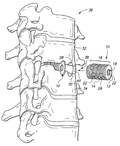
2o Fig. 1 is a perspective view of a vertebral structure showing a
a pair of vertebral interbody implants embodying this invention, one
11 inserted and the other positioned for insertion. A solitary implant (not
z3 shown) may be used in certain cases;
z.!
BA9-OOS.P03 A2795I21911ION g PAT USIAP-00
219330b
r II Fig. 2 is a side elevational view illustrating the vertebral interbody
1 implant of Fig. 1;
3 Fig. 3 is a leading end view taken generally on line 3-3 of Fig.
II 2~
s II Fig. 4 is a trailing end view taken generally on line 4-4 of Fig.
6 II 2~
' II Fig. 5 is an unrolled plan view of the outer peripheral surface
a II of the vertebral interbody implant of Figs. 1-3;
Fig. 6 is a cross sectional view taken generally on line 6-6 of Fig.
ro 3 of the vertebral interbody implant immediately after implantation;
rr Fig. 7 is a fragmentary and enlarged cross-sectional view taken
r1 generally on line 7-7 of Fig. 5 and shows initial surface through growth
r3 diagrammatically;
r.r Fig. 8 is a cross sectional view taken generally on line 8-8 of Fig.
rs 4 of the vertebral interbody implant subsequent to implantation and
r6 illustrating bone in-growth (diagrammatically) including the bone grafts
with interlocking;
rs Fig. 9 is a cross sectional view corresponding to that of Fig. 8,
r9 but taken later in time and illustrating advanced bone through-growth
zo (diagrammatically) beyond surface growth and ingrowth of Figs. 7 and
z1 8, and illustrates bone joining and connecting vertebrae;
11 Fig. 10 is a cross sectional view taken generally on line 10-10 of
z3 Fig. 2 illustrating bone through-growth at the same time as that
depicted in Fig. 9, but in histologic detail;
BA9-OOS.P03 A279511191110N 9 PAT USIAP-00
2193306
r Fig. 11 is a cross sectional view corresponding to that of Fig. 10,
1 but taken later in time and illustrating bone remodelling;
3 Fig. 12 is a fragmentary and enlarged centerline sectional view
a depicting a vertebral interbody implant having an alternative undercut
s thread construction with bony through growth;
Fig. 13 is a fragmentary and enlarged centerline sectional view
7 depicting a vertebral interbody implant having another alternative
undercut thread construction;
9 Fig. 14 is a fragmentary and enlarged centerline sectional view
depicting a vertebral interbody implant having a third alternative
m undercut thread construction;
m Fig. 15 is a perspective view illustrating a bridging vertebral
73 interbody implant embodying this invention for use in performing a
m corpectomy;
~s Fig. 16 is a front elevational view of a vertebral structure showing
l6 three vertebrae, with a midmost one having a visible cancerous or
benign tumor;
ra Fig. 17 is a front elevational view of the vertebral structure of
19 Fig. 16 depicting the cephalad and caudad vertebrae prepared to receive
zo the alternatively constructed interbody implant of Fig. 15;
z~ Fig. 18 is a front elevational view of the vertebral structure of
11 Fig. 17 after receiving the alternatively constructed interbody implant;
z3 and
1a I
BA9-OOS.P03 A1795I1191110N 1~ PAT USIAP-00
CA 02193306 2005-10-03
11
Fig. 19 is a front elevational view of the device of
Figs. 15 and 18 after removal of the mid-most vertebra,
insertion of a medial cruciate baffle, and prior to closure of
the surgical wound.
DETAILED DESCRIPTION OF THE PREFERRED ENBODIMENTS
In accordance with one aspect of the invention, a bone
joining implant is engaged between a pair of bone bodies to be
joined. The implant has a rigid, implantable base body with an
outer surface having at least one bone bed engaging portion.
The outer surface engages with a bone bed prepared in each
bone body to be joined. One or more splines are provided by
the bone bed engaging portion. The spline is constructed and
arranged to engage outwardly of the implant body. Furthermore,
the spline has an undercut portion configured to engage with
the bone bed provided in each bone body to be joined.
In accordance with another aspect, the invention further
provides a bone joining implant comprising a rigid,
implantable base body having an outer surface with at least
one bone bed engaging portion configured for engaging between
bone beds on a pair of bone bodies to be joined; and at least
one spline comprising a thread provided by the bone bed
engaging portion, the spline constructed and arranged to
extend outwardly of the body and having at least one tapering
undercut portion to produce a cross-sectional configuration
with a radial outer portion having a dimension sized larger
than a radial inner portion, the undercut portion configured
to engage the spline in interlocking relation with a bone
bed provided in each of the bone bodies to be joined.
In accordance with another aspect, the invention further
provides a bone joining member comprising a tubular body
having an outer dimension sized to be received between a pair
of adjacent bone bodies to be joined and a thread extending
about the body configured to engage in assembly with each
adjacent bone body, the thread being undercut along a radial
CA 02193306 2005-10-03
12
inner portion to engage with a correspondingly prepared bed in
each bone.
In accordance with another aspect of the invention, a
vertebral interbody implant is engaged between a pair of
adjacent vertebrae to be joined. The implant has a body sized
to be received between a pair of adjacent vertebrae to be
joined. The body forms an outer surface having at least one
bone bed engaging portion configured to be engaged with a bone
bed on each of a pair of vertebrae to be joined. At least one
thread is formed by the bone bed engaging portion that extends
radially outward of the body in a generally helical
configuration. The thread is configured to engage in assembly
with each adjacent vertebra. Furthermore, the thread has an
undercut portion provided on a radial inner portion for
engaging in interlocking relation with a bone bed in each
vertebra. In assembly, the implant engages in interlocking
relation with a pair of vertebrae joined there along.
In accordance with another aspect, the invention further
provides a vertebral interbody implant comprising an
implantable body sized to be received between a pair of
adjacent vertebrae to be joined; an outer surface formed by
the body having at least one bone bed engaging portion
configured to be engaged with a bone bed on each of a pair of
vertebrae to be joined; at least one thread formed by the bone
bed engaging portion, extending radially outward of the body,
and configured in a generally helical configuration, the
thread being configured to engage in assembly with adjacent
vertebra; and an undercut portion provided on a radial inner
portion of the thread, the undercut portion engaging in
interlocking relation with the bone bed in each vertebra;
wherein, in assembly the implant engages in interlocking
relation with a pair of vertebrae joined there along.
In accordance with yet another aspect of the invention, a
vertebrae bridging implant is received about a vertebral body
to be removed by a corpectomy. An implantable body on the
implant has an inner dimension sized to be received about the
CA 02193306 2005-10-03
12a
vertebral body to be removed. An outer dimension on the body
is sized to be received between a pair of vertebrae to be
joined adjacent thereto. The body has an outer surface with a
bone bed engaging portion configured to be engaged with bone
beds on each vertebra to be joined. An undercut spline extends
outwardly of the body, provided by the bone bed engaging
portion. In assembly, the spline mates in interlocking
engagement with the bone bed of each vertebra, joining the
non-adjacent vertebrae in interlocking relation.
In accordance with another aspect, the invention further
provides a vertebral bridging implant configured for use in
performing a corpectomy, comprising: an implantable body
having an inner dimension sized to be received about a
vertebral body to be removed, and an outer dimension sized to
be received between a pair of vertebrae to be joined adjacent
thereto; an outer surface provided by the body has a bone bed
engaging portion configured to be engaged with bone beds on
each vertebra to be joined; an undercut spline provided by the
bone bed engaging portion, the spline extending outwardly of
the body, the spline in assembly mating in interlocking
engagement with the bone bed of each vertebra; and a bridging
port ~on having a central chamber therein, providing the inner
dimension sized to be received about a vertebral body to be
removed, the central chamber constructed and arranged to
substantially encircle a vertebral body to be removed
therefrom, a top-most and a bottom-most portion of the
bridging portion defining at least one of a pair of the bone
bed engaging portions there along; upon implantation of the
spline within each prepared bone, the top-most and the bottom-
most portions being mated in interlocking assembly with bone
beds of neighbouring associated vertebra there along, wherein
the implant is mated in interlocking engagement with top-most
and bottom-most neighbouring vertebral while encircling a mid-
most vertebral body to be removed.
A preferred embodiment bone joining implant in accordance
with the invention is first described with reference to
CA 02193306 2005-10-03
12b
Figures 1 through 11. Such an implant is described further
below with respect to a threaded vertebral interbody implant
having an undercut thread portion. The undercut threaded
implant is designated in Figures 1-11 generally with numeral
10. A pair of implants 10 are depicted in this implementation.
2193306
1 Alternatively, a single implant could be used. As shown in Figure 1,
z such comprises a rigid, cylindrical base body 12 having a helically
3 configured spline or thread 14 configured on an outer surface 16 of
body 12. A central chamber 18 is formed within body 12 for receiving
s bone graft material 20 therein. Large and small fenestrations 22 and
s 24 extend through surface 16 into chamber 18 for facilitating bony
' ingrowth and through growth therethrough. Thread 14 has an undercut
26 which meshes in assembled engagement within bone beds 28 and 30
9 in vertebra 32 and 34. Each bed 28 and 30 forms complementary
to female threads for receiving the undercut thread in interlocking
m engagement there along. For purposes of this disclosure, a spline shall
r2 include any thread, web, strip, ridge, or portion of material formed from
m continuous material, or broken into fragments (interrupted).
- 1~ As shown in Figure 1, vertebrae 32 and 34 comprise neighboring
Is ~~ bone bodies of a vertebral column 36. A resilient articulation 38 or
16 II joint is formed between vertebra 32 and 34 by a disc 40 extending
1' II there between. Anatomically, the disc is made up of a central nucleus
la pulposus and an outer encircling annulus. The annulus and nucleus
l9 pulposus are composed of laminae of fibrous tissue and fibro-cartilage.
zo The nucleus pulposus, located at the center of the disc, comprises a
z1 soft, pulpy, highly elastic substance. The annulus is formed from
m laminae of fibrous tissue extending in criss-crossing fashion to encircle
z3 the nucleus pulposus. Additionally, the intervertebral disc is adherent,
za by its cephalad and caudad surfaces, to a thin layer of hyaline cartilage
BA9-005-P03 A279511191110N 13 PAT-USIAP-00
2193306
r that covers the top and bottom surfaces of adjacent vertebrae. In a
z healthy patient, adjacent vertebra 32 and 34 are spaced apart by the
3 disc 40. However, degenerative disc disease and localized trauma can
cause degradation ' or complete loss of the soft tissue components
s between neighboring vertebrae. For example, the annulus can partially
or completely tear which can seriously degrade the structural condition
7 of the articulation. Additionally, fluid can escape from the nucleus
s pulposus. When any of the above happens, vertebrae 32 and 34,
v loaded by the normal weight bearing of a patient, are pressed into
ro closer adjoining positions, which can result in pinching of nerves that
m extend from between vertebrae of the spinal column (not shown).
r1 Therefore, there is a need to recover the disc spacing provided
13 by a normal healthy disc 40 by way of inserting implants 10.
Furthermore, there is a_ need to provide implants 10 with a fixation that
rs instantly interlocks adjacent vertebra 32 and 34 together upon being
implanted. Furthermore, there is a need for such an implant 10 that
m facilitates staged stabilization resulting in arthrodesis to occur between
the vertebral bodies, following initial implantation.
19 As a result, implant 10 can be inserted, preferably in left and
zo right laterally positioned pairs, between adjacent vertebrae of patients
m who have bad, ruptured or degenerative discs. A solitary implant may
11 also be used in chosen cases. For example, the implant can be axially
z3 oriented anterior to posterior, or even laterally. In summary, implants
2 10 are adapted for implantation between prepared bony surfaces or beds
BA9-OOS.P03 A17951119111ON 14 PAT-USIAP-00
2193306
r 28 and 30 of articulation 38. A typical implantation might involve
1 placement of one or more implants 10 as required in order to stabilize
3 and fix the joint during bone ingrowth and through-growth of the
a implant structure. ~ Bone growth is also accomplished outside of and
s ~~ surrounding the implant.
Preparation of bone beds 28 and 30 is performed according to
7 well known techniques in the art, with the, exception that presently
a available tapping devices are modified according to one implementation
of this invention. During a surgical procedure, a prepared and exposed
ro vertebral column 36 receives a hollow guide tube (not shown) having
m teeth at its lower end. The tube (or sleeve) is mated with its lower
r1 end engaging across articulation 38, wherein the teeth engage vertebrae
13 32 and 34. A drill (not shown) is then guided down the tube in order
1.r to drill a pilot hole between the vertebrae, imparting the general cutout
is configuration to beds 28 and 30. Subsequently, a preliminary tapping
r6 device is received through the tube into the pilot hole where female
r7 threads of rectangular cross section are cut into beds 28 and 30. The
ra female threads are undersized in width, requiring further self-tapping by
rv the implant during insertion. Alternatively, the tapping device can be
1o configured to cut female threads having an undercut cross section sized
Ir and configured to conform with the undercut threads 14 on implant 10.
11 For the case where threads are cut with a rectangular cross section,
13 implant 10 has self tapping features provided by thread 14. The self
Ia tapping features enlarge the radial outermost portion of each
BA9-OOS.P03 A179511191110N 1S PAT-USIAP-00
2193306
II complementary corresponding female thread provided by beds 28 and 30,
~~ respectively.
3 An additional benefit is provided when implant 10 is self tapped
a into an undersized female thread in each bone bed 28 and 30.
s Namely, in practice it proves difficult to maintain precise spaced apart
positioning of vertebrae 32 and 34 following drilling of beds 28 and 30.
7 Therefore, even slight variations in spacing produce a misfit between the
a outer surface of implant 10 and each bed 28 and 30. However, where
9 implant 10 has self tapping features, the self tapping mitigates any slight
misfit condition by at least partially reforming the beds 28 and 30
rr during insertion. Furthermore, a laterally positioned pair of implants
r1 10 proves difficult to implant with a perfect fit up between the implants
r3 and the bone beds. Typically, a first site is prepared and one implant
r.s is inserted, after which a second site is prepared and a second implant
~s is inserted. However, the bone beds at each site tend to shift as each
implant is inserted since the pair of implants are not inserted
r7 simultaneously.
~a Figure 2 illustrates undercut threaded implant 10 in a side
m elevational view corresponding to a preferred rotationally positioned and
Zo implanted configuration within a patient. Large fenestrations 22 are
m formed along a pair of perpendicular axes that substantially bisect
z2 vertical and horizontal planes of a patient receiving the implant. Large
z3 fenestrations 22 are sized and located to allow for reorganization or
za II hardening of bone by maturity after initial bone healing. Figure 3
BA9-OOS.P03 A179511191110N 16 PAT USIAP-00
219330b
1 clearly illustrates such a configuration. In this manner, top most or
1 cephalad and bottom most or caudad portions of implant 10 present an
3 outer surface that is void of any large openings or fenestrations. Such
.r a surface substantially enhances load bearing there along, following
s implantation. However, such placement of large fenestrations 22 still
s provides a significant path for subsequent ingrowth and through growth
between adjacent vertebral bodies receiving implant 10. Additionally,
a small fenestrations 24 provide paths for ingrowth and through growth.
9 II Furthermore, the above-described orientation and size of fenestrations
l0 22 is well suited for staged fusion and subsequent bone remodelling,
I1 leading to structurally enhanced fusion between vertebrae 32 and 34.
m Implant 10 of Figure 2 is depicted with a plastic end cap 42 that
l3 mounts in engagement with an opening 44 formed in a trailing end 46
la (as shown in Figure 4). End cap 42 is optionally mounted to implant
Is 10 when performing a bilateral implantation by way of a posterior
16 approach. Such a posterior surgical approach presents a special
17 concern; namely, that the dura of the spinal cord might rub against the
la driving or trailing end 46 of implant 10. Therefore, end cap 42 is
l9 secured in opening 44 to protect the spinal cord, following implantation
zo of implant 10 and the inserted bone graph material therein.
11 Additionally, small fenestrations 24 are formed in implant 10.
11 II The small fenestrations 24 extend substantially throughout the walls of
13 II the cylindrical implant. Small fenestrations 24 offer avenues of
ingrowth
z.1 II of bone between vertebrae 32 and 34, which is stimulated by bone graft
BA9-OOS.P03 A179511191110N 17 PAT USIAP-00
2193306
1 material placed within central chamber 18 during implantation. In this
z manner, fenestrations 24 serve to facilitate earlier and more thorough
3 ingrowth of bone within implant 10. Furthermore, fenestrations 24
.1 enhance overall through growth of bone through implant 10.
s Large fenestrations 22 are preferably sized and configured to
interrupt no more than one complete width of thread 14. Hence, the
7 total amount of interlocking of undercut thread 14 with bone beds 28
a and 30 is optimized. According to this construction, fenestration 22
9 extends substantially between a first and a third adjacent portion of
to helically wound thread 14, while interrupting a second, or middle
11 portion. A taper 56, also provided along end 50 reduces the height of
Iz thread 14 substantially to a diameter of base body 12, immediately
m adjacent leading end 50. Placement of fenestrations 22 in close
I proximity with leading end 50 tends to interrupt thread 14, at least
Is partially providing a self-tapping feature there along. Furthermore,
16 fenestrations 22 allow for self depositing of bone chips during the self
I tapping process of implant 10. Bone chips are delivered into the large
rs and small fenestrations 22 and 24, as well as into central chamber 18
19 during self tapping of the implant. Such bone chips supplement bone
zo graft material 20 that is packed into the implant, which together
z1 encourage the early process of arthrodesis. These delivered bone chips
a produced in self tapping remain alive since they never leave the
z3 patient's body. Therefore, they tend to be more osteogenic than if they
z~ were nonviable.
BA9-OOS.P03 Az7951z191z10N 18 PAT USIAP-00
2193306
As shown in Figure 2, fenestrations 22 only partially provide for
1 complete self tapping. Therefore, cut-out portions 58 are also formed
3 along thread 14 in order to add further self-tapping features.
a Additionally, thread 14 within the region of taper 56 also forms an
s undercut 26 so that progressive formation of complementary
corresponding undercut female threads can be formed within bone beds
of adjacent vertebrae receiving the implant.
a Alternatively, fenestrations 22 can be provided immediately adjacent
to leading end 50, eliminating the need for cut-out portions 58
altogether when providing self tapping features on implant 10. For
rr cases where female threads 66 are preformed into beds 28 and 30 with
~z undercut, there is little or no apparent need for such self tapping
features. In such cases, fenestrations 22 can be placed anywhere along
ra implant 10, and cut-out portions 58 (Figure 5) can be eliminated (i.e.
~s No other self-tapping needed).
Referring to Figure 3, leading or advancing end 50 of implant 10
l~ is clearly shown. Cut-out portions 58 can be clearly seen in this view.
~a Additionally, taper 56 progressively decreases the height of thread 14 as
it extends helically toward leading end 50, until it merges into base
zo body 12. Furthermore, a plurality of axially extending through holes 60
zr cut through thread 14 are clearly shown. Through holes 60 facilitate
z1 early stage ingrowth of bone with implant 10 following implantation.
13 As shown in Figure 3, through holes 60 are also formed in thread 14
z~ in the region of taper 56, thereby interrupting thread 14 in the region
BA9-OOS.P03 A1795/1191110N 19 PAT-USIAP-00
2193306
of taper 56 so as to impart even further self-tapping features there
1 along. Alternatively, through holes 60 can be omitted in the region of
~~ taper 56.
a Opening 48 in leading end 50 is sized smaller in diameter than
s an inner wall 62 of the implant base body 12, as shown in Figure 3.
a In this manner, a structure enforcing bulkhead is provided at the
7 leading end. Additionally, a similar centrally positioned bulkhead 54 is
also provided intermediate of the advancing and driving ends 50 and 46,
respectively, within central chamber 18. Central bulkhead 54 has an
oval slot 52 extending completely therethrough for mating with a
m fingered end of a driver (not shown). The slot mates with the finger,
m allowing torquing of threaded implant 10 into the bony beds of adjacent
r3 vertebrae. Typically, such a driver construction is required in order to
impart large forces necessary to insert implant 10 into the lumbar
rs region of a patient's spine. Alternatively, a driver can be configured
16 to mate with trailing end 46 via a plurality of dowels on the driver.
The dowels are received in complementary corresponding receiving holes
(not shown) of the implant there along. Such a construction is well
19 suited to an implant that is sized to be received also in the cervical
zo region of the spine where less torque is required to insert implant 10
1~ II therein.
11 II A further alternative construction calls for formation of an oval
13 slot (similar to slot 52 of Figures 3 and 4) in place of opening 44 in
1~ trailing end 46. Provision of the siot in the trailing end will enable
BA9-OOS.P03 A17951119IIIDN 20 PAT USL1P-00
2193306
I driver engagement suitable to insert as well as remove the implant.
z Hence, the same tool can be used to remove the implant.
3 Furthermore, the leading end can have a similar slot, allowing for
a surgical removal of an implant where a posteriorly inserted implant
s needs to be removed from the anterior for surgical reasons.
As shown in Figures 3 and 4, axially extending through holes 60
are provided in implant 10 in locations that correspond with the vertical
and horizontal planes of a patient receiving the implant. Additional
through holes 60 are provided between the holes positioned along the
to vertical and horizontal planes, enabling further initial ingrowth there
11 along. For holes 60 that are positioned in locations along the vertical
m and horizontal planes, such holes are provided adjacent small
l3 fenestrations 24 which further facilitates ingrowth and through growth
there along.
is As shown in Figure 4, trailing or driving end 46 has opening 44
16 which is sized similarly to opening 48 in leading end 50 (of Figure 3).
m In this manner, opening 44 is sized smaller than inner wall 62, forming
la an enforcing flange from a portion of trailing end 46 extending there
19 along. Additionally, Figure 4 illustrates termination of undercut thread
zo 14 along trailing end 46 of implant 10. Preferably, axially extending
z1 through holes 60 are machined through the helically extending thread
zz 14 progressively from the trailing end 46 toward the leading end 50.
z3 In this manner, a drill bit received there along to cut holes 60 can be
za
BA9-OOS.P03 A1795111911ION 21 PAT USIAP-00
2193306
1 axially advanced, short of drilling through thread 14 in the region of
z taper 56 if such a construction is so desired.
3 According to Figure 5, outer surface 16 of implant 10 is shown
in an unrolled plan view to better depict layout of thread 14,
s fenestrations 22 and 24, through hole 60, and self tapping cutout
portions 58. The particular layout leads to the various above described
7 benefits. Various alternative layouts can be readily envisioned for
a thread 14, fenestrations 22 and 24, through hole 60 and self tapping
cutout portions 58, and fall within the claimed subject matter of this
to invention. Additionally, any one or more of such features can be
m eliminated, with the exception of an undercut spline, or thread, yet still
m remain within the claimed scope of this invention.
!s Figure 6 depicts implant 10 of Figures 1 through 5 immediately
m after implantation within prepared bone beds 28 and 30 of vertebrae
!s 32 and 34, respectively. According to the depicted implantation of
16 Figure 1, bone beds 28 and 30 are prepared by forming female threads
m 66 therein, prior to receiving implant 10. Subsequently, implant 10 is
is inserted by threading in into the beds with a driver.
19 As shown in Figure 6, a bore 68 is cut into vertebrae 32 and 34
zo (partially shown) by progressively enlarging the bore with a series of
z1 reamers received through a hollow guide tube (not shown).
zz Subsequently, a tap is inserted through the guide tube to cut thread 66
z3 into each vertebrae 32 and 34. In the embodiment of Figure 6, female
m threads 66 are cut to have a square cross-sectional configuration, after
BA9-OOS.P03 A179s1119I110N 22 PAT USIAP-00
2193306
' II which the self cutting features of thread 14 enlarge the square female
z threads during insertion of the implant. Hence, complementary
3 corresponding undercut female threads 66 are formed that are configured
to receive undercut thread 14 in snug and inter-locking engagement
s there along. By pretapping a square (or some other closely configured)
thread into the bone bodies, the self tapping implant 10 can complete
7 the tap, which interlocks the implant with each bone body. In this
s manner, implant 10 when inserted, interlocks vertebrae 32 and 34
9 together immediately upon implantation, instantly stabilizing articulation
'0 38. Subsequently, bone ingrowth and through growth occurs via through
" hole 60 and fenestrations 22 and 24, by way of enhanced development
m via bone grafts 20 packed into central chamber 18. Therefore, with
'3 such a construction, the prior art reliance on a wedging effect to
'~ stretch disc annulus 40 in order to stabilize the joint until bone fusion
's occurs there between is eliminated. However, it is still desirable to
'6 prepare beds 28 and 30 such that distraction occurs across disc 40,
'7 further stabilizing articulation 38 there between. Additionally, a bone
's distractor can also be used during preparation of bone beds and implant
'9 insertion to pull apart the vertebrae, putting the disc annulus on stretch
zo in the final implanted configuration.
z' Implant 10 according to the construction of Figures 1-5 facilitates
z~ staged stabilization and a bone fusion between vertebrae 32 and 34.
z3 Figure 6 illustrates the first stage of stabilization across articulation
40,
za wherein undercut thread 14 interlocks in female threads 66 of each
BA9-OOS.P03 A179511I9I1ION 23 PAT-USIAP-00
2193306
r ~~ vertebra, imparting instant fixation and frictional stabilization there
1 between. Furthermore, annulus 40 further cooperates with the implant
3 to control distraction. A second stage of stabilization is depicted in
Figure 7, wherein ~ new bone from the living female bone threads 66
s through grows into through hole 60. A third stage of stabilization is
depicted in Figure 7, wherein bone grows through small fenestrations 24
7 enjoins with bone growing from the same vertebra through an adjacent
s fenestration, and can further incorporate a portion of bone graft
material contained within the implant. Furthermore, such ingrowth can
to occur through large fenestrations (not shown). A fourth stage of
m stabilization is depicted in Figures 9 and 10, wherein initial bone growth
!1 occurs through the implant, connecting vertebrae 32 and 34. Such a
13 condition forms an initial bone fusion. A fifth and final stage of fusion
la is depicted in Figures 11 and 12, wherein the fused bone of Figure 11
!s has reorganized according to Wolff's law. The trabeculae relocate
16 through large fenestrations 22 to form a mature strengthening of the
r7 trabeculae. Additional reorganization can be facilitated outside of the
18 implant by providing bone graft material thereabout at the time of
!v implantation. Such a staged stabilization enables instant fixation
between vertebrae 32 and 34, after which the articulation 38 is
1! stabilized and stress protected by shifting load-bearing from the implant
11 to newly formed and reorganized bone occurring there between. For
Is cases where staged stabilization and bone fusion is not allowed to occur,
stress protection osteopenia can result from normal physiologic stress
BA9-OOS.P03 A17951219lIION 24 PAT USIAP-00
2193306
1 through the bone being removed via the implant, which can cause
1 localized osteoporosis.
3 Figure 7 is a fragmentary and enlarged cross-sectional view taken
a generally on line 7-7 of Figure 5 illustrating an initial stage of bone
s ingrowth with arrows. Bone ingrowth occurs early on through axially
extending through holes 60 due to their short length and proximity to
7 healthy developed bone in living beds 28 and 30. Since holes 60 are
s cut in thread 14 immediately adjacent base body 12, holes 60 are
9 shallow, and bone growth from living bone beds 28 and 30 need only
to progress a short distance in order to extend completely through holes
11 60. Therefore, early through growth can occur through holes 60, which
r1 further fixes and stabilizes implant 10 to vertebrae 32 and 34. Such
13 early ingrowth within holes 60 provides fixation above and beyond that
la provided by the interlocking fit of undercut thread 14 within female
Is threads 66. Additionally, through growth of bone into holes 60 prevents
Is any loosening or "unscrewing" of implant 10 from within female threads
17 66 of beds 28 and 30. Holes 60 impart a cotter-pin effect, locking
Is implant 10 within threaded beds 28 and 30. A typical time for
19 complete through growth of holes 60 in an active and healthy patient
Io ranges from about two weeks to four weeks.
a Figure 8 schematically depicts a cross-sectional view of implant 10
11 taken generally on line 8-8 of Figure 4. The arrows of Figure 8
z3 schematically represent bone ingrowth in the form of arrows that occurs
m through adjacent fenestrations 24 by way of the same vertebral body 32
BA9-OOS.POj A2795I2191210N 25 PAT USIAP-00
2193306
r or 34. The ingrowth through fenestrations 24 of Figure 8 occurs
1 somewhat later in time than the ingrowth through holes 60 of Figure
3 7. The typical time for through growth is from one month to 3
a months. Ingrowth~ by way of fenestrations 24 further ensures fixation
s of implant 10 to vertebrae 32 along a top portion, and to vertebrae 34
along a bottom portion. At this stage of stabilization, implant 10 still
7 holds vertebrae 32 and 34 together in structural relation. Furthermore,
s the ingrowth depicted in fenestrations 24 in Figure 8 also occurs
through fenestrations 22 (not depicted).
!o Figure 9, taken later in time than Figures 7 and 8, depicts
11 complete through growth (arthrodesis) of implant 10 by healing bone
Iz following earlier ingrowth. According to Figure 9, a simplified and
13 somewhat schematic cross-sectional configuration shows large fenestrations
m 22 and omits small fenestrations 24. for clarity. Furthermore, through
!s growth is depicted in the form of arrows extending between vertebra 32,
16 along a top portion, and vertebra 34, along a bottom portion. At this
stage of bone fusion, bone graft material 20 that was initially placed in
rs chamber 18 has fused with healing bone from vertebrae 32 and 34.
l9 The fused bone provides a continuous path of structural bone there
ao between. Furthermore, an additional pathway of bone not depicted in
a Figures 7-9 is bone growth outside of and around the implant. Such
z2 an initial bone fusion tends to have a somewhat random or directionally
z3 uniform bone cell orientation. The bone cells lack any optimized or
m reorganized structural orientation there through, other than that dictated
BA9-OOS.P03 A17951119l110N 26 PAT USiAP-00
219330b
1 by passageways that originally allowed for the bone growth. As shown
z in Figures 9 and 10, the general characteristics of such initial bone
fusion lacks any substantial reorganization. Hence, bone cell geometry
.1 has not been structurally oriented to optimize structural loading.
s Figure 10 depicts histologic bone cell geometry in greater detail,
6 corresponding in time to that depicted in Figure 9. Lacunae and canals
7 or voids 69 are formed between the bone 67. The lacunae represent
the voids between the bone where soft tissue, blood vessels, and fatty
9 deposits reside.
to According to Figure 11, reorganization of fused bone material
m through implant 10 is shown generally occurring between vertebrae 32
IZ and 34. The fused bone consists of bone ingrowth and through growth
l3 of holes 60, fenestrations 22 and 24, and openings 44 and 48.
1.r According to this simplified schematic representation depicting
Is fenestrations 22, the bone cells of Figure 11 have remodeled so as to
Is optimize the substantially vertical loading. Such loading represents the
17 vertical weight bearing that a patient will impart on the implant system
Is as surgically placed in the configuration of Figures 10 and 11. In such
19 a manner, bone cells have remodeled to form a definite elongated
zo configuration extending between vertebrae 32 and 34 through
2l fenestrations 22. Such remodelled bone through growth can be seen
m between fenestrations on same sides of a patient, occurring from
z3 cephalad to caudad, as well as between fenestrations along a diagonal
za configuration of the patient, from cephalad to caudad. The large
BA9-OOS.P03 A179511191110N 27 PAT UStAP-00
2193306
r fenestrations 22 allows nature (by Wolff's law) to locate and strengthen
z large trabeculae. The latter also occurs around the outside of the
s implant between the vertebrae, as shown in Figures 10 and 11.
a Figure 12 depicts an alternatively constructed bone joining implant
s 70 implanted between vertebrae 32 and 34, but in a totally fused
together and remodelled bone configuration. Such a final state of
fusion transfers loading through the remodelled bone resulting from
s arthrodesis and remodelling. Hence, the need for implant 70 to carry
9 load is greatly diminished, or even eliminated. In some cases. the
ro implant can be subsequently removed where arthrodesis has occurred
rr along the outside of the implant without reversing the bone fusion.
rz According to Figure 12, bone union can be clearly seen around the
J3 outside of the implant.
- r.! Implant 70 of Figure 12 has a modified form of thread 72 having
rs an undercut 74 formed by enlarging the radial outermost portion of
r6 thread 72. Alternatively, a bead having a cylindrical, oval, or semi-
circular cross-sectional configuration can be affixed to the base to form
ra thread 72. Fenestrations 24 are also depicted on implant 70.
J9 Additionally, large fenestrations 22 (not shown) substantially similar to
zo those depicted in the device of Figures 1 through 11 are also present.
zr I~ Figure 13 is a fragmentary and enlarged center line sectional view
zz of a further alternative construction for a thread 76 on implants 78
z3 substantially similar to that depicted in Figures 1 through 11. However,
z.! thread 76 is constructed to have a substantially vertical first face 80
BA9-OOS.P03 A1795J1J91110N 2g PAT-USIAP-00
2193306
r and a substantially undercut second face 82. When implanted, faces 80
1 and 82 cooperate to engage in interlocking relation with a
s complementary corresponding female receiving thread. Upon
.1 implantation of the' device, the resulting undercut thread construction is
s trapped in the female receiving thread. The construction of Figure 13
s provides the benefits of undercut, while decreasing the cost of producing
7 a thread having an undercut along both faces.
a Figure 14 is yet another alternative construction of a thread 84
v having undercut features 86 on an implant 88. Undercut 86 is provided
ro by through holes substantially similar to through hole 60 of implant 10
rr pursuant to Figures 1 through 11. However, the undercut 86 of Figure
r1 14 is less preferred since immediate fixation is difficult, if not
r3 impossible to impart immediately after implantation of implant 88 within
ra a bone bed. One technique for enabling immediate fixation is to also
rs provide bone cement, such as polymethyl methacrylate (PMMA) which
r6 is inserted into the prepared bed as well as into holes 86 so that
r7 immediate fixation can occur via undercut 86 with the bone bed upon
ra implantation of implant 88 there along. Additionally, or alternatively,
r9 undercut 86 can take on the form of a localized, or discreet undercut
z0 that extends into a base portion of thread 84 along only a discrete
Ir portion(s). Even furthermore, through holes 60 can alternatively extend
z1 between a side face of thread 84 and the radial outermost face of the
13 thread. Such a configuration will provide for undercut fixation.
2J
BA9-005-P03 A179511191110N 29 PAT-USIAP-00
2193305
' II Alternatively, the thread constructions depicted in Figures 1-14 can
be formed by any of a number of cross-sectional configurations of one
.' or more splines extending from a base body, and having an undercut
.r portion. Further examples would include a spline having a vertical first
s face and an opposite undercut face. The undercut can have! a f~rP
that is flat, concave, convex or some combination thereof. Furthermore,
7 the first face can be sloped, essentially the opposite of undercut,
s thinning the spline in locations away from the base body. However, a
corresponding undercut must be provided on the second face, in order
to to ensure interlocking between the implant and a bone bed instantly
m upon fixation. Another alternative construction has a pair of threads
'z extending about the implant. With such a dual thread construction, the
's threads start 180 degrees apart, such that cutting forces produced by
'.r self tapping threads will substantially offset one another. Hence, a self -
's tapping implant will tend to rotate into a pilot hole in a balanced
'6 manner, tending to follow the pilot hole. Furthermore, the balanced
17 forces lead to a compact and stable implant that is more readily
Is implanted in close relation with a solid bone bed. Such a relation
'9 enhances osteogenic effect, and allows a surgeon to deliver the implant
zv on a long and thin driver. Hence, a smaller incision can be used to
z' perform the surgery (via laparoscopy and thoroscope), resulting in much
a less trauma and less recovery time to a patient.
z3 Figure 15 illustrates a bone joining implant 106 suitable for use
m in performing a vertebral corpectomy. A corpectomy involves removal
BA9-OOS.P03 A179511191110N 3~ PAT USIAP-00
2193306
1 of a vertebral body from the posterior arch of a vertebra. Implant 106
1 has splines 118 and 120 configured on a top and bottom most outer
3 surface for interlocking in bone beds of vertebrae adjacent to the
vertebra to be removed. Implant 106 is first inserted into the bone
s beds of the vertebrae above and below the corpectomy after they are
prepared. This option allows stabilizing the two healthy vertebrae 90
7 and 94 before removing the vertebral body of vertebra 92. Otherwise,
s vertebra 92 would be dangerously destabilized. The implant has an
open central receiving cavity 114 that envelopes the vertebra to be
to removed. Following implantation, the vertebra is remove, after which
m a reinforcing brace, or medial cruciate baffle 136 is mounted within
r1 implant 106 to strengthen the structural bridging resulting there between.
13 Figure 16 illustrates a portion of vertebral column 36 wherein a
1., _ middle vertebra 92 has a cancerous tumor 96. Such a condition is a
Is suitable candidate for a corpectomy, which requires complete removal
Is of the vertebral body to extract cancerous tumor 96, and removal of
17 discs 98 and 100. However, it is very difficult to perform such an
Is operation without stabilizing the healthy vertebra first due to the
r9 tendency of surrounding muscles and soft tissue to compressively set the
zo spine, driving vertebral bodies 90 and 94 closer together. The main
z1 body portion of vertebra 92 is removed, and a posterior portion forming
a the arch is left intact, thereby removing significant protection to the
Is spinal cord and nerve structures. Therefore, it becomes necessary to
z~ maintain the positions of vertebrae 90 and 94 during removal of
BA9-OOS.P03 A179511191110N 31 PAT-USUP-00
2193306
I vertebra 92 until suitable implant material and/or implants can be
z inserted there between.
3 According to Figure 17, vertebrae 90 and 94 are cut to prepare
a bone beds 102 and 104 for receiving the bone bridging implant 106
s (Figure 16) of this invention. Each of beds 102 and 104 is formed
s from a single, centrally located and axially extending undercut groove
108 and a pair of laterally positioned and axially extending smaller
s grooves 110. Grooves 108 and 110 are preferably cut into vertebrae
9 90 and 94 with a saw or milling head, forming beds 102 and 104,
to respectively. Undercuts 112 in each groove ensure interlocking of
II implant 106 as discussed below. Preferably, beds 102 and 104 are
Iz prepared while vertebra 92 is left in position.
13 According to Figure 18, implant 106 is inserted into interlocking
Lr and fixed relation with vertebrae 90 and 92, along beds 102 and 104.
Is Subsequently, vertebra 92, which is contained within the receiving cavity
16 114 of implant 106 is cut from the arch extending posteriorally there
17 along, and removed from the implant.
Is Implant 106 of Figure 18 has a hoop shaped somewhat-rectangular
19 base body 116, defining receiving cavity 114 therein. An elongated and
zo axially extending large spline 118 and neighboring adjacently disposed
zr small splines 120 are configured on a top most 122 and bottom most
a 124 face of body 116. Splines 118 and 120 both have an undercut 126
constructed and arranged to engage in dove-tailed relation with undercut
m grooves 108 and 110 respectively. In this manner, implant 106 is mated
BA9-OOS.P03 A179511191110N 32 PAT USIAP-00
2193306
1 ~~ in immediate fixed relation between vertebrae 90 and 94, forming a
1 ~~ structural bridge there between. Subsequently, the vertebral body of
vertebra 92 is removed therefrom.
Implant 106 also has horizontally extending through holes 128
s formed in each spline 118 and 120. Additionally, vertically extending
fenestrations 130, as well as horizontally extending fenestrations 132 are
provided in base body 116 for facilitating bony ingrowth and through
growth there through. Furthermore, a plurality of brace receiving
grooves 134 are provided, one at each corner of cavity 114 for
to receiving a reinforcing brace (Figures 15 and 19) subsequent to removal
11 of vertebra 32 there from.
11 II According to Figure 19, reinforcing brace 136 is inserted within
13 ~~ implant 106 so as to structurally enforce the box-shaped base body
1~ configuration. Preferably, brace - 136 includes fenestrations 138 for
!s facilitating bony ingrowth and through growth there along. Preferably,
16 bone graft material 20 is also inserted in the remaining portions of
cavity 114, along brace 136. The bone graft material further facilitates
18 ingrowth and through growth between vertebra 90 and 94, and with
19 graft material 20. Additionally, a cavity 139 is shown optionally in
1o Figure 19, in which bone graft material 20 can be packed to facilitate
z1 ingrowth and through growth.
z1 Figure 15 also discloses an interrupted configuration for each of
13 splines 118 and 120. Interruptions 140 prevent inadvertent axial
Lr ~~ displacement of implant 106 subsequent to implantation. Interruptions
BA9-OOS.P03 A1~9511191I1ON 33 PAT USIAP-00
2193306
r 140 in splines 118 and 120 serve to prevent axial motion of each spline
1 within each groove 108 and 110, respectively. Additionally, or
3 alternatively PMMA can also be inserted between the splines and
a grooves during implantation. The PMMA, or bone cement facilitates
s fixation between the implant and grooves 108 and 110. PMMA can
s partially fill through holes 128 and/or fenestrations 130 and 132, further
7 joining with the vertebral bodies 90 and 94 to prevent motion there
a between.
In compliance with the statute, the invention has been described
to in language more or less specific as to structural and methodical
II features. It is to be understood, however, that the invention is not
II limited to the specific features shown and described, since the means
m herein disclosed comprise preferred forms of putting the invention into
effect. The invention is, therefore, claimed in any of its forms -or
Is modifications within the proper scope of the appended claims
16 appropriately interpreted in accordance with the doctrine of equivalents.
17
l8
19
Il
11
13
Lf
BA9-OOS.P03 A179511191110N 34 PAT-USIAP-00