Note: Descriptions are shown in the official language in which they were submitted.
219913
-L-
METHOD AND APPARATUS FOR FITTING A PROSTHESIS TO A BONE
FIELD OF THE INVENTION
This invention relates to prostheses which are
implanted in bone and, in particular, to an improved
system for preparing a cavity for receiving the prosthesis
and to improved prosthesis configurations for use with
such cavities.
BACKGROUND OF THE INVENTION
A variety of prosthesis configurations have been
proposed and used to implant prostheses in bone. See, for
example, Noiles, U.S. Patent No. 4,219,893 (see Figures 7-
9); Meyer, U.S. Patent No. 4,549,319; Noiles, U.S. Patent
No. 4,790,852; Penenberg et al., U.S. Patent No.
4,808,185; Noiles, U.S. Patent No. 4,846,839; Luman, U.S.
Patent No. 5,002,578; and the C-2 Conical Collar' Hip
System of the Kirschner Medical Corporation, Timonium,
Maryland, 21093.
In overview, one or more cavities are prepared at the
end of the bone for receiving the prosthesis. The
prosthesis is then inserted into the prepared cavity and
held in place by a mechanical fit or in some cases by bone
cement. The initial fit between the prosthesis and the
2193913
-2-
bone is critical to the long term success of the
prosthesis especially with a mechanical fit.
Two fundamental criteria which a prosthesis and its
cavity should meet are: (1) proper transfer of force from
the prosthesis to the bone (see Meyer, U.S. Patent No.
4,549,319); and (2) ready formation of the cavity so as to
achieve an accurate fit with the prosthesis (see Noiles,
U.S. Patent No. 4,790,852).
With regard to the first criterium, force needs to be
effectively transferred from the prosthesis to the
cortical (hard/strong) bone and, in particular, to the
cortical bone at the extreme end of the bone, e.g., the
proximal end of the femur in the case of the femoral
component of a hip joint, the distal end of the femur in
the case of the femoral component of a knee joint, and the
proximal end of the tibia in the case of the tibial
component of a knee joint.
In the case of knee joints, instead of directly
transferring force to the cortical bone, it is also common
practice to have the prosthesis abut primarily cancellous
bone and have the cancellous bone transmit force to the
cortical bone. In such cases, the cancellous bone must
have sufficient structural strength to sustain the loads
imposed on it.
2193913
-3-
The application of forces of physiological magnitudes
to bone fosters bone growth in the region where the forces
are applied. The transfer of force to the cortical bone
at the extreme end of the bone thus leads to bone growth
in this critical region. If the end of the bone is not
loaded, bone resorption can occur in this region. This
leads to a diminished amount of bone which is undesirable
in its own right and is particularly troublesome should
the prosthesis fail and need to be replaced.
With regard to the second criterium, for repeatable
success, the cavity for the prosthesis must be created in
a precise and reproducible manner. The cavity preparation
procedure preferably accommodates the anatomical variation
between patients. Moreover, the surgical site does not
favor complex procedures for preparing a cavity for
implantation of a prosthesis in bone.
Prior techniques have achieved these two criteria to
greater or lesser extents. Loading at the extreme end of
the bone has not been a natural consequence of the
prosthesis' configuration in many cases. With regard to
bone preparation, many prostheses require cavities whose
configurations do not lend themselves to precise
machining. The loading and bone preparation criteria have
often led to compromises regarding other desirable
criteria. For example, the ability to provide a one piece
219393
-4-
prosthesis which can be used in either the right or left
hand bones of the patient has been difficult with prior
prostheses.
SUN~1ARY OF THE INVENTION
In view of the foregoing state of the art, it is an
object of the invention to provide a prosthesis and cavity
configuration which maximize the loading of the patient's
hard bone at the extreme end of the bone in which the
prosthesis is implanted.
It is an additional object of the invention to
provide a cavity whose geometry can be readily machined in
the patient's bone with a high degree of precision and
which at the same time is a relatively close match to the
shape of the patient's hard bone at the end of the bone,
as for instance, a close match to the shape of the wall of
the femur anterior to the calcar for a femoral hip
prosthesis.
It is a further object of the invention to provide a
one piece neutral (symmetric) prosthesis which can be used
with both right and left bones. It is an additi~na~
object of the invention to achieve this goal with the
removal of a minimum of hard bone.
It is a still further object of the invention to
provide a prosthesis which can be implanted in either an
anteverted, neutral, or retroverted orientation. In
CA 02193913 2005-07-04
-5-
connection with this object, it is a further object to
minimize the removal of bone for each of these
orientations.
To achieve these and other objects, the invention
provides a prosthesis for implantation in bone which has
a bone engaging surface which comprises at least two
cone-like bodies, the axes of which are non-collinear. In
certain embodiments, the axes are parallel to one
another, while in others the axes intersect.
The invention also provides surgical instruments for
use in preparing the patient's bone to receive a
prosthesis having the inventive configuration of its bone
engaging surface.
As discussed in detail below, prostheses having the
inventive configuration readily achieve the twin goals of
high force transfer to the end of the bone and precise
fit to a prepared cavity within the bone.
In one aspect, the present invention provides a
prosthesis for implantation in bone, said prosthesis
comprising an outer surface at least a portion of which
is adapted to engage bone, said portion comprising: a
first region which is a cone-like surface of revolution
about a first axis; and a second region which is a cone-
like surface of revolution about a second axis; wherein:
the first and second axes are parallel but not collinear,
and the first and second regions intersect one another
and are adapted to be implanted in one end of a single
bone.
In another aspect, the present invention provides a
prosthesis for implantation in bone, said prosthesis
CA 02193913 2005-07-04
-5a-
comprising an outer surface at least a portion of which
is adapted to engage bone, said portion comprising: a
first region which is a cone like surface of revolution
about a first axis; and a second region which is a cone
like surface of revolution about a second axis; wherein:
the first and second axes are parallel but not collinear,
and for at least one transverse cross section, the two
regions have radii, said radii have a sum, and the two
axes are separated by a distance such that said sum is
greater than said distance.
In a further aspect, the present invention provides
a prosthesis for implantation in bone, said prosthesis
having a first end and a second end, said first end being
adapted to support a joint motion surface, said
prosthesis comprising an outer surface at least a portion
of which is adapted to engage bone, said portion
comprising: a first region which is a cone-like surface
of revolution about a first axis, said first region
expanding in a direction from the second end towards the
first end; and a second region which is a cone-like
surface of revolution about a second axis, said second
region expanding in a direction from the second end
towards the first end; wherein: the first and second
axes are parallel but not collinear, and the first and
second regions intersect one another.
In another aspect, the present invention provides a
prosthesis for implantation in bone, said prosthesis
having a first end and a second end, said first end being
adapted to support a joint motion surface, said
prosthesis comprising an outer surface at least a portion
CA 02193913 2005-07-04
-5b-
of which is adapted to engage bone, said portion
comprising: a first region which is a cone-like surface
of revolution about a first axis, said first region
expanding in a direction from the second end towards the
first end; and a second region which is a cone-like
surface of revolution about a second axis, said second
region expanding in a direction from the second end
towards the first end; wherein: the first and second
axes are parallel but not collinear, and for at least one
transverse cross-section, the two regions have radii,
said radii have a sum, and the two axes are separated by
a distance such that said sum is greater than said
distance.
In another aspect of the invention there is
described a prosthesis for implantation at the end of a
bone which has a longitudinal axis, said prosthesis
comprising a first cone-like region whose axis is adapted
to be substantially parallel with the longitudinal axis
when the prosthesis is implanted and a second cone-like
region which intersects the first cone-like region and
which has an axis which is parallel with the axis of the
first cone-like region, but not collinear with that axis,
said first and second cone-like regions being surfaces of
revolution and being adapted to be implanted in one end
of a single bone.
In a further aspect of the invention there is
described a prosthesis for implantation in bone
comprising a stem which has a longitudinal axis and which
comprises a first cone-like region having an axis which
is substantially collinear with the longitudinal axis and
CA 02193913 2005-07-04
-5c-
a second cone-like region which protrudes from the first
cone-like region and which has an axis which is parallel
to, but not collinear with, the axis of the first cone-
like region, said first and second cone-like regions
being surfaces of revolution and being adapted to be
implanted in one end of a single bone.
In a f final aspect , the present invention provides a
prosthesis comprising a bone-engaging surface which
comprises at least two cone-like surfaces of revolution
which intersect, one of said surfaces being
longitudinally longer than the other surface, each of
said surfaces having an axis; said axes being parallel
but not collinear.
BRIEF DESCRIPTION OF THE DRAWINGS
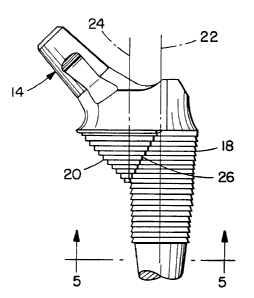
Figure 1 is a perspective view of a hip femoral
prosthesis constructed in accordance with the invention.
Figure 2 is a side view of the prosthesis of Figure
1. This view corresponds to an anterior view when the
prosthesis is implanted in the left femur of a patient.
Figure 3 is a medial view of the prosthesis of
Figure 1.
219391
-6-
Figure 4 is a superior view of the prosthesis of
Figure 2.
Figure 5 is an inferior view of the prosthesis of
Figure 2, partially in section along lines 5-5 in Figure
2. This figure illustrates that the sum of the radii R~
and RZ of the bone-engaging surfaces of bodies 18 and 20
is greater than the distance D between axes 22 and 24 for
at least one transverse cross-section through the
prosthesis.
Figures 6 and 7 are superior views of the prosthesis
of Figure 1 implanted in a typical orientation in the left
(Figure 6) and the right (Figure 7) femur of a patient.
The views shown in these figures are referenced to the
posterior aspect of the femoral condyles 10 of the knee.
Figures 8 and 9 are superior views of a patient's
left femur which have been prepared to receive the
prosthesis of Figure 1. Figure 8 corresponds to the
normal preparation of the bone so as to provide
approximately 15° of anteversion of the neck of the
femoral prosthesis relative to the femoral condyles 10.
Figure 9 corresponds to a preparation of the bone which
provides approximately 7° of retroversion of the neck of
the femoral prosthesis relative to the femoral condyles
10.
2193913
_7_
Figure 10 shows an alternate construction of the
prosthesis of the invention in which the axes of the two
cone-like, bone-engaging bodies of the prosthesis
intersect.
Figure 11 shows an alternate construction of the
prosthesis of the invention in which one of the cone-like,
bone-engaging bodies of the prosthesis has a concave
profile .
Figure 12 shows a construction of the prosthesis of
the invention suitable for use as the tibial component of
a knee joint.
Figure 13 shows an instrument for use in creating a
cavity in a patient's bone for receiving the prosthesis of
Figure 1.
Figures 14 and 15 are superior views of a patient's
left femur. These figures compare the configurations of
prepared cavities for receiving a prior art prosthesis
(Figure 14) and the prosthesis of Figure 1 (Figure 15).
Figures 16 and 17 are perspective views of the
cavities of Figures 14 and 15, respectively.
Figure 18 is a superior view of a patient's left
femur prepared for orienting a prior art prosthesis so
that it has a greater degree of.anteversion than would be
provided by the normal orientation of the prosthesis with
respect to the anatomy of the patient's bone.
2193813
-8-
Figure 19 is an anterior view of the femur of Figure
18 along lines 19-19 in Figure 18.
Figure 20 is a superior view of a patient's left
femur prepared for orienting the prosthesis of Figure 1 so
that it has a greater degree of anteversion than would be
provided by the normal orientation of the prosthesis with
respect to the anatomy of the patient's bone.
Figure 21 is an anterior view of the femur of Figure
20 along the same direction as lines 19-19 in Figure 18.
The foregoing drawings, which are incorporated in and
constitute part of the specification, illustrate the
preferred embodiments of the invention, and together with
the description, serve to explain the principles of the
invention. It is to be understood, of course, that both
the drawings and the description are explanatory only and
are not restrictive of the invention.
The reference numbers used in the drawings correspond
to the following:
10 femoral condyles of the knee
13 femoral hip prosthesis
14 neck
16 stem
18 first cone-shaped (cone-like) region of proximal
bone-engaging surface
21~39a3
_g_
20 second cone-shaped (cone-like) region of
proximal bone-engaging surface
22 axis of first cone 18
24 axis of second cone 20
26 line of intersection between cone 18 and cone
20
28 femoral bone
30 longitudinal axis of femoral bone
32 instrument for cutting cavity 200
34 body of instrument 32
36 bearing member of instrument 32
38 shaft of instrument 32
40 conical cutter of instrument 32
42 stop collar of instrument 32
44 calcar region of femur bone 28
46 line tangent to condyles 10
48 line through center of calcar region 44
50 line parallel to line 46
52 bone removal region of posterior wall of femur
54 bone removal region of anterior wall of femur
56 bone removal region of anterior wall of femur
58 concave profile of cone-like body
60 cone-like body of tibial prosthesis
62 cone-like body of tibial prosthesis
64 cone-like body of tibial prosthesis
180 conical cavity for cone 18
2~93~13
-10-
200 conical cavity for cone 20
DESCRIPTION OF THE PREFERRED EMBODIMENTS
Although the invention can be practiced with a
variety of prostheses, a preferred application is to
femoral hip prostheses. Accordingly, the initial
description of the invention will be in terms of such a
prosthesis, it being understood that this description is
not intended to limit the scope of the invention.
Figures 1-5 show the structure of a femoral hip
prosthesis 13 constructed in accordance with the
invention. The prosthesis includes a neck 14 for
receiving the ball (not shown) of the prosthesis and an
elongated stem 16 which extends into the shaft of the
patient's femur when the prosthesis is implanted. Neck
14's orientation with respect to prosthesis 13 is
preferably neutral with regard to anteversion/retro-
version. That is, the prosthesis is preferably symmetric
with regard to a longitudinal plane through the neck. As
discussed below, this allows the prosthesis to be used
with various anteversion/retroversion angles as well as
with right and left femurs, thus reducing inventory
requirements, i.e., there is less need to separately
manufacture, ship, and store left, right, and special
circumstance prostheses.
21991 ~
-11-
The bone-engaging surface of prosthesis 13 includes
a first cone-shaped (cone-like) portion 18 and a second
cone-shaped (cone-like) portion 20. For ease of
reference, these portions will be referred to herein as
first cone 18 and second cone 20.
As shown in Figure 2, first cone 18 has an axis 22,
which corresponds in this case to the longitudinal axis of
the prosthesis as defined by stem 16, and second cone 20
has an axis 24 which is parallel to, but not collinear
with, axis 22. When this prosthesis is implanted, axis 22
is essentially aligned with longitudinal axis 30 of
femoral bone 28 (see Figure 17).
As shown in Figure 3, cone 18 has an apical cone
angle cx and cone 20 has an apical cone angle ~. The cone
angles and spacings of axes 22 and 24 in Figures 1-5 are
such that cones 18 and 20 intersect along line 26.
A variety of cone angles and axis spacings can be
used in the practice of the invention. In the case of a
hip femoral prosthesis, cone angle a is preferably about
6°, cone angle ~i is preferably in the range between about
60° and about 120°, and the spacing between axes 22 and 24
is preferably chosen so that the apex of cone 20 lies in
the vicinity of the surface of cone 18.
As discussed fully below, one of the advantages of
the invention is that it allows flexibility in the angular
2193913
-12-
orientation of prosthesis 13 about the longitudinal axis
of the patient's bone. Cone angle ~3 is selected with this
orientation feature in mind.
Specifically, larger cone angles produce a shallower
cone 20 which allows more flexibility in angular
orientation without sacrificing the integrity of the
patient's bone. Smaller cone angles, on the other hand,
provide more purchase into the end of the patient's bone,
which may be required for some applications. Such smaller
cone angles give less flexibility with regard to angular
orientation.
The particular cone angles for any specific
application can be determined by persons skilled in the
art from the disclosure herein and the specific
requirements of a particular application of the invention.
In Figures 1-5, cone 20 is shown as having a
representative cone angle of 90° which provides a
substantial level of angular orientation flexibility in
comparison to prior art prostheses (see discussion of
Figures 14-21 below).
Implantation of prosthesis 13 in a patient's bone
requires the preparation of two adjacent conical cavities
180 and 200 (see Figures 8 and 17) to receive cones 18 and
20, respectively. Cavity 180 is aligned with the
longitudinal axis 30 of femoral bone 28 and is prepared
2193913
-13-
using a conventional conical reamer (see, for example,
Figures 4-5 of U.S. Patent No. 4,790,852). The
longitudinal location of cavity 180 along axis 30 is
chosen with the ultimate location of prosthesis 13,
including cones 18 and 20, along that axis in mind. Thus,
the conical reamer used to prepare cavity 180 preferably
includes means for indicating the depth of the reamer
relative to the end of the patient's bone.
Although the foregoing discussion has been in terms
of geometrical cones, it should be understood that cone
like bodies 18 and 20 are not limited to such shapes.
Rather, each of these bodies needs to be generally cone
shaped and to have a form such that a cavity to receive
the body can be generated by a cutting tool rotating about
a fixed axis.
The cone-like shape is important because it allows
the prosthesis to reach out toward the hard bone in the
region of the end of the patient's bone. That is, it
gives the prosthesis a longitudinal cross-section at the
end of the bone which is similar to the longitudinal
profile of the hard bone at that end. A spherical shape
of the type used in U.S. Patent No. 4,808,185, does not
have this property.
The ability to be received in a cavity formed by a
cutting tool rotating about a fixed axis is important
2193913
-14-
because it means an excellent fit can be achieved between
the prosthesis and the cavity under the real world
conditions which exist in the operating room.
Figure 11 illustrates a body 20 having such a cone
s like shape. Body 20 of this figure has a concave
longitudinal profile 58, which can even more closely
correspond to the inside surface of the hard bone in some
cases than a true geometrical cone. This would not be
true for a convex longitudinal profile. Accordingly, the
prostheses of the invention have cone-like shaped bodies
or regions whose longitudinal profiles are either straight
or concave.
For the more general case of cone-like bodies, as
opposed to bodies which are true cones, the relative
shapes of the bodies can be describe in terms of their
overall longitudinal profiles, rather than their cone
angles. In general terms, cone-like body 20's surface
area and diameter decreases faster than those of cone-like
body 18 in moving away from the end of the bone in which
they are implanted.
In most cases, the transverse cross-section of the
prosthesis and the cavity in the region of the end of the
bone will include two intersecting circular parts with
displaced centers. An alternate transverse cross-section
comprises a circle and an ellipse. This cross-section
219391.
-15-
arises when axes 22 and 24 of cone-like bodies 18 and 20
intersect, as opposed to being parallel, as shown in
Figure 10. These cross-sections can be characterized as
having a wasp-waisted configuration, a nipped in the waist
configuration, or a configuration which includes a cusp.
The cusp can be rounded out if desired.
Cavity 200 is preferably prepared using instrument 32
shown in Figure 13. The instrument has a body 34 whose
outer surface includes a comically shaped portion which
seats in conical cavity 180.
Body 34 carries bearing member 36. Shaft 38, which
carries cutter 40 at its distal end, is rotatable and
slidable within bearing member 36. Shaft 38 is rotated
and advanced into the patient's bone by conventional
means, such as, a T-handle (not shown). Stop collar 42 is
mounted on shaft 38 and defines the end point of the
advance of cutter 40 so that the spatial relationship of
cavities 180 and 200 matches that of cones 18 and 20.
The instrument of Figure 13 can be used to prepare a
cavity in the patient's bone for various orientations of
the neck 14 of the prosthesis relative to the remaining
calcar region 44 of the patient' s f emur . Figures 8 - 9 , 15 ,
17, and 20-21 illustrate some of the possibilities.
In each of these figures, the remaining calcar region
44 is shown having a degree of anteversion of about 7°
2193913
-16-
relative to line 46 which is tangent to condyles 10. That
is, line 48 which passes through the center of calcar
region 44 and intersects longitudinal axis 30 is rotated
7° counterclockwise relative to line 50 which is parallel
to line 46 and also intersects longitudinal axis 30.
(This geometric construction is for purposes of
illustration only since, as is well known in the art,
there is a considerable variation in version angles and
anatomy in human hips.) For ease of reference, a
prosthesis whose neck 14 is aligned with line 48 will be
referred to as having an anteversion of 7°.
The average anteversion of the natural femoral head
of the femur is greater than 7° because the natural neck
turns in a forward direction as it rises from the calcar
region. For many patients, the anteversion of the natural
femoral head is in the range of about 12° to about 15°.
Accordingly, in practice, it is generally desired to
orient the neck 14 of prosthesis 13 at some greater amount
of anteversion than that of the remaining calcar region
2 0 44 , a . g . , between about 12 ° and about 15 °
counterclockwise
from line 50 for a left femur.
This generally preferred orientation of the neck 14
of prosthesis 13 is illustrated in Figures 6-8.
Specifically, Figure 6 shows implantation of prosthesis 13
in the patient's left femur at 15° anteversion and Figure
2193913
-17-
7 shows implantation in the right femur, again at 15°
anteversion. Figure S is the bone preparation for the
implantation of Figure 6. The bone preparation for the
implantation of Figure 7 is the mirror image of that of
Figure 8.
A cavity for use in providing a relatively extreme
orientation of neck 14 of prosthesis 13 is shown in Figure
9. In this case, the neck of the prosthesis when
implanted is retroverted by 7° with respect to line 50.
Although such an orientation is generally unlikely, it may
be needed for some patients. It should be noted that some
removal of the posterior wall of the femur is likely to
occur during preparation of the bone for this orientation
of the prosthesis (see region 52 in Figure 9). However,
due to the shallowness of cone-like cavity 200, the
remaining bone still provides a strong structural support
for the prosthesis.
A cavity for use in providing another relatively
extreme orientation of neck 14 of prosthesis 13 is shown
in Figure 20. In this case, the neck of the prosthesis
when implanted is anteverted by more than 15 ° with respect
to line 50. Again, some removal of the wall of the femur
is likely to occur during preparation of the bone for this
orientation of the prosthesis, specifically, removal of a
small portion of the anterior wall is likely to occur (see
2193913
-18-
region 54 in Figures 20 and 21). Again, however, the
remaining bone still provides a strong structural support
for the prosthesis because the flare of the bone in region
54 is in the same direction as the flare of the cone 20.
This is particularly so because the force from the
prosthesis to the bone in the calcar region is directed
posteriorly where the external wall of the femur is still
intact. It should be noted that the orientation of Figure
20 will be more common than the orientation of Figure 9.
Significantly, a single neutral prosthesis can be
used for all of the orientations shown in Figures 6-9 and
20, as well as for a variety of orientations within and
beyond those illustrated. As discussed above, prosthesis
13 is preferably~symmetric with respect to neck 14, i.e.,
the prosthesis has neutral version. Through the use of a
cone 20 which is relatively shallow, such a neutral
version prosthesis can be used for both the right and left
femurs as illustrated in Figures 6 and 7, and for the
relatively extreme orientations of the prosthesis as
illustrated by Figures 9 and 20. Specifically, the
shallow cone 20 allows for angular variation about axis 30
of the placement of the prosthesis in the calcar region of
the bone without compromising the bone's structural
strength or the fixation of the prosthesis.
2193913
-19-
This "shallowness" aspect of the invention is
illustrated in Figures 14-17 which show a prepared femur
for receiving the prosthesis of the invention (Figures 15
and 17) and a prepared femur for receiving a prior art
prosthesis (Figures 14 and 16). As illustrated in these
figures, the bone-engaging surface of the prosthesis of
the invention in the calcar region tends to be as much "on
the bone" as "in the bone" because of the shallowness of
cone 20. This geometry further encourages the favorable
loading of the bone at the end of the bone.
For the prior art prosthesis, on the other hand, the
bone-engaging surface of the prosthesis in the calcar
region is clearly "in the bone". As a result, rotation of
this part of the prior art prosthesis to provide
additional anteversion causes the removal of an
unacceptable amount of the anterior wall of the femur as
shown in Figure 19 (see 56 in Figure 19).
Put another way, if the geometry of the prior art
prosthesis were to be used in a one piece neutral
prosthesis and that prosthesis were to be oriented in a
more anteverted orientation than that of the calcar
region, more critically placed bone would have to be
removed than for the comparable prosthesis constructed in
accordance with the geometry of the present invention.
2193913
-20-
The ability to use a single neutral prosthesis for a
variety of orientations is a significant advantage of the
invention because it reduces the costs of manufacture,
shipping, and storage relative to the use of right-handed
and left-handed prostheses. Moreover, the prosthesis of
the invention provides greater latitude of orientation
compared to single orientation left and right hand
prostheses.
Further, the invention provides this multiple
orientation advantage in a single piece prosthesis, as
opposed to a modular prosthesis (see U.S. Patent No.
4 , 790 , 852 ) . It should be noted that the two cone geometry
of the invention can be used with modular prostheses, if
desired.
Although it is not preferred, the present invention
can be used in connection with left and right handed
prostheses with anteverted necks if desired.
Figure 12 shows application of the invention to a
tibial knee prosthesis. In this case, the prosthesis
includes three cone-like bodies 60, 62, and 64. As shown
in this figure, bodies 62 and 64 have the same shape.
Different shapes can be used for these bodies if desired.
In addition to hip joints and tibial components of
knee joints, the invention can also be used for various
other joints, such as, the humeral component of a shoulder
2193913
-21-
joint prosthesis, the femoral component of a knee
prosthesis, and the like.
The prosthesis can be constructed of various
biocompatible materials suitable for implantation now
known or subsequently developed. For example, it can be
made of a cobalt-chromium-molybdenum alloy (see ASTM-F75
and ASTM-F799) or a titanium alloy such as Ti-6A1-4V
(ASTM-F136). The cone-like, bone-engaging bodies of the
prosthesis can include surface texturing, such as the
steps shown in the figures. These surfaces can also be
porous coated, plasma sprayed, chemically modified, or the
like to enhance fixation. Similarly, the surfaces can be
coated with bone growth stimulating materials such as
hydroxylapatite.
Although preferred and other embodiments of the
invention have been described herein, additional
embodiments may be perceived by those skilled in the art
without departing from the scope of the invention as
defined by the following claims.