Note: Descriptions are shown in the official language in which they were submitted.
METHODS FOR HARVESTING ADIPOSE TISSUE CONTAINING
AUTOLOGOUS MICROVASCULAR ENDOTHELIAL CELLS ~~ 9 3 9 4 5
Field of the Invention
The present invention is directed to a method for the
harvesting of microvascular endothelial cells. More
particularly, the present invention relates to a method
for the collection of adipose tissue and the initial
refinement of microvascular endothelial cells for
deposition on the surface of synthetic prosthetics.
Backcrround of the Invention
Arteriosclerotic vascular disease is a leading cause
of death throughout the world. While sophisticated
medical techniques such as arterial endarterectomy and
percutaneous balloon dilatation are being applied more and
more often to treat pathologic stenotic occurrences, quite
often the most effective therapy is the surgical removal
of the occluded section of the vessel. In such cases, the
restoration of blood flow to ischemic tissue depends on
the implantation of a vascular graft.
Although autologous vascular tissue is the most suit
able material for use in such grafts, prior surgical
intervention and advanced vascular disease often limit the
availability of such tissue. Accordingly, it has become
common in recent years to implant vascular grafts fabri-
cated of synthetic materials. While commercially avail-
able synthetic grafts are extremely durable and may be
used to successfully restore blood flow to occluded
tissue, associated thrombogenic complications reduce their
effectiveness. In particular, smaller diameter vascular
grafts tend to become dysfunctional as they are blocked by
the normal clotting mechanisms. Specifically, the syn-
thetic surface of the graft promotes the deposition of
fibrin leading to associated cellular adhesion and occlu-
sion of the vessel. Consequently, the long term prognosis
for non-coated synthetic grafts is relatively poor.
2
2 ~ 93945
To circumvent the problems associated with non-coated
synthetic vascular grafts, procedures are being developed
for lining prosthetics with human endothelial cells to
produce a non-thrombogenic cell surface such as exists in
native human vessels. The endothelial lining of natural
blood vessels is a highly complex, multifunctional cell
surface which interacts with both the blood and the under-
lying vessel wall components to maintain physiological
homeostasis. Tests with animals have shown that the depo-
sition of a functional large vessel endothelial cell
lining on the interior surface of synthetic vascular
grafts decreases the formation of thrombogenic occlusions
and minimizes the disruption of blood flow through the
vessel. However, harvesting a sufficient number of large
vessel cells from a donor is difficult at best.
Recent advances in molecular biology and tissue cul-
ture have allowed the isolation and subsequent propagation
of large vessel endothelial cells. In practice, the use
of cultured large vessel endothelial cells is expensive,
complicated and subject to inherent limitations. One
problem is that cell culture techniques are highly tech-
nical requiring trained personnel and the use of special-
ized equipment under laboratory conditions. Yet, even
under the best of conditions, the yield of cultured large
vessel endothelial cells may be low. Moreover, typical
seeding procedures using cultured cells require the use of
specialized media under complex conditions to assure the
complete and even deposition of endothelial cells on the
synthetic surface of the graft.
In addition, cultured cells are generally not derived
from the patient receiving the graft and, accordingly, may
precipitate a wide range of immunological complications.
If the immune response of the patient is not attenuated,
the transplanted endothelial cells will likely be attacked
and stripped from the surface of the graft by the body's
3
2 ~ 93945
defenses. Conversely, if the patient's immune system is
artificially suppressed it may lead to life-threatening,
opportunistic infections.
In view of these and other complications associated
with the use of large vessel endothelial cell treatments
of prosthetic devices, alternative methods of reducing the
inherent thrombogenicity of synthetic materials have been
developed. In particular, it was quickly recognized that
human microvascular endothelial cells could be effectively
used to render synthetic grafts non-thrombogenic.
Microvascular endothelial cells are derived from
capillaries, arterioles and venules and are present in an
abundant supply in most body tissues. While endothelial
cells may be isolated from tissues such as brain, lung,
retina, adrenal glands, liver and muscle tissue, the use
of fat tissue as a source for these cells is preferred due
to its abundance, availability and because its removal
should not adversely affect the patient being treated.
Quite often, microvascular endothelial cells are present
in concentrations of 106 cells per gram of fat or higher,
providing an ample source of materials for high density
deposition procedures. Moreover, as the microvascular
cells used to treat the synthetic graft are usually
autologous, that is, taken from the recipient of the
vascular prosthesis, immunological complications may be
obviated.
Typically, microvascular endothelial cells are iso-
lated from autologous adipose tissues such as perinephric
fat, subcutaneous fat, omentum, or fat associated with the
peritoneal cavity. Harvesting usually takes place under
sterile conditions with the required amount of fat removed
in one procedure. The collected tissue may then be washed
before being transferred to a buffered digestive solution
generally containing proteolytic enzymes such as collagen-
ase, papain, trypsin, and mixtures thereof.
CA 02193945 2005-07-27
4
The adipose tissue is digested at 37°C for a selected
period to disrupt the connective matrix and disperse the
cellular components including microvascular endothelial
cells. Following digestion, the cellular components may
be separated by low speed centrifugation to provide a
cell-rich pellet. The pellet may be washed and used in
the deposition procedure or purified further using a
continuous gradient. In either case, purified cells are
diluted in buffer and subsequently incubated with the
synthetic prosthesis to provide endothelialized surfaces.
Commonly, collection of the desired adipose tissue
involves the use of a suction pump connected to a collec-
tion apparatus having a needle or cannula. For example,
U.S. Patent Nos. 5,035,708 and 4,834,703, disclose the
collection of adipose tissue using a suction pump to
provide the necessary vacuum. However, such collection
devices and associated methods tend to employ strong,
uncontrollable suction that is extremely rough on the
microvascular cellular components of the collected
tissue. The resulting disruption of the relatively
fragile cellular membranes can substantially lower the
viability of the harvested cells. This, in turn,
dramatically reduces the efficiency of the deposition
process. While such collection procedures may provide
sufficient adipose tissue, samples collected using such
techniques generally require several additional labor-
intensive preparatory steps to assure an adequate
concentration of relatively pure microvascular
endothelial cells for eventual deposition.
Further, source tissue collected using suction pumps
is often relatively dirty, contaminated with unwanted
body fluids and non-adipose cellular debris. Rather than
obtaining translucent, white samples as seen in
relatively pure adipose tissue, samples collected using
3 5 pump~ener
5
2193945
ated vacuums often appear bloody, with concentrations of
connective or membrane tissue dispersed within the fat.
The incorporated contaminants interfere with each step
of microvascular endothelial cell isolation including the
initial homogenization and preparation of the collected
sample for digestion. Moreover, such contaminants
directly inhibit the enzymatic activity of the proteolytic
enzymes leading to incomplete digestion of the sample and
a corresponding reduction in the yield of non-adipose
cellular components subsequently obtained by centrifuga-
tion. Finally, those cells which are collected and
pelleted contain increased level of non-endothelial
components. The use of such contaminated pellets further
lowers the efficiency of the cell deposition procedure and
interferes with the homogeneous layering of endothelial
cells on the prosthetic surface. Consequently, the
patient may have to endure more extensive liposuction than
would otherwise be required in order to provide a suffi-
cient number of microvascular endothelial cells.
As the efficiency of the endothelialization process is
lowered at each step along the way by contaminants, the
importance of starting this procedure with a relatively
clean sample is evident. That is, a small increase in the
amount of contaminating materials initially collected can
dramatically reduce the yield of viable microvascular
endothelial cells available for deposition~on the surface
of the synthetic graft. In addition to increasing the
amount of adipose tissue which must be initially col-
lected, the inevitable reduction in cell viability due to
contaminating materials must be compensated for by longer
deposition times or additional purification steps, both of
which reduce the operating efficiency of the entire pro
cedure. This can be particularly detrimental if the cells
are to be collected immediately prior to the implantation
of the prosthetic device.
2193q45
Accordingly, a need exists to improve the yield of
viable endothelial cells recovered from adipose tissue
collected from a patient preparatory to implantation of a
synthetic prosthesis. That is, microvascular endothelial
cells which are present in a fat specimen should be more
efficiently separated from the fat cells, blood cells,
connective tissue, and other materials that are present in
the specimen, so that a larger number of such endothelial
cells are available to be deposited onto the synthetic
graft.
In addition to the actual problems associated with the
collection of material, the use of a suction pump compli-
cates the operating environment and interferes with the
surgeon's ability to freely maneuver the adipose tissue
collection apparatus. More particularly, the collection
apparatus is usually attached to the vacuum source via
thick, unwieldy hoses that severely compromise the man-
euverability of the collection tip. Such pumps often do
not allow the precise, real time control of the strength
of the vacuum at the collection tip, making it difficult
to maintain constant, even harvesting of the desired
source tissue. This lack of convenience and precise
control inevitably results in the aspiration of undesir-
able tissue, thereby increasing the contaminant level of
the samples or resulting in the collection of less prefer
able adipose tissue containing lower levels of microvas
cular endothelial cells. Further, vacuum sources, es
pecially those approved for use in medical procedures, are
generally complicated instruments that are relatively
expensive to maintain.
In view of the deficiencies of the related technology
as outlined above, it is an object of the present inven
tion to provide an efficient, cost effective method for
the collection of adipose tissue containing microvascular
endothelial cells.
2193945
It is another object of the present invention to
provide a reliable convenient method for the collection of
substantially pure adipose tissue containing high levels
of microvascular endothelial cells with a minimum of blood
cells, connective tissue and other contaminants.
It is still a further object of the present invention
to provide a reliable convenient method for the rapid
homogenization of adipose tissue to facilitate the subse-
quent separation of microvascular endothelial cells.
SUMMARY OF THE INVENTION
These and other objectives are achieved by the present
invention which, in a broad aspect, is directed to effi-
cient, reliable and cost effective methods for the har-
vesting of adipose tissue containing identifiable cellular
components such as microvascular endothelial cells. More
particularly, the present invention is directed to methods
of harvesting adipose tissue so as to preserve an
increased population of viable endothelial cells using a
collection apparatus generally comprising a variable
volume container, typically a syringe assembly, attached
to an elongated cannula. The elongated cannula, in fluid
conducting communication with the variable volume
container or syringe, preferably includes apertures
appropriately sized and configured to minimize stress
placed on cellular components while disrupting the
connective matrix of the adipose tissue. That is, by
collecting adipose tissue using specifically configured
cannular apertures, the yield of endothelial cells may be
substantially increased. Further, the collection
apparatus is inexpensive, lightweight, easy to manipulate
and allows accurate control of the applied suction.
The tissue harvesting methods of the present invention
generally begin by inserting at least a portion of the
cannula of the collection apparatus into the patient and
8
2193945
'- directing the cannula tip to the area where the adipose
tissue is to be collected. Preferably the harvesting
procedure is carried out under aseptic conditions.
Optionally, a saline solution or other biocompatible
liquid may be injected into the collection area of the
patient prior to harvesting to loosen the adipose tissue
matrix. Following the insertion and positioning of the
cannula tip, sub-ambient pressure is generated in the
central bore of the syringe by drawing back a displaceable
piston affixed to a plunger. If desired the piston may be
retained in this withdrawn configuration by a locking
mechanism attached to the plunger and designed to interact
with the body of the syringe. The locking mechanism frees
the hands of the operator and, when combined with the
I5 light weight of the collection apparatus provides enhanced
maneuverability. In any case, the sub-ambient pressure in
the central bore suctions the adipose tissue from the
selected collection area, into the disruptive apertures of
the cannula, through the cannula body and into the syringe
assembly. As the central bore of the syringe fills with
collected tissue, the sub-ambient pressure slowly
equilibrates. Once the central bore of the syringe i,s
substantially filled with relatively homogeneous adipose
tissue the cannula tip is removed from the patient.
Another aspect of the present invention allows for the
collected adipose tissue to be readily homogenized and
washed with aqueous solutions to remove contaminating
matter. Following removal of the cannula tip from the
patient, the cannula may be detached from the syringe
assembly. A filter, contained in a filter hub, may then
be attached to the syringe assembly where the cannula was
previously affixed. A second syringe assembly, preferably
the same size as the first is then attached to the oppo-
site side of the filter hub. When so joined, the piston
of the first syringe assembly is substantially rearward in
CA 02193945 2002-06-12
9
the syringe and the piston of the second syringe assembly
is in a substantially forward position. By using the
plungers to displacing the two pistons, the collected fat
may be rapidly homogenized as i.t is forced through the
filter which transects the flow path of the sample
tissue. Optionally, rinse solutions may be added during
the homogenization to separate contaminants from the
endothelial cell rich adipose tissue homogenate. After
homogenization and rinsing, collected adipose tissue, now
f0 substantially free of intact connective tissue and other
contaminants, may be transferred to appropriate
containers for digestion and further purification.
According to one aspect of the invention, there is
provided an endothelial cell collection system,
comprising:
a hollow tubular body defining a central bore along
a longitudinal axis, the body having a proximal end
defining an opening, and a distal end defining an
entrance port into the bore;
a piston sealingly disposed within l~he bore;
a plunger operatively connected to the piston and
extending through the proximal end opening of the body,
the piston being movable to define a variable volume
chamber within the central bore i.n commu.nication with the
entrance port;
an elongated cannula removably attached to the
distal end of the body having a lumen extending
therethrough in communication with the chamber, the
cannula having a plurality of distal collection apertures
opening transverse to the lumen axis and. having sharpened
edges, the plunger and piston combining to allow manual
control of the pressure in the chamber to create sub- or
supra-ambient pressure in the cannula lumen, thus
CA 02193945 2002-06-12
9a
enabling gentle collection of fat tissue comprising
microvascular endothelial cells from a body cavity
through the collection apertures and into the chamber via
the lumen;
a hub assembly attachable to the distal end of the
body in place of the cannula; and
a homogenizing member mounted within the hub
assembly, the homogenizing member comprising a plurality
of apertures, wherein the fat tissue collected in the
chamber can be homogenized by ejecting it from the
chamber through the distal port and homogenizing member.
According to another aspect of the invention, there
is provided a kit for homogenizing fat tissue obtained
from a patient prior to digestion of the fat tissue, the
kit comprising:
a collection syringe having a first port and within
which is disposed fat tissue comprising microvascular
endothelial cells;
a receiving syringe having a second port;
a hub assembly attached to both the first and second
ports and coupling the collection and receiving syringes;
and
a homogenizing member mounted within the hub
assembly, the member comprising a plurality of apertures
having cutting edge surfaces, wherein the fat tissue can
be homogenized by injecting it from the collection
syringe through the homogenizing member to the receiving
syringe, and then from the receiving syringe through the
homogenizing member to the collection syringe.
According to a further aspect o:E the invention,
there is provided a method of collecting fat tissue from
a patient, the method comprising the steps of:
providing an elongated tubular cannula having a
CA 02193945 2002-06-12
9b
distal tip end and an opposite proximal end, a lumen
extending in the cannula, and an aperture opening
outwardly from the lumen on the cannula adjacent to the
distal tip end and defining a tis:~ue cutting edge
surface;
inserting the cannula into a patient so that the
aperture contacts fat tissue;
cutting the fat tissue with the tissue cutting edge
surf ace ;
creating a suction force in the lumen to draw the
fat tissue from the patient through the aperture into the
lumen;
selectively controlling the amount of suction to the
lumen; and
collecting the fat tissue from t:he lumen into a
collection chamber, wherein the collection chamber
includes an entrance port through which the fat tissue
passes from the lumen in the step of collecting, the
method further comprising the step of discharging the fat
tissue from the collection chamber through the entrance
port and through a homogenizing member having apertures
with cutting edge surfaces and sized to lower the
viscosity of the fat tissue by disrupting a connective
matrix without exposing cellular components within the
fat tissue to excessive shearing forces.
According to another aspect of the invention, there
is provided a method for homogenizing fat tissue obtained
from a patient prior to digestion of the fat tissue, the
method comprising the steps of
obtaining fat tissue from a patient, the fat tissue
comprising microvascular endothelial cells;
disposing the fat tissue in a collection container
comprising a first port;
CA 02193945 2002-06-12
9c
positioning a planar homogenizing member comprising
a plurality of apertures adjacent to the first port,
wherein the plurality of apertures comprise at least one
tissue cutting edge surface;
discharging the fat tissue from the collection
container first port in a first direction generally
normal to the planar homogenizing member through the
plurality of apertures, so that the tissue cutting edge
surface of the plurality of apertures cuts the fat tissue
so as to free the endothelial cells;
receiving the fats tissue discharged through the
homogenizing member in a receiving container through a
second port; and
discharging the fat tissue received in the receiving
container through the second port in a second direction
opposite the first direction and generally normal to the
planar homogenizing member through the plurality of
apertures into the collection container.
Additional objects and advantages of the present
invention will be apparent from a reading of the
following detailed description and exemplary preferred
embodiment of the invention taken in conjunction with the
appended drawing figures in which like reference numerals
denote the same feature or features which are analogous
in structure.
BRIEF DESCRIPTION OF THE DRAWINGS
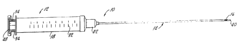
Fig. 1 is a perspective view of an adipose tissue
collection apparatus showing attachment of the cannula to
a syringe assembly with the plunger in an inserted
position;
Fig. 2 is a partial sectional view of an adipose
tissue collection apparatus showing the plunger held in a
CA 02193945 2002-06-12
9d
withdrawn position by an exemplary locking mechanism;
Fig. 3 is a partial sectional view of two syringe
assemblies interconnected by a filter hub illustrating a
configuration of the present invention used for homogen-
ization of harvested adipose tissue;
Fig. 4 is a cross-sectional view taken across line
4-4 of Fig. 3 showing a homogenization filter according
to the present invention;
,,
2193945
Fig. 5 is a partial perspective view of a tip of a
cannula used to provide high yields of microvascular
endothelial cells in accordance with the teachings of the
present invention;
Fig. 6 is a cross-sectional view taken along line 6-6
of Fig. 5 showing the positioning of the collection
apertures adjacent to the tip of the cannula;
Fig. 7 is a partial perspective view of a tip portion
of an embodiment of a cannula used to harvest adipose
tissue;
Fig. 8 is a cross-sectional view taken along line 8-8
of Fig. 7 illustrating the elliptical shape and position-
ing of the collection apertures adjacent to the tip of the
cannula;
-15 Fig. 9 is a partial perspective view of a tip portion
of another alternative embodiment of a cannula used to
harvest adipose tissue.
DETAILED DESCRIPTION
Although this invention may be embodied in many
different forms, there are shown in the drawings and will
be described in detail specific embodiments thereof with
the understanding that the present disclosure is to be
considered as an exemplification of the principles of the
invention and is not intended to limit the invention to
the specific embodiments illustrated. In particular, it
must be emphasized that the present invention provides for
the association of a wide variety of syringe bodies and
cannulas beyond those shown in the figures.
Moreover, the present invention may be used to harvest
any identifiable cells associated with the adipose tissue
matrix and is not limited to the harvesting and isolation
of microvascular endothelial cells. As used herein the
term "identifiable cellular components" refers to those
11 2? 93945
cells which may be recognized by commonly used immuno
genic, chemical or physical separation methods or tests.
Similarly, while the endothelialization of vascular
prosthetics is an important object of the present inven
tion, those skilled in the art will appreciate that the
cellular products collected may be used to treat other
implantable devices. Implants which can be treated to
produce an endothelial cell lining or covering include,
but are not limited to, intravascular devices such as
artificial hearts, valvular prosthetics, and natural or
artificial valve leaflets. The collection apparatus and
methods of this invention for harvesting endothelial cell
rich adipose tissue may be used in the treatment of
surfaces comprised of known synthetic materials such as
polyester, polytetrafluoroethylene, or fixed and unfixed
naturally occurring materials such as veins, arteries,
heart valves and other tissues from animal sources,
including humans.
Turning now to the figures, Figs. 1 and 2 show an adi
pose tissue collection apparatus 10 essentially comprising
a variable volume container in the form of syringe
assembly 12. Syringe assembly 12 is in fluid tight
communication with an elongated cannula 14 having a lumen
26 and at least one collection aperture 16 appropriately
configured for the relatively homogeneous collection of
adipose tissue containing microvascular endothelial cells.
Syringe assembly 12 generally comprises a hollow tubular
body 18 defining a central bore 20 having a displaceable
piston 22 sealingly disposed therein. Preferably piston
22 is affixed to an elongate plunger 28 which extends
through an opening in the rear of hollow tubular body 18.
By manually displacing plunger 28, piston 22 may be
reversibly moved along the length of central bore 20.
Sealing rings 31 ensure that piston 22 maintains good
contact with the interior of tubular body 18 as it moves
2193945
longitudinally. An entrance port 24 at the forward end of
hollow tubular body 18 provides for fluid conducting
communication between cannula lumen 26 and central bore
20.
Preferably syringe assembly 12 is a Toomey-type
syringe (Sherwood Medical Co., St. Louis, Missouri) having
a tapered tip 30 defining entrance port 24. While a
Toomey-type syringe is preferred due to the ease of
switching cannulas or other attachments, syringe
assemblies having other types of connection mechanisms
such as threaded connectors, catheter tips or luer locks
are compatible with the present invention. Typically,
hollow tubular body 18 of syringe assembly l2 is formed of
an inexpensive, non-reactive material such as
polypropylene or other rugged polymer composition. Of
course, those skilled in the art will appreciate that the
size and fluid capacity of syringe assembly 12 may vary
based on~the amount of adipose tissue to be collected.
However, for obvious reasons it is preferable that syringe
assembly 12 be of sufficient volume to harvest the desired
amount of adipose tissue in one collection procedure. As
the volume of adipose tissue needed to provide the neces-
sary amount of endothelial cells for an average cell depo-
sition procedure is on the order of 20 mls to 50 mls, a
preferred syringe volume is approximately 60 mls. Grada-
tions 32 on the sides of most commercially available syr-
inges provide an easy method for monitoring the amount of
fat collected.
Optionally, syringe assembly 12 is provided with a
locking mechanism 34, attached to plunger flange 48 which
reversibly engages annular flange 38 at the rear of hollow
tubular body 18 to retain plunger 28 and attached piston
22 in a withdrawn configuration. This configuration is
illustrated in Fig. 2. When plunger 28 and piston 22 are
in a substantially forward position, as shown in Fig. 1
2193945
the inner surface of hollow tubular body 18 will act on
shoulders 38 of locking mechanism 34 to maintain it in a
closed position. However, when plunger 28 is withdrawn
past a certain predetermined point where shoulders 38 are
no longer restrained, locking mechanism 34 will spring
open due to the elastic memory of the mechanism material.
When in an open position, shoulders 36 of locking
mechanism 34 will be positioned to engage annular flange
38 as well as plunger flange 48 thereby preventing plunger
28 from reentering hollow tubular body 18. However,
plunger 28 may be easily moved to a forward position by
manually compressing locking .mechanism 34 to reduce the
diameter of shoulders 36 where they disengage annular
flange 38 and slide easily into hollow tubular body 18.
Although various locking mechanisms are compatible with
the present invention, one particularly suitable device is
sold under the trade name Grazer-Grams Lock (Gram Medical,
Costa Mesa, California). In addition to being obtainable
for a variety of syringe sizes, these locking mechanisms
are also available in a single shoulder embodiment.
The last major constituent of collection apparatus 10
is cannula 14 having a lumen 26, a proximal end and a
distal cannula tip 40. Many commercially available can-
nulas having different lengths and diameters are compat-
ible with the present invention and may be used with
various syringe assemblies. Cannulas which are particu-
larly compatible and provide relatively high yields when
used in accordance with the teachings herein are sold
under the trade name Mercedes (Grams Medical, Costa Mesa,
California) and have inner diameters ranging from approx-
imately 1 mm to 8 mm. Particularly preferable inner
diameters range from 1.5 mm to 4 mm. While generally
formed out of metal alloys such as stainless steel for
ease of resterilization and reuse, the surfaces of these
219945
cannulas may be coated with ,biocompatible polymers to
reduce stress on the collected cellular components.
While the general configuration of cannula 14 is
relatively consistent for the different embodiments i.e.
generally elongated with at least one lumen 26, other
characteristics of compatible cannulas may vary markedly.
For example cannula connection 42, shown in Figs. 1 and 2
at the proximal end of cannula 14, is adapted to seat on
and releasably engage a syringe having a tapered tip 30.
Those skilled in the art will appreciate that, as with
syringe assemblies, many types of cannula connections are
compatible with the teachings of the present invention as
long as they are adapted to engage selected syringe
assembly 12. For example, cannulas having luer
connectors, catheter connectors, threaded connectors and
compression fittings may be used for the harvest of
adipose tissue as long as they are compatible with the
connector of the selected syringe assembly. Moreover,
cannulas permanently affixed to a syringe assembly to form
a collection apparatus are also within the scope of the
present invention and may be used with comparable results.
Another important feature of the cannulas of the pres
ent invention which may vary depending on the desires of
the operating physician are the conf iguration and position
of the collection apertures. For example, Figs. 5, 6, 7,
8 and 9 all show different configurations of collection
apertures. In accordance with the present invention is
desirable that the shape and configuration of the
collection apertures impose stresses during harvest which
disrupt the macro structures and connective components of
the adipose tissue to provide a relatively homogeneous
yield. Further the collection apertures should be large
enough to resist blocking by any non-disrupted tissue
thereby necessitating the removal of the cannula from the
patient and interruption of the harvest procedure. Based
293945
on such considerations, collection apertures preferably
range from 1 mm to 4 mm and more preferably from 1.5 mm to
3 mm.
The aperture configuration of Fig. 5 displays
elongated tissue cutting edges on apertures 42 which
result in substantially increased cell yields. In
contrast, the rounded or less abrupt aperture edges such
as those shown in Figs. 7 and 9, do not appear to provide
tissue cutting edges sufficiently disruptive to the
connective matrix of the tissue resulting in less
homogeneous sample composition and lower cell yield.
While tissue cutting aperture configurations disrupt the
macro connective structure of the harvested tissue, they
do not unnecessarily place stress or shearing forces on
the delicate cellular components dispersed within the
adipose tissue matrix. While the rounded edges of
collection apertures 42 and collection apertures 46 assist
in reducing these undesirable shearing forces, they do not
provide for the collection of substantially homogeneous
tissue and therefore lower the overall cell yield.
Just as various configurations and sizes of collection
apertures are compatible with the methods of the present
invention, so to are different aperture placement schemes
and cannula shapes. For example there is no requirement
that the collection apertures be limited to locations near
distal cannula tip 40. While such placement may promote
sample purity as the collection area can be gauged more
accurately, apertures placed further away from the tip may
work equally well. Similarly, there is no requirement
that cannula 14, and by extension lumen 26 be cylindrical
in nature. For example, other, more elliptical, shapes
may provide the same cell yield as the perfectly
cylindrical shape illustrated in Fig. 6. Accordingly, as
with the syringe assemblies, a wide range of cannula
shapes, sizes and configurations are with in the scope of
16
219945
the invention and may be chosen based on the preferences
of the individual operator.
In any event, once a collection apparatus is selected
and assembled, the actual harvesting of the adipose tissue
and identifiable cellular components may begin. Prefer
ably the entire procedure is carried out under aseptic
conditions. Collection apparatus 10 may be pre-assembled
and sterilized ahead of time or may be assembled in the
operating area just prior to use. Typically, syringe
assembly 12, minus locking mechanism 34, is commercially
available in a disposable, presterilized and prepackaged
form. Conversely, cannula 14 is typically reusable and
has been cleaned, packaged and resterilized on site.
Accordingly, following the selection of compatible
components which may be releasably engaged, syringe
assembly 12 and cannula 14 are usually mated to form
collection apparatus 10 just prior to insertion in the
patient. At approximately the same time as syringe
assembly 12 is attached to cannula 14 optional locking
mechanism 34 may be affixed to plunger 28 via plunger
flange 48. Preferably, locking mechanism 34 is affixed
prior to engaging cannula 14 to reduce the chances of
inadvertent sample contamination.
As an optional preliminary step, saline or .other
biocompatible solutions may be injected into the adipose
collection area prior to harvesting the desired material.
The introduction of liquids into the area appears to
disrupt the adipose matrix and reduce the cohesion of the
connective tissue. To assist this disruption the fluid
injected tissue may be massaged vigorously or subjected to
other external forces. As those skilled in the art will
appreciate, the actual volume of saline injected, area of
injection and the time of injection before harvesting will
depend on the circumstances of the operation such as age
and health of the patient, amount of tissue to be har-
17
2193945
vested and the location of the adipose collection area.
Typically several milliliters of solution will be injected
approximately thirty minutes to an hour before harvesting
is undertaken. A standard syringe and injection needle
are used for the procedure. While it appears to improve
the homogeneity of the sample recovered, adequate micro-
vascular endothelial cell yields may be obtained without
the addition of fluids or the application of external
forces prior to harvesting.
When the adipose tissue is considered ready for har-
vest at least a portion of cannula 14 is inserted in the
patient near the adipose tissue to be taken. Given the
typical size of cannula 14 and its relatively blunt distal
tip 40, a small incision is usually made in the skin of
the patient for the insertion. Following insertion,
distal cannula tip 40, and more particularly aperture or
apertures 16, is maneuvered to the area where the adipose
tissue is~to be harvested. As previously discussed, the
adipose tissue is usually taken from perinephric fat,
subcutaneous fat, omentum, or fat associated with the
peritoneal cavity. Given the light weight and relatively
small size of collection apparatus 10 the operating
physician will have little trouble guiding cannula 14 as
desired and precisely positioning it in the proper
location.
It is important to note that, during the insertion and
positioning of cannula 14 in the body of the patient,
plunger 28 and displaceable piston 22 are fully inserted
in hollow tubular body 18. That is, forward surface 50 of
displaceable piston 22 is seated flush against the forward
end of hollow tubular body 18 adjacent to entrance port
24. At the same time optional locking mechanism 34 is
retained in a closed position by the inner surface of
hollow tubular body 18. With displaceable piston 22 in a
fully forward position, substantial amounts of fluid and
~ ~ 9945
other bodily material are prevented from entering cannula
14 and syringe assembly 12. Displaceable piston 22 is
retained in this configuration until cannula 14 is prop-
erly positioned and the physician is ready to begin
harvesting the adipose tissue surrounding apertures 16.
To initiate harvesting of the microvascular endo-
thelial cell rich adipose tissue sub-ambient pressure is
applied to cannula 14. Typically plunger 28 and affixed
piston 22 are slowly drawn back through hollow tubular
body 18 by the operator. If desired, optional locking
mechanism 34 may be engaged with annular flange 38 to
maintain the withdrawn configuration and sub-ambient
pressure. The increase in sealed volume defined by
forward surface 50 of piston 22 and the inner surface of
hollow tube body 18 creates sub-ambient pressure in
syringe assembly 12. This, in turn, creates a suction in
lumen 26 of cannula 14 which is in fluid tight
communication with central bore 20 through entrance port
24. Unlike prior art suction pumps which maintained a
uniformly high suction force at the collection tip, the
present invention provides a gentle sub-ambient pressure
which is easily and instantaneously adjustable. Judging
by the "feel" of collection apparatus 10, or the appear-
ance of the adipose tissue being harvested, the operator
can attenuate the suction applied at apertures 16 by ad-
justing the amount plunger 28 is withdrawn from hollow
tubular body 18. By pushing plunger l8 the suction will
be reduced while withdrawing it further (maintaining the
sealable deposition of piston 22 hollow tubular body 18)
will rapidly increase the suction at the aperture. Alter-
natively, the operator may simply rely on locking mechan-
ism 34 to maintain a steady suction at apertures 16.
The easily controllable sub-ambient pressure, combined
with the favorable tissue cutting characteristics of aper
tures 16 provide cleaner more homogeneous adipose tissue
z ~ 93945
for processing. Obstructing connective tissue is prefer-
ably disrupted while preserving the integrity of the cell-
ular components. Moreover, as the operator is able to
easily and efficiently adjust the position of cannula 14,
regions of higher contamination may be avoided further in-
creasing the purity of the tissue obtained. Should there
be a problem with obstruction of apertures 16 or cannula
14, the operator may simply push plunger 28 slightly for-
ward to apply positive pressure to the cannula and aper-
tunes thereby clearing the obstruction. Finally, as the
plunger is under direct control of the operator, the
amount of adipose tissue collected may be controlled more
closely.
Following the harvest of the desired amount of homoge
neous adipose tissue and pressure equilibration of collec
tion apparatus 10, cannula 14 is removed from the patient
through the initial insertion site preferably maintaining
aseptic conditions. As previously discussed the actual
amount of adipose tissue collected will depend on a number
of factors including number of microvascular endothelial
cells needed and the capacity of collection apparatus 10.
Typical volumes range from approximately 10 ml to approxi-
mately 100 ml with average volumes ranging from about 40
ml to about 60 ml. Of course those skilled in the art
will appreciate that smaller or larger volumes may be
collected for the purification of microvascular endo-
thelial cells or other cellular components using the
methods of the present invention. After cannula 14 is
removed from the patient, it is usually disengaged from
syringe assembly 12 containing the harvested tissue for
cleaning and resterilization.
At this point, the harvested tissue may be processed
further or stored for later use. For storage, the tissue
is usually removed from syringe assembly 12 by ejecting it
through harvesting port 24 into a separate container which
CA 02193945 2005-07-27
may then be chilled. For processing, the collected tissue
may be similarly transferred to microvascular cell isola-
tion apparatus such as the one described in United States
Patent No. 5,409,833. The desired identifiable cellular
components will then be separated from the adipose tissue
using digestion and the other procedures previously
discussed.
Alternatively, in accordance with the teachings of
the present invention the harvested adipose tissue may be
processed further using syringe assembly 12 for rinsing
and homogenization. For example, water or other aqueous
solutions could be introduced into central bore 20 with
the collected sample and shaken. Afterward the mixture is
allowed to settle, preferably in a syringe stand (not
shown), and separate. The adipose cells and associated
tissue including the overwhelming majority of microvas-
cular endothelial cells will float while connective
tissue, red blood cells and other contaminants sink or
our solubilized in the aqueous solution. The rinsed
adipose cells and associated tissue may then be decanted.
Of course the process may be repeated as many times as
necessary.
In another procedure the harvested tissue may be
homogenized and rinsed at the same time. Referring now to
Fig. 3, syringe assembly 12, containing the harvested
tissue, is releasably attached to filter hub assembly 60.
A second syringe assembly 212, preferably the same size
as syringe assembly 12, is releasably attached to the
opposite side of filter hub assembly 60. For the purposes
of clarity reference numerals previously used for syringe
assembly 12 will be used in the following discussion.
Corresponding components of syringe assembly 212 will use
the same reference numerals with the prefix 2.
_21
2i~~9~5
._
In the embodiment shown, syringe assemblies 12 and 212
are Toomey-type syringes having tapered tips 30 and 230
positioned at their respective front ends. However, as
previously discussed, many types of connectors are compat-
ible with the teachings of the present invention. Accord-
ingly, any type of tip which is releasably engageable with
filter hub assembly 60 may be used.
Filter hub assembly 60 comprises a male hub 62 and a
female hub 64 which may be mated using releasably engage
able male threads 66 and female threads 68. When so
mated, male hub 62 and female hub 64 cooperatively define
passage 70. Passage 70 traverses filter hub assembly 60
with openings on opposite faces adapted to releasably
engage tapered tip 30 and tapered tip 230 thereby placing
syringe assembly 12 and syringe assembly 212 in sealed
fluid conducting communication with each other. Filter
member 74, shown more clearly in Fig. 4, is positioned
axially with respect to passage 70 transecting it as the
filter is held in place by compression forces imposed by
mated male hub 62 and female hub 64. By transecting
passage 70, filter 74 interrupts any flow of tissue or
fluid therethrough. Elastic grommet 72, adjacent to
filter member 74 ensures that hub assembly 60 is sealingly
engaged.
Filter member 74 is a flat, radial disk-like structure
having a central portion indicated by arrowed line 78.
Multiple filter apertures 80, positioned in central
portion 78 traverse the thickness of filter 74 thereby
allowing material pass through. Exemplary embodiments use
a filter member 74 having an outer diameter of 24 mm with
filter apertures 80 having a diameter of approximately 1
mm. Fig. 4 also shows female hub 64 surrounding filter
member 74. Of course those skilled in the art will
appreciate that other aperture diameters may be employed
depending on the amount of homogenization desired.
22
~ ~ 93945
Typically, filter member 74 is formed of a tough,
resterilizable metallic alloy such as stainless steel.
However, as previously discussed, the use of metallic
components to process adipose tissue may be detrimental to
the yield of viable microvascular endothelial cells as
metallic alloys have inherently high surface energy.
Accordingly, it is preferable if filter 74 is formed of a
material having a low surface energy or, if metal is used,
that it is coated with a material such as parylene. By
low surface energy, it is meant that the materials have a
lower electrochemical energy in comparison with metals.
Examples of materials having low surface energy and good
biocompatability which may be used to practice the present
invention include, but are not limited to, polyethylene,
parylene, polypropylene, nylon and other fluoropolymers.
While the size and configuration of selected apertures
16 yield a relatively homogeneous sample of adipose tis-
sue, further homogenization to disrupt connective tissue
in the adipose matrix may improve cell yield if done
gently. As indicated above, collected adipose material is
retained in syringe assembly 12 following harvesting. The
material may be in its natural, harvested state or rinsed
as previously described. Optionally, liquids may be added
to the collected material. Filter hub assembly 60, having
filter member 74 positioned across passage 70, is releas-
ably engaged to tapered tip 30. Syringe assembly 212 is
similarly attached to filter hub assembly 60 on the side
opposite syringe assembly 12. When connected in this
manner, syringe assembly 12 is in sealed fluid conducting
communication with syringe assembly 212 through passage
70.
Plunger 28 is then pushed forward into hollow tubular
body 18 to discharge the harvested adipose tissue and any
added liquids from harvesting port 24 defined by tapered
tip 30. The ejected material then traverses passage 70
23 2 ~ 93945
passing through filter apertures 80 of filter member 74
before being received by syringe assembly 212. As the
adipose tissue is forced through the appropriate size
filter apertures 80, the connective matrix is disrupted
without exposing the associated identifiable cellular com-
ponents to excessive shearing forces. This, in turn,
lowers the viscosity of the collected material allowing
contaminants to be more easily removed as well as improv-
ing the subsequent digestion of the sample and increasing
the ultimate yield of endothelial cells. As the filtered,
harvested adipose tissue enters syringe assembly 212
through tapered tip 230 positive displacement forces
plunger 228 toward the rear of hollow tube 218. Of
course, this procedure may be repeated by reversing the
sequence of events to move the tissue from syringe
assembly 212 to syringe assembly 12.
The improved yield of microvascular endothelial cells
provided by the methods of the present invention is
illustrated in the following nonlimiting example.
Example 1
Human adipose tissue was collected from the thigh of
a female Caucasian. Four different commercially available
cannulas having apertures of various sizes and configura-
tions were used to withdraw the fat from the thigh with
the harvesting procedure taking place approximately thirty
to sixty minutes prior to the experiment. Then samples
collected by each type of cannula were then processed
separately.
In each case the adipose tissue was briefly rinsed
with Dulbecco's phosphate buffered saline. One of the
samples of the harvested material was then homogenized by
running the tissue between two syringes having a filter
member with 1 mm filter apertures interposed between them.
10 grams of the respective fat sample and 10 mls of a
24
collagenase solution (4 mg/ml, Boehringer Mannheim) were
then combined in 50 ml Erlenmeyer flasks and placed in a
shaker to incubate at 37°C for twenty minutes at 100
cycles per minute.
The resulting digestion slurry was then poured into
ml conical centrifuge tubes and spun at 1800 rpm for
seven minutes.
The endothelial cells and red blood cells precipitated
at the bottom of conical centrifuge tubes. Dark collagen
10 ase solution formed a middle layer and the nonsoluble fat
and associated adipose tissue formed a plug on top of the
centrifuge tube. Both the dark collagenase solution and
the fat were discarded.
The endothelial cell pellets were resuspended using
15 10 ml of 0.1% bovine serum albumin in Dulbecco's phosphate
buffered saline, pooled in a new sterile conical centri
fuge tube and spun at 1800 rpm for four (4) minutes. The
supernatant was discarded and the endothelial cell pellets
were resuspended with 14% human serum in Plasma-Lyte~
(Baxter Healthcare Corporation) an FDA-approved medium for
sodding with human blood serum.
The final volume of this solution in each case was
approximately 9 ml. 0.2 m1 of each resulting endothelial
cell suspensions were diluted to 20 ml with Isoton~ solu-
tion (Baxter Scientific Products). The cell yield and
cell sizes.in each suspension were determined using a
Coulter Multisizer II. The cell yield was defined as the
number of cells (larger than 7.78 Vim) recovered per unit
gram of fat. The yield of cells was used as the index of
suitability of the design although cell viability was not
studied in detail at this time. The adherence of the iso-
lated cells on the well plate was examined only occasion-
ally with reasonably good results.
The results of different syringe and cannula configu-
rations are shown in Table 1 immediately below.
25 2193945
TABLE 1
Type of Syringe/ Cell Yield
Cannula (No. of Cells/ fat)
Catheter tip/ 6
3.7 mm Mercedes 1.12X 10
Catheter tip/ 6
3.7 mm Mercedes, 4X filter l.6oX 10
Toomey-type/ 6
2.13X 10
3.0 mm Mercedes
Luer lock/ 6
1.58X 10
1.5 mm luer lock
Toomey-type/
1.15X 10
3.00 mm Curret S ecial
These data clearly show the improvement in identifi-
able cell yield through, the use of the methods of the
present invention. In particular, the results illustrate
that the selection of an appropriate aperture size and
configuration can increase the yield of microvascular
endothelial cells from a given source of adipose tissue.
For example, use of the Curret Special having a 3 mm
diameter and rather rounded apertures only yielded
approximately half of the viable cellular components
obtained using a Mercedes having a 3 mm diameter and
apertures with well-defined tissue cutting edges. More-
over, it is important to note that the diameter of the
cannula alone is not the determinative criteria for
increasing cell yields. This is illustrated by the fact
that the use of either a 3.7 mm Mercedes or a 3 mm Curret
Special only provided approximately 70% of the viable
cells provided by a 1.5 mm Luer lock cannula having aper-
tures with better tissue disruptive capabilities.
Finally, the data show that homogenizing the harvested
adipose tissue in accordance with the teachings of the
present invention can substantially increase microvascular
26 2193945
endothelial cell yields. For example, when samples are
collected using identical 3.7 mm Mercedes cannula, homog-
enizing the adipose tissue by passing it four times
through a filter member having 1 mm filter apertures
increased the yield of viable cells by over 40%. Such
increases in cell yields can easily prove to be the dif-
ference between successful endothelialization and incom-
plete coverage of the synthetic graft which may lead to
the formation of life-threatening clots.
Those skilled in the art will further appreciate that
the present invention may be embodied in other specif is
forms without departing from the spirit or central attri-
butes thereof. In that the foregoing description of the
present invention discloses only exemplary embodiments
thereof, it is to be understood that other variations are
recognized as being within the scope of the present inven-
tion. Accordingly; the present invention is not limited
to the particular embodiments which have been described in
detail herein. Rather, reference should be made to the
appended claims to define the scope and content of the
present invention.