Note: Descriptions are shown in the official language in which they were submitted.
wos6/002ss 2 1 ~ 3 ~ 5 4 Pcr/usss/0~8s~
TAR~k~ GENE DELIVERY SYSTEM
BACKGROUND OF THE INVENTION
A variety of techniques have been used to introduce foreign genes into
cells. Physical m~thods include co-plcc;p;l~;on with c~lr~illm phosph~te,
elecl.u~ ;on; and particle bom~aldlllent. While these direct transfer techniques
are ade~luat~ in vitro, they are impr~ti~l in vivo. Promising in vivo gene therapy
relies on a carrier such as viral vectors or liposo,lles for delivery. There are still
lingering safety col-c~ ..c for viral vectors. Another limitation is the size of the
DNA seq~ ces, usually limited to 7-8 kb, that can be incol~atcd into the viral
vector. Lipos~l--es, on the other hand, have low lo~ ng level in general. In both
cases, there is the issue of cell or tissue spe~ificity for these gene delivery systems.
Controlled drug delivery has signifi~ntly improved the success of many
drug the.ap;es (Langer, R., 1990, New nl~tho~ls of drug delivery, Science,
249:1527-33; po7n~nsLy~ et al., 1984, Biological approaches to the controlled
delivery of drugs: a critical review, Pharrnacol. Rev., 36:277-336). A major goal
of drug delivery is to loc~li7e the drug to the target site. These targete~delivery
systems often take the for n of injectables colnposed of liposo..~es (Gr~oliadis,
W O 96/00295 2 1 9 3 9 5 4 PCT~US9S/078S7
-2-
G., 1988, Liposo,l,f s as Drug Carriers, New York: Wiley; Lit inger, et al., 1992,
Phosphatidylethanolamine liposomes: drug delivery, gene transfer and
immunofli-qgnostis applications, Biochimica et Biophysica Aaa., 1113:201-27) andmicr~a~,h~.es made of proteins (Cumming~, et al., 1991, Covalent coupling of
doxorubicin in protein micr~s~hl .es is a major determinant of tumor drug
depositiorl, Biochem. Pharm., 41:1849-54; VerriJik, et al., 1991, Polymer-coatedalbumin micr~aph~s as carriers for intravascular tumor targeting of cisplatin,
Cancer Chemother. and Pharm., 29:117-21; Tabata, et al., 1988, Potf.---l;a~;on of
qnt;~.J...;)r activity of ll,acloph~f s by ,~..-binqnt inte.Ç~,oll alpha A/D Coht~;~u ~
in gelatin micloa~hf/l~s~ Jpn. J. Cancer Res., 79:63~646), polys~ h~ s
(Rongved, et al., 1991, Crossed-linked, de~ ' le starch Illic~*,hc~s as carriersof parqmq.~nP*c ,f~nqnc~ imqging: synthesis, de~rq.~q*on, and relqYqtion
plu~lies~ Carboh:ydrate Res., 145:83-92; Carter, et al., 1991, The cornl~inq*on
of degradable starch Illicr~a~h&~s and angiotensin II in the manipulation of drug
delivery in an animal model of colorectal lllC't~ ';C, Bn~ish J. Cancer, 65:37-9),
and synthetic polymers (Davis, et al., 1984, Mic~o~h~es and Drug Therapy,
Amsterdarn; Fl~iri~, et al., 1991, Riode~radable rnic,ùal.h~,s as a vaccine
delivery system, Molec. ~mmunology, 28:287-94; Pappo, et al., 1991, Monoclon-q-lantibodr-di,~t~ ~g~,ling of fluo,~nt polystyrene mic,.,.~,hcr~s to Peyer's
patch M cells, Immunology, 73:277-80). Polymeric s~Lellls share some of the
advantages of liposomal systems such as altered pharmacokinetics and
bio~ tribution. While lipos~ es mighthavebetter plOa~:ta of bioco",pa~ibility
and poter,tial for fusion with cells, polymeric micros~hc.es have more controllable
Wo 96100295 2 1 9 3 9 5 4 pcrtusgs/0~857
--3--
release kineti-~s, be~ter stability in storage, and higher drug-loading levels for some
classes of co...pounds~
We have previously synth~i7~d microsphe~s by the complex coacervation
of gelatin and chondroitin sulfate (Truong, et al., 1993, A target-spe~ific
mi~osphe~s drug ddivery system made of enzym~,tit~lly degr.q-d-q-hle gelatin and
chondroitin sulfate coacervates, Controlled Release Society, Abstract #1336;
Azhari, et al., 1991, Protein release from enzym~ti~-qlly d~grq~q.~le chondroitin
sulfate/gelatin micfosphe~ s, Intern. Symp. Control. Rel. Bioact. Mater., 18).
These mic~hc~s could be st~ili~d by cross-linking with glutq~qld~hyde, the
extent of which controls the ~e~ qtion and drug release rate. riode~;lddability
of these lllic~h~,~cs in serum is errc~t~d by pr~nce of metallop~ n~s such
dS g~ ;n~ collagenase, and trypsin.
Thus there is a need in the art for a ~t~ DNA delivery system which
can provide controlled rdease, is simple to make, is stable, is cost effective, has
a high DNA loading level, and is relatively non-i.. l-nGg~nic.
SUMMARY OF 1~ INVENTION ~
It is an object of the invention to provide polymeric particles for delivery
of DNA to cells.
It is an object of the ill~cntion to provide a method of ma~ing polymeric
p~îicles for delivery of DNA to cells.
It is ~lotllc object of the invention to provide a method of delivering DNA
to cells using polymeric p~licles.
These and other objects of the invention are provided by one or more of
the e--lbo~ en~ desr-rib~i below. In one çmbo(~ en- a mi~lo~licle for gene
WO 96/0029S 2 1 9 3 9 5 4 PCI/US5S,G l~5 1
--4--
ddivery to spe~-ific targets is provided. The mic~o~a~Licle comprises gelatin and
DNA, and a linking molecule or a targeting ligand is ~qttqc~ed to the- surface of
said mi~o~Licle.
In another e-mbo~imçnt of the invention a method of forming mic,opal~icles
for gene delivery to s~-ific targets is provided. The method comprises the steps
of: fol"~ing micn,palLicles by coacervation of DNA and gelatin; and ~lh~rine a
linking molecule or a targeting ligand to the surface of the microp~licles.
In yet another embo~ ent of the invention a method for introdu~ing genes
into cells is provided. The method c~mrri~Ps incubqting cells to be l.~n,r~
with solid microp~licles ~ y.;~;ng gelatin and DNA, wherein a talge~,ng ligand
is qtt~h~ to said mi~;lo~Licle's su~ , said ~geling ligand binding to the
surface of said cells.
Thus the present invention provides the art with an attractive DNA delivery
system which is simple to pl~p~e, is cost effective, has controlled release ability,
is storage stable, and is bioco r_ ~k
BRIEF DESCRIPIION OF ~lm; I~RAWINGS
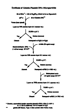
Figure 1. Schematic diagram showing the synthesis of gelatin-DNA
coacervates.
Figure 2. Gel el~r~phGles~s of cDNA before and after encars~ nn.
(std=standard; Sup=su~Y.-~I~nt, Pellet=micro~clespellPtç~d by
centrifugation) .
Figure 3. Controlled release of intact LAMP-l cDNA was de~l~on~ ted in
vitro. The miclop~licles were cross-linked with gluta~dehyde at
various glut~ldphyde conc~ ions then degr~ with trypsin.
WO 96/0029S 2 1 ~ 3 9 5 4 pCTlUS95/078S~
_5_
Pigure 3A shows the time course of DNA release at various
gllJtsr~sl~ehyde-cros-linking levels. Figure 3B shows (on gels and
~f~. c;10~ tracing) the DNA which was released from the
microparticles at various times and at various levels of
~ll)tsrslAPhyde-cross-linking-
Figure 4. Fluo~ t images of U937 cells transfected by controls and
LAMP-l cDNA-loaded l,licro~icles (at day 3 post-transfection).
Figure 4A: anti-DC44 mic~pallicles without cDNA; ~igure 4B:
- cgl~ m pho~phst~ tr~sn~ferti~n; Figure 4C: LAMP-l mic opal~cles
without allhbo l~, Figure 4D: LAMP-l micl~pa,licles coated with
anti-CD-44 mAB. LAMP-l ~ ion is ~..ani~sl~d a gr.snlll~s
(in l~s~sc,-~cs) in the cells.
Figure 5. Flow c~ h ;c analysis of the transfection efficiency of U937 cells
by antilymphocyte function associated antigen-l coated
iclopa licles and controls. The actual mean fluol~nc~ int~,lsily
(l~:I) is shown in the insert. Msp= mic~ her~s, MRK= a
mism~tche~ anti-P-glycoprotein antibody, PLM=anti-LFA
antibody.
Figure 6. Te-m~r~l ~A~ ssion of LAMP-1 in 293s cells ~r,sr~d by anti-
CD44 coated micr~p~licles.
DETAILED DF.~CRIPrION OF T~IE PREFERRED EMBODIMENTS
It is a discovery of the present invention that nucleic acid n~ol~~ s of
various chain lengths can CG rlex with polymeric cations in aqueous con~itiQn~
to form solid microparticles ~n~ng from submicron tO microns in size. These
W09~0C25S 21 93954 PCT/US95/078S7
~_ --6
nucleic acid-loaded ...icro~ icles, when app~l,.iately targeted, can transfect cells
with phagocytic activity. The rate of microparticle degradation and nucleic acid
release can be dç~ig~ a priori by varying the e~tent of cross-linking. The loading
levd of nucldc acid can be as high as 30~ (w/w), with an enc~rsul~tio
effiriçncy of > 95 % .
Accol~ling to the present invention, gelatin or other polymeric cation having
a similar charge densiq to gelatin, is used to complex with nucleic acids to form
miclopallicles. The source of gdatin is not thought to be critical; it can be from
bovine, pCilC~ne, human, or other animal source. Typically the polymeric cation
has a mqle~ qr weight of ~t- ~ 19,000 30,000. Poly-L-lysine may be
particularly useful as the poly.l-c~ic cation of the present invention. Desirably
sodium sulfate is used to induce the coacervation of polymeric cation and nucleic
acids. Fth~nol can also be used at a conc~nl.alion of about 40 to 60% to induce
- coacervation. Chondroitin sulfate can also be incol~.~led into the mic~pa~Licle,
which is ç~i~lly b~Pfi~i~l if one desires other ,ubs~nces such as drugs to be
incolllola~ in the ~,icropa~licle. Typically the conc~ t;on of chondroitin
sulfate is l~h. OC~'~ about 0.005 % and 0.1% .
T~ ing ligands can be directly bound to the surface of the Inicr~p~licle
or can be ind~ tl -cl-~l using a "bridge~ or "spacern. Rec~Jse of the amino
groups provided by the lysine groups of the gelatin, the surface of the
mi~r~pa.licles can be easily d~i~dtiz~xl for the di~ect coupling of targeting
moieties. ~lt~ tivdy, spacers (lin~ng mo!eculPs and derivatizing moieties on
targeting ligands) such as avidin-biotin can be used to indirectly couple targeting
ligands to the mic~l~nides. Biotinylated antibodies and/or other biotinylated
W096/00295 2 1 939 54 pCI'/US9S/078S7
--7
ligands can be co~,pled to the avidin-coated microparticle surface e-Mci~ntly
b~l~ of the high affinity of biotin (k,--10'5 M-') for avidin ff~:~7U~I~, et al.,
1990, ~oc~-cs;~g of p,~ulsor int~ -le~.kin 1 beta and innZ~ ol~ ~se, J. Biol.
Cnem., 265:6318-22; Wilchek, et al., 1990, Introduction to avidin-biotin
t~c.h. ology, Methods In Enymology, 184:5-13). Orient~tinn-selective ~tt~chmçnt
of IgGs can be a~h;e.cd by bioli,lylalin~5 the antibod~ at the oligGs~c(h~ e groups
found on the Fc portion (O'Sh~nn~ss~, et al., 1984, A novel p~dul~ for l~eling
i,"..,u--oglobulins by conjugation to oligos~ch~rides moieties, Immlu20l. Lett.,
8:273-27'7). This design helps to p~ e the total number of available binding
sites and renders the ~ h~A antibodies less immunogenic to Fc .~epLor-bearing
cells such as l..~ophaees Spacers other than the avidin-biotin bridge can also
be used, as are known in the art. For ~Y~mplr Staphylococ~l protein A can be
coated on the ,-,iclo~licles for binding the Fc portions of immunoglobulin
mol~c~lPs to the "licç~p~licles.
Cross-lin ing of linking molecules or targeting ligands to the mic~ licle
is used to promote the s~bili~ of the "liclu~licle as well as to covalently aff~7c
the linking ~ e or l~g~,ling ligand to the l,lic~p~licle. The degree of cross-
lin_ing dil~ly affects the rate of nucleic acids release from the miclosh~s.
Cross-linl~ng can be acco...pli~h~ using glllt~ hyde, c~l,od;imi~s such as
EDC (1-ethyl-3-(3-dimethylaminopropyl)-carbodiimide, DCC (N,N'-
dicycloh~,Aylc~~ e),c~hl~Ayls(peptidebond)linlGIge,bis(su~os~ccinimi~yl),-"t~., dimethyls~ -;n~;~at~ etc.
Ta~gcling ligands acco~ing to the present invention are any rnolrJules
which bind to a~-;r.c types of ce!ls in the body. These may be any type of
WO 9~ ~29S 2 1 9 3 9 5 4 pCl'lUS951078S7
" --8--
mo1 xule for which a cellular r~epLor exists. Preferably the cellular receptors are
e..p.~s~d on specifi~ cell types only. Examples of targeting ligands which may
be used are hoi,l,one~s, antibollius, cell~ hPcion mole~culPs, c~ s, drugs, and
n~ l, ns~
The microp~Licles of the present invention have good loading prope.hes.
Typically, following the method of the present invendon",.iclo~ ides having
at least 5 % (w/w) nucleic acids can be ach~eved. P~ fe.ably the loading is greater
than 10 or 15% nucleic acids. Often mic~p~licles of greater than 20 or 30%
nucleic acids can be acbie~od. Typically loading effi~i~P-n~ s of nucleic acids into
iCÇ~p~hCl~S of greater than 95% c~n be achieicd.
The method of the present in~renhon involves the coacervadon of polymeric
cations and nucleic acids. ~ se this pr~cess dep~n~ls on the intr"~ n of the
positively chalged polymeric cadons and the negatively ch~E,~d nucleic acids it
can be collsidered as a comple~ coacervadon process. However, sodium sulfate
(or ethanol) induçe.s the coacervation reaction by induçing a phase n~l;C;~;on, and
th" .~fo~ it could also be COfi' 'lC~Cd as a simple coacervation 1~ n Nucleic
acids are present in the coacervation ~~Lul~ at a c onc~nl ~I;on of between 1 ng/ml
to 500 ~glml. ~i~ly the nucleic acids are at least about 2-3 kb in length.
Sodium sulfate is present at bet . ~ll 7 and 43 mM. Gelatin or other polymeric
cation is present at between about 2 and 7~G in the coacervation l-~lu~.
An attractive .,uc~ icle delivery system l~u~,S a ~iP-lic~tp balance
among factors such as the simpi~y of ple~l;ol-, cost effectivene s, nucleic
acids loading levd, controlled release ability, storage stability, and ~ nvnogenicity
of the co",ponçnts. The gene delivery system described here may offer advantages
WO 9GI~25~ 2 1 9 3 9 5 4 PCTtUS95tO7~57
_g_
co~ d to other par~culate ddivery systems, including the liposomal system.
The problems of instability, low loading level, and controlled release ability are
better resolved with the polymeric microparticle systems. Gelatin has received
incl~s;ng biologic use ranging from surgical tissue adhesive (WP-incche1b~)m, et
al., 1992, Surgical treatment of acute type A dissecting aneurysm with
preservation of the native aortic valve and use of biologic glue. Follow-up to 6
years, J. Thorac. Cardiovasc. Surg., 130:369-74) to quantitative
immunohictoc-hemit~l assays (Izumi, et al., 1990, Novel gelatin particle
~lul;n~l;on test for sero~ia~nocic of leprosy in the field, J. ~'~inicn~ Microbiol.,
28:525-9) and as drug delivery vehicle (Tabata, et al., 1991, Effects of
l~l,-binant alpha-intc.Ç~.on-gelatin conjugate on in vivo murine tumor cell
growth, Cancer Res., 51:5532-8), due to its biocG~ atibility and ehL~IIIatiC
de~ hility in vivo. CGIIIP&'~Od to other ~ynlhe~c polymeric sy~."s, such as the
e~tensively studied pol~,l~;tic/polyglycolic copolymers, the mild con~i~io~c Of
microp~licle forrn~ on are ~?ling. Unlike the solvent evaporation and hot-
melt techniques used to formuhte S~llh~,l~C pol~",~.ic micç~ icles, complex
coacervation 14UUC'S neither contact with organic solvents nor heat. It is also
p~ul~l~ svit~llk for p ~p~ ting bio-macromo~ es such as nucleic acids not
only through passive solvent capturing but also by direct cha~ge charge
int~ ;o~.c
Unlike viral vectors, which cannot deliver genes larger than 10 kb, the
~"ic~p~licle delivery system of the present invention does not have such size
li...;li~l;onc Nucleic acid mole ~llPs of greater than about 2 kb can be used, and
nucleic acid mol~Jles even greater than 10 kb may be used.
WO 96/00295 2 1 9 3 9 5 4 pCIlUS95107857
--10--
In general, the range of possible targets is dependent on the route of
injection e.g. intravenous or intraarterial, subcutaneous, intra-peritoneal,
intrth~AI, etc. For systemic injections, the speeifir~ty of this delivery system is
affected by the ~r~ihility of the target to blood borne microparticles, which in
turn, is affected by the size range of the particles. Size of the particles is affected
by (~ AI-- e~ CG~pone~t COnCent~tiQn~ and pH in the coacervation Illixlule.
The particles can also be size-fractionated, e.g., by sucrose gradient
ult~c~nl~.~ug~;on. Particles with size less than 150 nanometers can access the
inte.~itial space by traversing through the f~e,l AI;on~ that line most blood vessels
walls. Under such c~.. cl; nc~s the range of cells that can be ~ge~d is
extensive. An abbreviated list of cells that can be ~,e~d includes the
p~nch~nal cells of the liver sinusoids, the fi~loblas~ of the c~nn~;ve tissues,
the cells in the Isleu of TAngr.l.~nc in the par,~ as, the cardiac myocytes, the
Chief and parietal cells of the il~t,~ e, osteocytes and chrondocytes in the bone,
l~ratinocytes, nerve cells of the p~ . ;ph~ ,Al nervous system, epithe~ cells of the
kidney and lung, Sertoli cells of the testis, etc. The targets for particles with sizes
greater than 0.2 I-,;el~ns will be cQ~r.~e~ largely to the vascular coll,p~~ nl.
Here, the ~g_t~ble cell types include erythrocytes, leukocytes (i.e. monocytes,
mac~phages, B and T l~lnphoc~tes, ncu~l)hils, natural killer cells, progenitor
cells, mast cells, 6Gs;nophils), plAtPlpts~ and endoth~-liAl cells.
For ~.~u~;~n~us injection~, the targetable cells incllJdes all cells that
resides in the cQnn~l;ve tissue (e.g. fibroblasts, mast cells, etc.), ~ ~n~" .I.~n~
cells, ~;no.;y~s, and muscle cells. For innO~ 1 injections, the targetable
cells include n~ ns, glial cells, a~LIocytes, and blood-brain barrier e-ndoth~liAl
WO96/00295 2 1 9 3 9 5 4 PCI/liS95/078S7
--11--
cells. For int~A~. ;ton~l injection, the targetable cells include the macrophages
and n~ ophil.
E.~amples:
Matrix Malerials: Gelatin (60 bloom, type A from porcine skin),
chondroitin 4-sulfate, glutaraldehyde (25%, grade 1), 1-ethyl-3-(3-
dimethylz.,..l-oplop,)~l)-carbor~iimi~ehydrochlori~e(EDChydro~-hlori~le),andultra-
pure sucrose were puf~ha~d from Sigma Chemic~l Co. (St. Louis, MO). Biotin
LC hy~r~7;~e, and NeutrAvidin, and Coo...assie protein assay ~ nts were from
Pierce (Roc~old, IL) . Centric~n micloc4~-c~ tc.-~ were from Amicon (Beverly,
MA).
Monoclonal an~iho~iP~: mAb PLM-2, a BALB/c mouse anti-porcine LFA-
1 (IgG",) which also cross reacts with murine LFA-l, was icol~t~A and purified as
previously described (Hildreth, et al., 1989, Monoclol-~l antibodies against porcine
LFA-l: species cross-reacdvity and function~l effects of b-subunit-specifi~
antibodies, Molec. Immunol., 26:883-895). IB4B, a rat anti-mouse LAMP-1
ascite fluid and a mouse anti-human CD44 mAb were icol~ted as previously
;k~ (de Wet, et al., 1987, Firefly lucif~.~ gene: sllu~:lu~ and cA~ on
in ,.. z.. Ali~n cells, Mol. ~ Cell. Biol., 7:725-37). CHA is a IgG, that does not
l~cognlze any known in viw mouse epilopes ~Hybritech Inc., San Diego, CA).
Affinity-purified FlTC and Texas Red-labeled polyclonal anti-rat IgGs were
obt~ined from Sigma.
W096100295 21 93954 PCT/US95/078S7
--12--
Genes: Two genes were used to demonstrate the feasibility of this delivery
system. The LAMP-a cDNA is a 6.4 kb circular supercoiled plasmid cDNA with
a mouse LAMP-l gene (2.4 kb) inserted into an Invitrogen pl~cmid cDNA with
a CMV pro"l,ot~. (G~ ..;e-;, et al., 1993, J. Biol. Chem., 268:1941). Detçction
of LAMP-l c.~p~saion was done by st~ining cells with anti-LAMP-l mAb and
with swondsl~ anti-IgG mAb conjugated with Texas Red. The gene coding for
lucirc~aae enzyme is widely used in cell biology for the study of gene eApl~ssion
bcca~ of the hlgh sensitivity of the assay, its s.mplicity, and low cost. In
~iitiQn, the ~ ""e is a good rtpol~r of gene eA~.ession because it is a cytosolic
protein that does not require post-trAnsl~tionAl plocess;ng for enzymatic activity (de
Wet, et al., 1987, Firefly luciferase gene: structure and c.~ ssion in mAmm~ n
cells, Mol. ,t~ Cell. Biol., 7:725-37; Wood, et al., 1989, Introduction to beetle
luciferases and their appli~-Ations, J. of Biolumin. ~ Cherrulum., 4:289-301). The
prcsence of luçiferA~e can be readily dct~ct~ by an enzymatic reaction that involve
the oxidAti~n of beetle lucif~in with con~o,..;l~nl productiol- of a photon (in the
form of ch~-mil~J,.-ine~c~-nce ) The assay was carried out using an assay kit
pulchaaod from ~o,nega Corp. (~A~lisQn~ WI).
Synthesis of mic,~s~,heres: A det~i4d sc-h~ nAI;s tlia~Am for the synthesis
of the gelatin-DNA coacc~ t~S is shown in Pigure 1. All con~ ns described
are final conc~ntrations in the reaction ~ ule set at 67~C unless otherwise stated.
Gelatin/p~mid DNA micr.pa,licles coated with avidin were synth~i7~ by first
pl~p~ing a 3.5 mg/ml solution of plasmid DNA en~ nP a lysoaol"al A~ tf~d
membrane protein-l (LAMP-l) (6.7 Kb, circular supercoiled) in 42 -.IM sodium
sulfate (Na~SO4). Coacervation was initiAt~ by the addition of gelatin (5%) to
wo 96/0029s 2 1 9 3 ~ 5 4 pcI/USgS/078S7
--13--
the DNA/Na2SO4 solutiQn at equal volume while vortexing at high speed for l
minute. Co~ qr,.~lqti~n of drugs and other agents can be achieved-by adding
directly to the DNA/Na2SO4 soluti~n before initi~ting coacervation with gelatin.
Avidin (5 mg1ml) was added to the lluc~us~,h~,~ sl~cp~n~ n at a final con~nt.~;on
of 75 ug avidin/ml mic~s~e e solutiom The microspheres ~ U~ was layered
onto a layer of 70% sucrose (w/v) and cent-;rugod at 6,000 x g for 4 minutes
(Rrinkm~n Ina~ ta Inc., Westbury, NY, model L8-75). Micn~allh~; fractions
recovered from the sucrose layer was diluted 5-fold with water then cross-linked
with glutaraldehyde (12.5 mM final con~ tiQn) for lO ~;n~tes at room
~n~ .~ Un~ t~l glllpra~ hyde was qu~ ~fh~i by adding eth~rlQl~mine (l
M) for lO ...;n.lt~s The u~i,lOaphc~s were dialyzed by sucroâe centrifugation as
d~e-;~ed above.
Atlachment of bioti~ylated mAbs to avidin-coa~ed mic,~eres: 30 ug of
anlibodies (biotinylated acc~ld--lg to established plocedule.s (O'Sh~nneccy, et al.,
1984, A novd p ~lu-~ for labeling immunoglobulins by conjugation to
oligos~hq~ s moieties, Irn~nunol. Lett., 8 273-27n) was added to l ml of
avidin-coated IlliC~Oa~ s ~s~ cion (1 1 mg/ml) for l hour with gentle ~git~ltion
Unbound mAb was removed from l.lic.uaphe~s by dialysis.
Cha,~t~,~on of microsphere and binding pe~orrnance: DNA loaded
mic~s~hc.es exhibited pol~r--,o.l.hic colloid shape with a polydispersed particle
size of less than 3 .,li.;.uns as d~ ncd by light micloacopy. Purified
micro~b~cs werc stablc for at least one month without apl.r~iable degr~tiûn
The loading level for LAMP-l plasmid DNA was 20~ (w/w). The enc~ps~ tion
effi~ency was typically ~95%. The mobility of the free LAMP-I DNA and the
21 93954
WO 3~ 5S PCT/IJS9~/078S7
--14--
leleased DNA (from the microsphere) in 1% agarose gel electrophoresis were
identi~l (Fig. 2),'s~r,l;ng that the encapsulated DNA was released in its
original form. Rdease rate of the cDNA from the micro~he.cs was dependent
on the crosclinking density and on thc enzyme level (Figure 4). Sust~inPA release
of up to weeks can be readily obt~ned.
We tested the ability of the LAMP-l DNA loaded mic~us~hel~s to bind and
subs~lcrlll~ nsÇ~cl a human histiocytic lymphoma cell line (U937j in tissue
culture. When coated with either anti-LFA or anti-CD44 monoclonal antibody
- (both protein targets were cA~l~sed in high amount of U937 cell surface),
eA~I~s~ion LAMP-l protein was detP~tPd by day 3 (Fig. 4A, fluol~nt granules)
when stained with antibodies l~cogni2ing LAMP-l. The st~ining pattern of U937
cells inc~b~t~d with LAMP-l mic~os~he,~s was identical to the calcium phosphate
method of tran~lion (Figure 4B). Miclus~Jhe~s that were either coated with
avidin or non-s~ific CH~ mAb showed no granular st~ining p~tterns, and were
idPn~ l to ~mL~t~ cells (Fig. 4C).
Using flow cyl~ .A~, we showed that the eAp~s~ion of LAMP-l was
~t~h~ in up to 5% of U937 cells in culture (Figure 5). None of the controls --
blank .,licr~h~ s""ic~ù ~he.~s with cDNA but no ~ntibodies, ,llicros~ e.~s
with cDNA and coated with a ~ n.~ Pd anti-P-glycoplu~n antibody (MRK-
Msp.), and free cDNA at a concen~ ion six time higher than entrapped in the
microspheres showed any evidence of transfection. The effiriency of
tr~ncfection ~ to be dose-n s~nsi~e. In general, the tPncf~P~irJn effirier~cy
of tnis particular gene and cell type is comparable between the proposed
~llicr~aphc.ic delivery system (1-10~) and the c~lriurn pho;,~h~tP pl~;pit~tion
wo 9~ G25S 2 1 9 3 9 5 4 PCT/US95/078S7
--15--
method (2-15%). Figure 6 demonstrates the concept in a different cell type using
a dirÇ~ent molloclQn~l antibody. Again, free cDNA could not tTansfect the cells.
Eventually the LAMP-l e~l~ssion ~ ppc~cd after several passages. Positive
results were also ob~intd for the luciferase repo~ gene system. Transfection
was clearly ~e~t~l by ,..~s.-~.,-~nt of luciferase enzymatic activity, in 293s cells
inC~ ~ with lu~-;f~-~, gene-loaded ll.icr~sl.hercs.