Note: Descriptions are shown in the official language in which they were submitted.
N'O 96/04543 PCTlUS95/09509
2 i X3971
' - .' , 4,
PSEUDO TELECENTRIC OPTICAL DESIGN FOR FLOW CYTOMETRIC BLOOD
CELL ANALYZER
BACKGROUND OF THE INVENTION
This invention relates to a multi-dimensional optical
design. More particularly the invention relates to an
optical design of a multi-dimensional system which can
simultaneously detect five or more distinct properties of
particles or cells when the design is applied to a flow
cytometric analyzer.
Particle analysis, known generally as flow cytometry,
consists ofpassing particles one at a time through a
sensing region of a-flowcell, and detecting the properties
or characteristics, of each particle. These specific
properties, which are sometimes referred to as dimensions,
are usually combinations of multi-angle light scatter and
multi-color fluorescence.
Flow cytometry has become a particularly important
method for analyzing blood cells in the hematology
laboratory where patient test load is an important metric.
This is because the method is rapid, enabling as many as
five to ten thousand cells per second to be analyzed, and
because it is much more statistically accurate than the -
manual microscope inspection method. It is important,
however, to the hematology laboratory, that the entire -
process, both sample preparation and analysis, be automated.-
A large number of products exist today which feature
such multi-dimensional capability, but only a few automate
the entire process. Two of the most well known such
products in which the entire process of blood cell analysis,
or differentiation is fully automated are the Cell-Dyn~
series 3000 and 3500 analyzers manufactured by Abbott
1
VVO 96104543 P~'IUS95/09509
~. I .~''.t ';,i
21~397~
Diagnostics.- Each of these instruments measures
simultaneously four dimensions which include three angles of
laser light scatter, and a fourth dimension which is
depolarized light scatter.
A number of products exist which measure several
simultaneous dimensions of fluorescence and scatter in which
only the analysis is automated. One of the most well known
of these is the Becton Dickinson FACSCan~ flow cytometer.
This instrument is capable of simultaneously detecting one
dimension of fonaard scatter, one dimension of side scatter,
and three colors of fluorescence.
However, in none of these multi-dimensional products
which combine several colors of fluorescence and light
scatter, is the entire process automated. Part of the
reason for this is the complexity of building a system which
is stable enough to maintain proper alignment for many
simultaneous dimensions while at the same time, assuring the
measurement integrity of each cell or particle in the sample
stream for all dimensions.
Among the prior art contributions, is the Auer et al.
U. S. Patent (4,038,556 which describes a two-dimensional
system with a flowcell, a laser light source, and two
simultaneous optical paths, a side angle collection system
for measuring cell fluorescence, and a forward angle system
for measuring light scatter. The patent teaches that by
placing the forward angle detector in the back focus of a
light collectinglens, an important and practical
simplification of system alignment results; the precise
relationship o~ the forward angle optical system, with
respect to the remaining elements of the system, is greatly
relaxed. Although the side angle beam focus, the laser beam
focus, and the stream focus must be established to be
mutually collinear in the Auer et al. teachings, it is not
reguired for theforward angle path. This is due to design
of the fonaard path system which transforms the two
2
VVO 96104543 PCTlUS95/09509 _
z i 9'39T ~
,, ,. .
dimensional distribution of intensity vs angular
distribution in the flowcell space to intensity vs spatial
distribution at the detector.
Hirako, in U. S. Patent #4,953,979, describes a side =
angle collection system for flow cytometry which has the PMT -
front surface conjugate with the condenser exit pupil while
the flow stream (containing the particles or cells) is
conjugate with an external aperture located between the
condenser and the PMT. The external aperture, which limits
unwanted background light, is located at the front focus of
a second lens, which functions to image the condenser exit
pupil on the PMT. The patent teaches that as the stream
position, or cell position within the stream varies, the
effect on cell coefficient of variant (mC.V.") of detector
sensitivity variations are eliminated.
Hirako, ignores the C.V. effect of stream or cell
position variations within the flowcell upon the angular
integrity of the scattered light with respect to the laser
beam.
It is one object of this invention to maintain the
angular integrity of the scattered light with respect to the
laser beam in both the forward and side angle light paths.
It is another object of this invention to improve the
stability of, and at the same time simplify, the alignment
and tracking requirements of a multi-dimensional flow
cytometer.
It is yet another object of this invention to combine
this design approach with a multi-element array detector and
a simple laser beam translating mechanism, to assure beam to
stream tracking simplification, while at the same time
assuring the measurement integrity of each particle or cell,
independent of cell location in the stream, or the precise
stream location within the flowcell.
3
W 0 96/04543 PCTIUS95I09509
. . ' ~ = ~,19~97i
It is another object of this invention to maximize
these advantages W at least two separate light paths
simultaneously.
These and other advantages will become more apparent in
the following detailed description.
SUMMARY OF THE INVENTION
The present invention is directed to a flow cytometric
optical system for the simultaneous detection of several
characteristics o~ particles suspended in a flowing medium
wherein the system comprises a flowcell through which the
particles pass substantially one particle at a time, an
optical system for-directing light from a light source onto
the flowing particles in the flowcell, a side angle optical
collection system-for receiving light from the flowing
particles and for directing the light to one or more of a
first set of detectors, and a forward angle collection
system for receiving light from the flowing particles and
for directing the light to one or more of a second set of
detectors. The side angle optical collection system
comprises a condenser lens for directing light toward the
first set of detectors with an exit pupil of the condenser
lens located at the back focal plane of the condenser lens;
a photosensitive surface of one or more of the first set of
detectors is located at conjugate points of the back focal
plane of the. condenser lens such that an image of the exit
pupil is positioned at the photosensitive surface of one or
more of the first set of detectors. The forward angle
optical collection system comprises a collecting lens for
directing light toward the second set of detectors where the
collecting lens ex~a pupil is located in the back focal
plane of the collecting lens and a photosensitive surface of
one or more of the second set of detectors is located at the
back focal plane of the collecting lens.
4
VI'O 96104543 PCT/US95109509
._ ;, X193911
BRIEF DESCRIPTION OF THE DRAWINGS:
Figure 1 is a schematic of the optics of a simple
microscope.
Figure 2 is an optical plan view layout of a preferred
embodiment of the invention.
Figures 3a and 3b are a schematic-of a forward scatter
optics system of a preferred embodiment of the present
invention.
Figure 4 is a thin lens equivalent schematic
illustrating the principals of the side angle optical
collection system of~the present invention.
DETAILED DESCRIPTION OF THE INVENTION:
Although geometric imaging doesn't have the same
significance in a flow cytometer system as in a diffraction
limited system such as an optical microscope, the
performance of a flow cytometer system is best understood by
means of simple geometric image analysis.
In all properly designed systems, there are two system
stops which function to limit the ray paths through the
system. At any point along the optical-path, these stops,
or images of these stops, determine the extreme ray paths
which are admitted through the system. In classical -
geometric optics, the one stop can be referred to the
"field" stop, and the other the "pupil" stop. Figure 1 is a
schematic of a simple microscope which illustrates this.
The lens in Figure 1 is designed to satisfy a condition
referred to by designers of microscope systems as the
"telecentric condition". A general understanding of the
performance of a "telecentric" design is useful in
understanding some of the key aspects of this invention.
W O 96/04543 P~1YUS95/09509
~~ '~~,v ~:~, 2193973
In Figure 1, a two dimensional object normal to axis
110 of lens 101, is located at field stop 100 which is
positioned at the,front focal point of lens 101. Object
point 102 lies on-xhe lens axis and is thus coincident with
the front focal point of lens 101, while point 105 is
displaced laterally some small distance from lens axis 110.
At the same time, the lens exit pupil 103, is located at the
back focal point 104 of lens 101. Object 100. can be thus
expressed as a two dimensional distribution of intensity vs
linear distance from the lens axis. This object is
transformed into an intensity vs angular distribution after
passing through lens 101. This same visualization can be
used in the reverse direction. Exit pupil 103 can be
described as an object located in the back fecal plane 103
with an intensity vs linear distance dimensional
distribution, which after passing in a reverse direction
through lens 101 is transformed into an intensity vs angular
distribution.
The unique aspect of a telecentric design is that each
discreet point in the field is transformed into a collimated
ray bundle with a discreet trajectory in the.space of the
exit pupil. Conversely each discreet point in the exit
pupil is transformed into a collimated ray bundle with a
discreet trajectories in the space of the field. Thus in
Figure 1, rays 105 and 107 which are diverging from field
point 105, are parallel to each other upon leaving lens 101.
Similarly, rays 108 and 109 which are diverging from field
point 102, are parallel to each other upon leaving lens 101,
but at a slight angle relative to the parallel rays which
came from point 105. In the same sense in the reverse path,
rays 106 and 107 which diverge slightly from pupil point 1I1
are traveling parallel to each other as they leave lens 101.
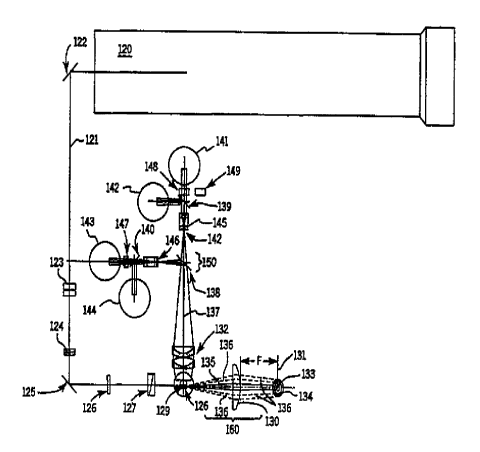
Figure 2 is an optical plan view layout of a preferred
embodiment of the invention. Beam 121 from laser 120 is
directed to flowcell 128 by means of mirrors I22 and I25,
6
,. - ~ CA 02193971 1999-08-18
WO 96/04543 PCT/US95/09509
beam shaping lenses 123 and 124, focusing lens 126, and a
vernier fine adjust element 127. The direction of flow of
sample stream 129 is normal to the plane of the Figure. In
a preferred embodiment, a side angle optical collection
system 150 consists of a compound condenser lens 132 which
collects scattered and fluorescence light from particles
within sample stream 129, and directs this light 133 to
photomultiplier detectors 141, 142, 143. In the preferred
embodiment, lens 132 is a 9.0 mm focal length which is
optically coupled to the flowcell with a resulting numeric
aperture of 1.2. Dichroic beam splatters 138, 139 and 140-
function to spectrally partition the optical beam 133 as is
appropriate for each detector. Optical filters such as
illustrated by 147, 148, and 149 are inserted automatically
as required by the particular test protocol. It should be
understood that the paths which are folded by means of
dichroic beam splatters 138, 139, and 140, are optically the
equivalent to the unfolded beam, and for the sake of
clarity, the principals of the side angle optical collection
system 150 is more simply understood by referring to the
thin lens equivalent schematic of Figure 4.
In Figure 4 the compound lenses with curvature,
. thickness, and air spaces, are replaced with thin lens
equivalents, which enables a clearer understanding of the
imaging properties of the invention Exit pupil 151 of
condenser 132 is located in the back focal plane of
condenser 132. Fu=ther, an image of exit pupil 151 is
conjugate with the nominal photosensitive surface 152 of
detector.141. Note that point 155 at detector
photosensitive surface 152, is conjugate with point 154 at
the outer edge of exit pupil 151, and that because of the
telecentric nature of the design, the rays emanating within
the flowcell which pass through these points, 156 and 157
are mutually parallel in the laser/flowcell space. This
combination assures that all rays arriving at a given point
7
CA 02193971 1999-08-18
WO 96104543 PCT/US95I09~~~ "
at the detector correspond to a particular scatter angle
relative to the laser, independent of where the particle is
located within the flowcell. Thus the C.V. of particles
within the flowcell is substantially independent of location
within the stream, stream location within the flowcell, or
spectral sensitivity of the photodetective surfaces.
An additional feature of side angle collection system
150 is that an image of the stream is placed at external
aperture~242 which is located very near Field lens 145.
Aperture 242 functions to limit excess background light from
the detectors, however it's size is not critical, and thus
it is sized to be large enough to prohibit any sample light
from being vignetted in cases where the stream image at.the
aperture is defocused due to stream wander along the beam
axis 133 of the side angle collection system. This system
overcomes the usual problem of the requirement to realign
the side angle optical path whenever a flowcell or nozzle
problem occurs. Additionally, the system intrinsically
assures consistent angular integrity of the scattering
particles relative to the laser illumination source.
Forward angle collection system 160, is also
illustrated in Figures 2 and 4. Photodiode detector 131 is
placed in the back focal plane of lens 130. Figure 4 again
illustrates the principal that all rays arriving at a
discreet point on the detector emanate from the flowcell
with a specific angular trajectory. In the reverse path
sense, points in the detector space correspond to collimated
rays in the flowcell space. In the pref erred embodiment,
detector 131 is an array detector in which the dimensional
extent of each array element becomes the exit pupil of
forward angle collecting lens 130.
Thus, so long as lens 130 and detector 131 are properly
aligned with respect to each other, outer element 134 which
is a circular ring with inner diameter 3.6 mm and outer
diameter of 12.3 mm, will accept only scattered light from
8
WO 96!04543 PGT/US95I09509
- ~ ~~~~'~' ' '~ 219 3 9 71
the flowcell with a range of scattering angles between 3
and 10 degrees relative to the laser axis. This signal is
referred to as Intermediate Angle Scatter iIA$). Inner
element 133 is rectangular in shape to match the beam
divergence of the laser in the ~lowcell space. In the
preferred embodiment the dimensions of element 133 are 1.5 - _
mm x 0.4 mm which corresponds to the vertical beam
divergence of 37 mrad, and a horizontal divergence of 9.7
mrad. The equation which relates the pupil radial dimension
to the angular divergence is:
Y = F
where Y is the radial dimension at the pupil, and ~ is the
scattering angle relative to the laser axis.
Inner element 133 detects a signal generally related to
particle size, which is referred to as Axial Light Loss
(ALL). In the ALL system, detector 133 collects only light
within an incident cone of laser illumination. The signal
of interest is a negative signal subtracted from the steady
state laser signal.
From an alignment perspective this configuration of
forward angle collection optics is a substantial
simplification over prior art. The usual requirement that
the forward angle system be precisely collinear with the
side angle system, the stream, and the laser, is
unnecessary. Additionally, the usual beam blocking and
corresponding adjustment is not required, since the laser
signal is used instead of blocked. Finally, once the proper-
positional relationship has been established between lens
130 and detector 131, the alignment, due to the back pupil
aspect, is simply to adjust the detector for maximum steady
state signal in the absence of any particle in the sensing
zone. Thus, the telecentric aspect of this design in
combination with the laser ALL measurement assures the
9
WO 96104543 PCTIUS95I09509
v~~~~'~ 2193971 1
absolute angular integrity-of detector-131, and the
lithographic process establishes the relative integrity of
array 134 and 133.
In Figure 3; the laser is brought into maximum
coincidence with the stream by means of fine adjust
mechanism 127. This consists of a pair of wedge prisms
located between laser focusing lens 126 and flowcell 128.
The wedge prisms are positioned so that change in the air
space laterally displaces the laser beam in flowcell 128
without any change in the illumination angles. The
mechanism is extremely easy to control in order to
accommodate micron beam displacements in the flowcell for
maximum signal sensitivity. Since the adjustment is lateral
rather than angular, the alignment of the fonaard angle
collection system 7.,60 as well as side scatter system 15~
remain unaffected is affected.
While certain representative embodiments-and details
have been shown for the purpose of illustrating the
invention, various changes and modifications can be made
herein without departing from the essence and scope of the
invention defined in the claims.
1o