Note: Descriptions are shown in the official language in which they were submitted.
~~~~~~J~
R'O 96/01611 PCT/U595/08151
I
-1-
Implantabie device containing tumor ce77s for treatment of cancer.
" Field of the Invention .. __ ,
S This invention relates to cancer prevention and treatment through the
implantation of tumor cells into the patient where the tumor cells are
contained in a
chamber which segregates the tumor cells from the patient's tissues.
B~~?und of the Invention
Currently accepted therapies for most tumors are surgery, chemotherapy,
radiation therapy, bone marrow transplants or various combinations of these
therapies. In general these treatments are aimed at the destruction of the
tumor cells
by mechanisms independent of activation of the patient's immune system. In the
course of radiation and chemotherapy significant damage to the immune system
is
an unfortunate side effect. Moreover, the long term effectiveness of these
treatments for some tumors is questionable.
a. Activation of the Lm_rt,_une S c em
Within the last decade therapeutic approaches have been developed based on
the activation of the immune system to mediate anti-tumor activity. Generally,
a
normal host response to tumor cells begins with T-cell recognition of tumor
associated antigens on tumor cells or via antigen presenting cells.
Recognition via
T-cell antigen receptor triggers signal transduction pathways that mediate the
activation of the T-cells. This results in secretion of interleukin-2 (1L-2),
gamma
interferon; tumor necrosis factor-alpha, and other cytokines from the T-cells
and
accessory cells. The host immune system is thus mobilized to kill the tumor
cells.
However, for reasons that are poorly understood, for many tumors this host
response does not occur or is inadequate to kill tumor cells.
One therapy designed to activate the immune system is the systemic
administration of IL-2. However, the doses of IL-2 required to achieve
adequate
amplification proved to be toxic to the patient. Cellular immunotherapy
approaches
to activate the immune system have focused on two types of cells: LAK cells
and
TIL cells. LAK (lymphokine activated killer) cells are cells of the immune
system
which have been non-specifically activated through the use of cytokines such
as 1L-
WO 96101611 PC'fIUS95/08151
2'194555
_2_
2 (Lotze et al., Cancer Res. 41, p. 4420-4425 (1981); Grimm et al., J. Exp.
Med.
155, p. 1823-1844 (1982)) and/or the use of monoclonal antibodies such as anti-
CD3 (Ochoa et al., Cancer Res. 49, p. 693-700 (1989)). These cells can mediate
significant anti-tumor activity without the major histocompatibility complex
(MHC)
related restrictions characteristic of the T-cell receptor of classical
cytolytic T-cells
(CTL). In one recent study continuous infusion of IL-2 and LAK cells for
advanced tumors resulted in responses in 12% of patients with melanoma and 3%
of patients with renal cell carcinoma (Lotze, Cell Transplantation 2, p. 33-47
(1993)). While most LAK cells characterized to date consist of activated
natural
killer (NK) cells (Ortaldo et al., J. Exp. Med. 164, p. 1193-1205 (I986);
Ferrini et
al., J. Immunol. 138, p. 1297-1302 (1987); Phillips and Lanier, J. Exp. Med
164,
p. 814-825 (1986)), LAK cells can also be generated from a subset of T-cells
known as y8 T-cells (T'-cells which lack the classical a(J subunits of the T-
cell
receptor and instead express the y8 subunits) (Ochoa et al., Cancer Res. 49,
p. 693-
700 ( 1989)). LAK cells can also be generated from isolated CD4+ or CD8+ T-
cells
which have been cultured in the presence of IL-2 and anti-CD3 monoclonal
antibodies (Geller et al., J. Immunol. 146, p. 3280-3288 (1991)). TIL. (tumor
inf-Iltrating lymphocytes) cells are lymphocytes which have been isolated in
vitro
from tumors. Like LAK cells, these cells can be expanded by culturing in the
presence of cytokines such as IL,-2 or IL-4, but, unlike LAK cells, these
cells are
tumor specific. Using TIL cells, a 20-50% response rate has been observed in
patients with melanoma (Lotze, Cell Transplantation 2, p. 33-47 (1993)).
b. F h ncim~ IrxLmnnoQ_enicity of Tumors
Other approaches for tumor immunotherapy involve increasing the
immunogenicity of tumor cells rather than enhancing the activity of responding
lymphocytes. It is believed that many tumor cells lack a degree of
immunogenicity
required to induce an adequate immune response (Houghton and Lewis, "Cytokine
Induced Tumor Immunogenicity," ed. Forni et al., Academic Press, p. 35-54
( 1994)).
Generally, stimulator cells (such as tumor cells) activate T-cells by
engagement of the T-cell receptor with peptide associated with either Class I
or
Class II MHC molecules on the stimulator cell. These peptides can either be
taken
up from the external environment by the stimulator cell, in which case they
are
2194555
WO 96/01611 PCTIUS95/08ll51
~3-
processed and presented along with MHC Class II molecules or, they can be
peptides produced endogenously by the stimulator cell and then presented with
MHC Class I molecules. Presentation of exogenously derived peptides by the
stimulator cell is referred to as indirect presentation since the peptides are
not being
presented on the cell from which they were derived. In the case of direct
presentation, peptides are presented on the surface of the cells from which
the
peptides were derived. Figure 1 is a schematic diagram showing direct vs.
indirect
presentation.
In addition to the T-cell receptor and MHC antigens, a number of cell
surface antigens have been identified that may play a role in mediating
interactions
between antigen presenting cells and the responder T-cells. These co-
stimulatory
molecules include intercellular adhesion molecules (ICAMs), vascular cell
adhesion
molecule 1 (VCAM-1), lymphocyte function-associated antigen 3 (LFA-3), heat
stable antigen (HSA) and CD28 on lymphocytes, and the ligand B7 which must be
present on the antigen presenting cell (Pardi et al., Immunol. Today 13, p.
224-230
( 1992); Chen et al., Immunol. Today 14, p. 483-486 ( 1993)). Engagement of
the
T-cell receptor with the antigen presenting cell in the absence of
costimulatory
molecules can lead to T-cell anergy and failure of the immune response against
the
tumor (Gimmi at al., Proc. Natl. Acad. Sci. 90, p. 6586-6590 (1993)).
Unique tumor antigens have been defined for several tumors including the
MAGE (van der Bruggen et al., Science 254, p. 1643-1650 (1991)) and MART
(Kawakami, Proc. Natl. Acad. Sci. USA 91, p. 3515-3519 (1994); Boon et al.,
Ann. Rev Immunol. 12, p. 337-365 ( 1994)) antigens for melanoma and mucins for
breast and pancreatic tumors (F"mn, J. Cellular Biochem. 17D, p. 92 (1993);
Domenech et al., J. Cellular Biochem 17D, p. 108 (1993); Fontenot et al., J.
Cellular Biochem. 17D, p. 125 (1993)). See ,also Brown, J.P. et al., U.S.
Patent
No. 5,141,742 (melanoma associated antigen). It has been observed that tumor
cells do not efficiently present self-peptides (direct presentation) even when
the cells
do express MHC antigens, suggesting that there might be a defect/deficiency in
another molecule necessary for effective direct presentation of antigen by
tumor
cells. Many tumor cells have been demonstrated to express low levels of B7.
Accordingly, one therapeutic approach is to restore the immunogenicity of the
tumor cells by the introduction of the gene for B7 into the patient's tumor
cells, thus
promoting direct tumor antigen presentation (Chen et al., Cell 71, p. 1093-
1102
WO 96/01611 PCT/US95/08151
21 X4.555
-4-
(1993) and EPO 600591; Chen et al., J. Exp. Med. 179, p. 523-532 (1994);
Townsend and Alison, Science 259, p. 368 370 ( 1993); Baskar et al., Proc.
Natl.
Acad. Sci. USA 90, p. 5687-5690 (1993)). The introduction of the CD28 ligand
B7 to immunogenic lymphoma, mastocytoma, melanoma or sarcoma resulted in
increased CTL activity against the wild type tumor and protection against
subsequent injection with the wild type tumor (Chen et al., Cel171, p. 1093-
1102
(1993) (melanoma); Chen et al., J. Exp. Med. 179, p. 523-532 (1994)
(mastocytoma, fibrosarcoma, lymphoma, melanoma, carcinoma); Townsend and
Alison, Science 259, p. 368-370 (1993); Baskar et al., Proc. Natl. Acad. Sci.
USA
90, p. 5687-5690 (1993) (sarcoma)). Further, injection of EL4 lymphoma cells
expressing B7 resulted in a 60% cure rate in mice with established EL4 derived
tumors (Chen et al., J. Exp. Med. 179, p. 523-532 (1994)). 5imilarIy,
transfection
of marine colon adenosarcoma or fibrosarcoma with genes for marine Class I
molecules could mediate the regression of unmodified tumor, although tumors
were
not completely eliminated (Plautz et al., Proc. Natl. Acad. Sci. USA 90, p.
4645-
4649 (1993) (fibrosarcoma, colon carcinoma)). This approach is currently being
tested in human clinical trials (Nabel, Proc. Natl. Acad. Sci. USA 90, p. 94-
97
( 1993) (melanoma)). In both these examples the introduction of the foreign
genes
enhanced direct, Class I mediated recognition of the tumor cells by the
effector cells
of the host. The response is tumor specific. Treatment with the genetically
modified tumor had no effect on the growth of an unrelated tumor. This
response
is throught to require direct cell-cell contact. See also Hock et al., Gene
Therapy
Weekly, p. 22 (January 9, 1995) (marine neuroblastoma cells expressing Class
II
MHC).
A slightly different approach was taken by Trojan et al. for treatment of
glioblastoma. Glioma cells express high levels of insulin-like growth factor I
(IGF-
1). Treatment of glioma cells with an anti-sense gene for IGF-1 appears to
reverse
the tumorogenic phenotype rendering the cells immunogenic. In these studies,
injection of glioma cells expressing the IGF-1 anti-sense sequence resulted in
elimination of pre-existing tumor in all animals treated (Trojan et al.,
Science 259,
p. 94-97 (1993)). Although this response was shown to be mediated by CD8+ T-
cells, it is not clear whether they are activated directly by the modified
tumor cells or
indirectly via antigens picked up by antigen presenting cells or both.
~ 9~. ~~5~
R'O 96/01611 PCTIU595108151
_g_
Additional approaches for enhancing the immunogenicity of tumors involve
' engineering the tumor cells to express cytokine genes such as IL,-2, IL-4,
IL-6,
tumor necrosis factor, interferon-y or granulocyte macrophage colony
stimulating
' factor (GM-CSF) (Dranoff et al., Proc. Natl. Acad. Sci. USA 90, p. 3539-3543
(1993); Golumbek et al., Science 254, p. 713-716 (1991) (renal cell
carcinoma);
Gansbacher et al., Cancer Res. 50, p. 7820-7825 (1990); Gansbacher et al., J.
Exp. Med. 172, p. 1217-1224 (1990); Bannerji et aL, J. Immunol. 152, p. 2324-
2332 (1994) (fibrosarcoma); Fearon et al., Cell 60, p. 397-403 (1990) (colon
carcinoma); Columbo et al., J. Exp. Med. 173, p. 889-897 (1991)
(adenocarcinoma); Haddada et al., Hum. Gene Therapy 4, p. 703-711 (1993)
(mastocytoma); Lollini et al., Int. J. Cancer SS, p. 320-329 (1993) (mammary
adenocarcinoma); Watanabe et al., Proc. Natl. Acad. Sci. USA 86, p. 9456-9460
(1989) (neuroblastoma); Pardoll, Curr. Opin. Oncol. 4, p. 1124-1129 (1992);
Tepper and Mule, Hum. Gene Therapy 5, p. 153-164 (1994); Porgador et al.,
Cancer Res. 52, p. 3679-3686 (1992) (Lewis lung carcinoma); See also WO
92/05262 Hopkins/University of Texas. Here too, the genetically modified tumor
cells are able to stimulate an immune response in situations in which the
parent
tumor lines are non-immunogenic. Researchers in this area have observed that
the
immune response extends to destruction of unmodified tumor cells as well as
the
engineered tumor cells and can, in some cases result in complete regression of
pre-
existing tumor in experimental animals (Dranoffet al., Proc. Natl. Acad. Sci.
USA
90, p. 3539-3543 (1993); Golumbek et al., Science 254, p. 713-716 (1991);
Gansbacher et al., Cancer Res. 50, p. 7820-7825 (1990); Gansbacher et al., J.
Exp. Med. 172, p. 1217-1224 (1990); BannerPi et al., J. Immunol. 152, p. 2324-
2332 (1994) (fibrosarcoma); Fearon et al., Cell 60, p. 397-403 (1990); Columbo
et
al., J. Exp. Med. 173, p. 889-897 (1991); Haddada et al., Hum. Gene Therapy 4,
p. 703-711 (1993); LolIini et al., Int. J. Cancer S5, p. 320-329 (1993);
Watanabe et
al., Proc. Natl. Acad. Sci. USA 86, p. 9456-9460 (1989); Pardoll, Curr. Opin.
Oncol. 4, p. 1124-1129 (1992); Tepper and Mule, Hum. Gene Therapy S, p. 153-
164 (1994); Porgador et al., Cancer Res. 52, p. 3679-3686 (1992); Vieweg et
al.,
Gene Therapy Weekly, p. 20 (November 21, 1994) (prostrate cancer)).
In general these experimental protocols involve immunizing animals one or
more times with irradiated tumor cells that have been genetically engineered
to
express the exogenous gene. Irradiation prevents the cells from dividing but
does
W0 96101611 PCTIUS95/08151
-6-
not diminish their antigenicity. Anti-tumor responses are then tested in one
of three
ways: (i) the animals are challenged with unmodified tumor cells after the
immunization process is complete; (ii) the animals are challenged with
unmodified
tumor cells during the vaccination process; or (iii) small tumors are
established
before immunization with modified tumor cells.
The majority of the studies utilizing genetically modified tumor cells have
involved the introduction of cytokine genes into various tumors (see Pardoll,
Curr.
Opin. Oncol. 4, p. 1124-1129 (1992); Teppec and Mule, Hum. Gene Therapy 5, p.
153-164 (1994) for reviews). One of the most effective molecules is GM-CSF
(granulocyte macrophage-colony stimulating factor) which augments specific
immunity for several tumor types (Dranoff et al., Proc. Natl. Acad. Sci. USA
90,
p. 3539-3543 (1993) (B16 melanoma, colon carcinoma, lung carcinoma,
fibrosarcoma, renal carcinoma)). GM-CSF is unique in that it may be mediating
this anti-tumor effect by stimulating the proliferation and differentiation of
dendritic
cells which are extremely potent antigen presenting cells capable of
presenting
antigens to both CD4+ and CD8+ T-cells (Steinman, Ann. Rev. Immunol. 9, p.
271-296 (1991)). Metzinger has recently suggested that the only way the immune
system can be activated to respond to tumors is via shed antigens being picked
up
and presented by professional antigen presenting cells such as dendritic cells
(Metzinger, Ann. Rev. Immunol. 12, p. 991-1045 (1994)). Similarly Bannerji et
al. have recently suggested that the rejection of IL-2 secreting fibrosarcoma
cells is
not T-cell mediated although the subsequent systemic immunity is dependent
upon
the presence of both CD4+ and CD8+ T-cells (Bannerji et al., J. Immunol. 152,
p.
2324-2332 (1994)). They hypothesize that the destruction of the modified cells
is
mediated by NK cells resulting in the release of tumor antigens which can be
taken
up by antigen presenting cells expressing both Class I and Class II molecules
on
their cell surface. These cells would then be capable of activating both CD4+
and
CD8+ T-cells. A similar model has been discussed by Shoskes and Wood
(Shoskes and Wood, Immunol. today 15, p. 32-38 (1994)).
In recent experiments Cohen and co-workers have been able to prolong
survival of mice with pre-existing melanoma by injecting the animals with
allogeneic fibroblasts which have been transfected with the gene for 1L-2 and
DNA
isolated from melanoma cells (Kim and Cohen, Cancer Res. 54, p. 2531-2535
(1994)). By using allogeneic cells there is no need to irradiate the cells,
which
2194~5~
WO 96101611 PCTIUS95108151
"., ;
could affect cytokine expression. Since the transfected cells are fibroblasts
they do
not form tumors and since they are allogeneic they readily activate the immune
system. However, since they are readily rejected there is no long term
stimulation
of the immune system. Others have mixed cytokine expressing fibroblasts with
irradiated tumor cells and then administered the mixture as a vaccine (WO
93/07906, PCT US92/08999). Still others have coupled nontumorous fibroblast
cells to an adjuvant and administered the cells as a tumor vaccine (Eggers,
U.S.
Patent No. 5,208,022).
Yet another therapy for prevention and treatment of tumors is immunization
with tumor antigens (WO 93/06867 Pardon, Mulligan). Another vaccine protocol
is administration of irradiated tumor cells together with a bacterial adjuvant
(Pardon,
5 Cur. Opin. Immunology, p. 719-725 (1993). Others have irradiated unmodified
tumor cells and administered them alone as a vaccine (Dranoff et al., 90 PNAS,
p.
35393543, Figure 4A (1993)).
IS
c. Tu_n?or Evolut',_on
Most cancers are believed to be clonal in origin and that new subpopulations
arise continuously during evolution of a cancer due to Darwinian selection of
genetic variants that have a growth advantage. Some of the genetic variants
are
characteristic of a particular tumor type and in fact can serve as the basis
for
classifying the severity of tumor, in other cases the changes are idiotypic,
i.e.
specific to the individual's own tumor. Mutations giving rise to growth
advantage
include mutations in growth regulatory genes, changes in morphology, hormone
dependence, enzyme patterns, and surface antigens. Some of these changes may
avow the abnormal cells to escape either homeostatic controls of the patient
or
destruction by treatment. Conventional chemotherapies are often effective
initially
in slowing the progression of disease. However, with time, repeated treatments
become less effective, perhaps through evolution of successively less
sensitive
clones (G. Klein and E. Klein, PNAS USA 74, p. 2121 (1977)). See also
Schreiber, H., "Tumor Immunology;' Chapter 32 in Fundatmental Immunology
W. Paul, ed. (1993).
W096101611 ~ '~ PCT/US95I08151
8 S
;t
t
d. Diffusion Chambers _.
Diffusion chambers which prevent cell to cell contact have been used for
many years to study immunologic mechanisms. HIein et al. have used tumor cells
in a diffusion chamber as a model to study the host immune response to tumor
cells. They conclude that tumor cells produce soluble factors that promote
delayed
type hypersensitivity and also stimulate angiogenesis which promotes tumor
growth
(Tumor Biol., I5, p. 160-165 ( 1994)).
Stillstrom has implanted tumor cells in diffusion chambers in order to
induce immunity in rodents and found that after ten weeks the level of
immunity
induced by tumor cells in a diffusion chamber deposited subcutaneously for
seven
days was 10-I00 times lower than that achieved with directly inoculated cells.
In
other experiments he found no significant difference in the immune state of
animals
inoculated directly and those given diffusion chambers containing tumor cells.
He
also found that chambers were rejected if left subcutaneously for several
weeks
(Acta Path. Microbiol. Scand., Sect. B 82, p. 676-686 (1974)).
Biggs and Eiselein used diffusion chambers to show that a certain tumor cell
type releases a viral particle which diffuses out of the chamber providing
immunity
to subsequent challenge with the tumor cells. They also show that very low
porosity of the chamber can prevent immunization (Cancer Research, Vol 25, p.
1888-1893 (1965)).
Cochrum et al. U.S. Patent No. 5,015,476 discloses the use of
lymphokines or cytokines as an adjuvant when micro-encapsulating parasites and
implanting them to obtain immunization against parasitic infection.
Summary of the Invention
Applicants' novel cancer therapy cured 60% of experimental tumor bearing
animals. When used for the prevention of cancer the method was 100% effective.
No experimental animals developed tumors despite challenge with an injection
of
106 tumor cells.
Applicants' invention is a method to prevent or treat cancer in a patient '
comprising: administering a first set of tumor cells, wherein at least some of
said
first tumor cells have at least one tumor antigen corresponding to antigen
found on
the patient's tumor cells, wherein the tumor cells are contained in an
implantable
chamber, the chamber defined by wall means including a porous boundary means
219455
WO 96101611 PCT/US95108151
...: .>. g
between the patient's immune cells and the contained cells, said boundary
means
being pervious to subceIlular antigenic material, whereby the boundary means
prevents contact between patient immune cells and the contained tumor cells,
and
whereby the boundary means permits subcellular antigenic materials to exit the
chamber. The administered tumor cells may be unmodified or the tumor cells may
be modified to express and secrete an immunopotentiating molecule (e.g.
lymphokines).
Alternatively, instead of tumor cells, the administered cells may be
nontransformed somatic cells engineered to express tumor associated antigens
or
other antigens; and they may be further engineered to express cytokines. The
tumor
cells may be live or irradiated. The tumor cells may be administered
prophylactically to prevent tumors from developing or therapeutically to treat
existing tumors or metastases. The tumor cells may be autologus tumor cells
administered following surgical removal of a tumor. They may be allogeneic.
Or,
they may be from a tumor cell line developed from an allogeneic donor. The
tumor
cells may be administered before, during or after other cancer therapies such
as
chemotherapy or radiation therapy. They may be administered together with
local
administration of cytolcines using, for example, liposomes.
In accordance with the present invention, administered tumor cells are
segregated from the patient's cells using any suitable implantable cell-
containing
chamber which can retain the tumor cells while ,allowing subcellular material
to pass
to and from the chamber. The chamber prevents cell to cell contact between the
administered cells and the patient's immune cells. The tumor cells may be
implanted at the site of an existing tumor or at a site distant from a tumor.
The present invention further provides a chamber containing tumor cells.
In an, alternative embodiment irradiated tumor cells are administered in the
chamber, with or without live tumor cells also in the chamber. Preferably, the
chamber is such that it allows live tumor cells to survive following
implantation for
a period longer than they would survive if in contact with cells of the
patient's
immune system.
In another alternative embodiment the chamber is such that it allows
irradiated tumor cells contained in it to survive following implantation for a
period
longer than they would survive if in contact with cells of the patient's
immune
system. In other words, the chamber preferably delays or prevents rejection of
the
w0 96/01611 PCTIH1595I08151
~1~4555
- lo-
contained cells. In a preferred embodiment the chamber is of a type that
causes a
chronic wound healing inflammation at its surface which acts as an adjuvant to
enhance the patient's immune response to the implanted tumor cells.
The present invention further provides a novel cancer therapy comprising (i)
S the administration of tumor cells in an implantable cell-containing chamber,
in
combination with (ii) the administration of tumor cells which have been
rendered
nontumorigenic. The tumor cells which have been rendered nontumorigenic are
administered outside the chamber as an inoculation of "free" cells. They are
preferably rendered nontumorigenic by irradiation. Alternatively, any method
which renders them nontumorigenic may be used. For example, it has been
reported that administration of nonirradiated tumor cells in combination with
1L-2
renders them nontumorigenic. In accordance with the present invention tumor
cells
administered outside the chamber may be unmodified or they may be modified to
express an immunopotentiating polypeptide. Alternatively, instead of tumor
cells,
the cells administered outside the chamber may be nontransformed cells
engineered
to express tumor associated antigens or other antigens, with or without
cytokines.
They may be autologus or allogeneic. Or, they may be from a cell line
developed
from autologus or allogeneic cells.
The tumor cells implanted inside the chamber or outside the chamber may be
autologus, i.e. taken from an existing tumor of the patient. Alternatively,
they may
be allogeneic: taken from another individual having tumor cells which have
tumor
antigens corresponding to those found on the patient's tumor cells. Or they
may be
from a tumor cell line corresponding to the type of tumor to be treated or
prevented
in the patient. They may also be nontumor cells engineered to express tumor
associated antigens or other antigens, with or without concurrent expression
of
cytokines.
Brief Descri~j~on of the I~rawing_s
Figure 1 is a schematic diagram illustrating direct vs. indirect antigen
presentation.
Figure 2 is a diagram of the chamber used in a preferred embodiment of the
invention.
Figure 3 is a table showing the response of mice to tumor challenge
following treatment according to the present invention, as described in
Example 1.
2194555
W 0 96101611 PCTIUS95108151
=11-
Animals were implanted with two devices each containing 106 cells. One animal
received only one device. All animals were given the first challenge with 106
free
MCA-38 cells three weeks after implant. In Experiment I, a second challenge
was
at 8 weeks after implant; in Experiment II a second challenge was given at 11
weeks
after implant.
Figure 4 shows the number of days at which tumor was detected at the
challenge site in animals challenged with 103 MCA-38 cells at the time of
device
implantation, as described in Example 1. Control animals did not receive
devices.
Figure 5 shows the size of remaining subcutaneous masses in dog 142-3
following implantation of devices containing tissue from one excised mass as
described in Example 2.
Figure 6 illustrates the size of the remaining tumor mass in dog 4008
following surgical excision of >95% of the tumor. Excised tumor was used as
source of tissue to load devices which were implanted subcutaneously, as
described
in Example 2.
Figure 7 (from Example 3) shows the survival rate of C57B6 mice in
which preexisting MCA-38 tumor was treated by administration of irradiated MCA-
38 tumor cells both inside and outside the chamber. It also shows the survival
rate
for treatment by administration of unirradiated cells inside the chamber in
combination with irradiated cells outside of the chamber.
Figure 8 (from Example 3) shows the survival rate of C57B6 mice in
which preexisting MCA-38 tumor was treated by administration of irradiated and
nonirradiated MCA-38 tumor cells both inside and outside of the chamber.
Figure 9 (from Example 3) shows the survival rate of C57B6 mice in
which preexisting MCA-38 tumor in the dorsal subcutaneous space was treated by
administration of irradiated tumor cells both inside and outside of the
chamber.
Figure 10 shows the protocol for an experiment of Example 4.
Figure 11 (from Example 4) shows the survival rate of C57B6 mice in
which preexisting MCA-38 tumor was resected and then treated by administration
of chambers containing unirradiated MCA-38 tumor cells and no cells outside
the
chamber.
Figure 12 shows the protocol for the experiment of Example 5.
CA 02194555 2001-06-O1
-12-
Figure 13 (from Example S) shows the survival rate of C57/B6 mice
challenged with B 16 melanoma after first being immunized with B 16 melanoma
grown up in and transferred from syngeneic animals.
Figure 14 (from Example 6) shows the survival rate of C57/B6 mice
challenged with C57 ovarian tumor four weeks after first being immunized with
both
free irradiated C57 ovarian tumor cells and devices containing irradiated C57
ovarian
tumor.
Detailed Description of the Invention
A novel method of tumor therapy is described comprising the administration
of tumor cells to a patient while preventing cell to cell contact between at
least some
of the administered tumor cells and the patient's immune cells. As used herein
"tumor" shall include solid tumors, metastatic tumor cells and nonsolid
cancers of the
blood, marrow, and lymphatic systems. "Tumor" shall include: carcinomas
(cancers
derived from epithelial ce-lls), sarcomas (derived from mesenchymal tissues)
lymphomas (solid tumors of lymphoid tissues), and leukemias (marrow or blood
borne tumors of lymphocytes or other hematopoietic cells).
As used herein the "treatment" of or "therapy" for cancer shall include
applicants' methods which eliminate existing tumor, delay progression of
disease,
reduce the size of existing tumor, prevent tumor enlargement which would occur
without treatment or therapy, delay the onset of tumor formation, delay tumor
enlargement, and methods which prevent, reduce or delay metastases. As used
herein
"metastases" shall mean tumor cells located at sites discontinuous with the
original
tumor, usually through lymphatic and/or hematogenous spread of tumor cells.
In accordance with the present invention, the tumor cells are implanted in the
patient using chamber means that prevents cell to cell contact between the
tumor cells
and the patient's immune cells. Preferably this segregation is accomplished
using a
device as described in the published PCT Patent Application WO 92/07525. Use
of a
device of this type prevents cell to cell contact, allows subcellular material
to pass
through the chamber, and provides patient
WO 96101611 219 ~ ~ ~ 5 PCTlUS95108151
:~ n 13 -
vasculature at the implant site. In addition, it avoids formation of a classic
foreign
body capsule and it provides a chronic wound healing inflammation at its
surface
which acts as an adjuvant.
Any device for cellular segregation that allows the implanted tumor cells to
interact with the patient's immune system in any way other than through direct
cell
to cell contact will be suitable. Alternate means include hollow fibers, sheet
membranes or encapsulation of single tumor cells or groups of tumor cells in,
for
example, alginate macro- or micro- capsules or in liposomes. Use of a chamber
that can be retrieved intact from the patient is preferred, especially if
viable tumor
cells are administered in the chamber. This offers the advantage of being able
to
remove the contained tumor cells from the patient. Use of the preferred
chamber
also has the advantage of allowing one to administer live autologous tumor
cells,
live allogeneic tumor cells, or live nontumorous allogeneic or autologous
cells
engineered to express tumor antigens.
The prior art tumor cell vaccines administered to patients generally use
irradiated tumor cells or allogeneic cells without chambers or encapsulation
techniques. In accordance with an embodiment of the present invention,
nonirradiated living cells contained in the chamber are not rejected or
destroyed by
the host immune response and are believed to have a continuous
immunostimulatory effect for as long as they survive. In contrast, the
nonviable
tumor cells of the prior art provide only transient stimulation since they are
rapidly
cleared from the host. The chamber may be implanted in the patient and later
loaded
with cells or the chamber may be loaded prior to implantation.
Surprisingly, tumor cells administered in the chamber in combination with
the administration of free irradiated cells can provide a therapy superior to
either
technique used separately. Although applicants do not know the mechanism for
this result, it is thought that the use of free cells allows early cell to
cell contact to
initiate an enhanced immune response and the use of cells in a chamber allows
a
prolonged enhanced immune response thereafter.
In the case of treatment for existing tumor, the administered cells are
preferably autologus cells, and preferably comprise a mix of all the various
cells
which may be present in a heterologus tumor. Preferably the administered tumor
cells reflect the heterogenicity of the patient's own tumor. The bulk of the
tumors)
present in the patient are removed using conventional surgical techniques.
WO 96101611 ~ ~ (~ ~ ~ PGT/US95108151
-14-
Removed tumor cells are collected, mixed in a suitable medium, and loaded into
a
chamber or multiple chambers, depending upon the dose of cells desired. The
cells
in the chamber may be irradiated or not. The chambers may be implanted
subcutaneously, intraperitoneally, at or within the site of the tumor
regardless of
tissue type, or at any other suitable site. The loaded chamber may or may not
be
administered in combination with the administration of free nonviable
(irradiated)
tumor cells at the chamber implant site or distant from the chamber implant
site.
Multiple chambers and multiple sites may be used.
If allogeneic tumor cells are used they preferably are from a tumor cell line
which expresses at least one of the antigens expressed by the patient's tumor
as
determined by tumor biopsy. The allogeneic cells are administered in the
chamber
as described herein. The contained cells may be irradiated or not. In
addition, the
allogeneic cells may be irradiated and also administered as free cells.
Alternatively, in accordance with the present invention, the chamber
containing tumor cells may be administered with a dose of immunopotentiating
molecules (e.g. lymphokines). The dose may be administered using nontumorous
cells (e.g. fibroblasts) engineered to express and secrete immunopotentiating
molecules (e.g. lymphokines). The loaded chambers may be administered with or
without free irradiated tumor cells, with or without immunopotentiating
molecules.
The engineered cells may express more than one cytokine or immunopotentiating
molecule. Other sources for direct local administration of immunopotentiating
molecules may be used such as liposomes, microcapsules, time release capsules,
or
micro-pumps, all of which are known in the art of drug delivery.
As used herein "immunopotentiating molecule" includes any molecule that
stimulates or enhances the activity of the immune system when used in
combination
with the chamber and tumor cells of the present invention. Those skilled in
the art
will recognize that this may include cytokines as well as antigenic lipids
including
phosphoIipids, hormones, carbohydrates, nucleic acids, virus particle
components,
bacterial cell antigens, and proteins. Those skilled in the art will recognize
that to
be of use in the present invention, the immunopotentiating molecule must
present in
high enough quantities and with a degree of antigenicity adequate to enhance,
stimulate or elicit an immune response. The immunopotentiating molecule may be
secreted or shed from live or irradiated cells or may be a degradation product
from
dead cells; or it may be a synthetic or purified drug. Some immunopotentiating
2194555
W0 96101611 PCTlUS95/08151
.~ 15 -
molecules are described in Frost et al., WO 92/05262. The use of cytokines as
a
sophisticated immune adjuvants is known in the art and described by Houghton
and
Lewis in "Active Specific Immunotherapy in Humans" Chapter 5 of "Cytokine
Induced Tumor Immunogenity," Eds. Forni, G. et al. (1994). Golumbek, P.T., et
al. describe the co-injection of irradiated tumor cells plus GMC-SF contained
in
microcapsules, as a cancer vaccine in a murine model .(Cancer Research, 53, p.
5841-5844 (December 15, 1993)).
In determining protocols including appropriate doses, one skilled in the art
may refer to the many published protocols approved by the National Institutes
of
Health Recombinant DNA Advisory Committee for cancer vaccines using irradiated
modified or unmodified tumor cells. For example, see Human Gene Therapy April
1994 Vol. 5, p. 553-563 and references therein to published protocols. These
published protocols include: (i) Immunization of Cancer Patients Using
Autologous
Cancer Cells Modified by Insertion of the Gene for Tumor Necrosis Factor,
Principal Investigator S. A. Rosenberg, Human Gene Therapy 3, p. 57-73 (1992);
(ii) Immunization of Cancer Patients Using Autologous Cancer Cells Modified by
Insertion of the Gene for Interleukin-2, Principal Investigator S. A.
Rosenberg,
Human Gene Therapy 3, p. 75-90 (1992); (iii) A Pilot Study of Immunization
with
Interleukin-2 Secreting Allogeneic HLA-A2 Matched Renal Cell Carcinoma Cells
in
Patients with Advanced Renal Cell Carcinoma, Principal Investigator B.
Gansbacher, Human Gene Therapy 3> p. 691-703 (1992); (iv) Immunization with
Interleukin-2 Transfected Melanoma Cells. A Phase I-II Study in Patients with
Metastatic Melanoma, Human Gene Therapy 4, p. 323-330 (1993); (v) Gene
Therapy of Cancer: A Pilot Study of IL-4 Gene Modified Fibroblasts Admixed
with
Autologous Tumor to Elicit an Immune Response, Principal Investigators M. T.
Lotze and I, Rubin, Human Gene Therapy 5, p. 41-55 (1994) (melanoma, renal
cell carcinoma, breast, colon); (vi) A protocol was approved February 17, 1995
for
colon cancer which combines tumor cells plus fibroblasts engineered to express
1L-
2 (San Diego Regional Cancer); (vii) Phase I Study of Cytokine-Gene Modified
Autologous Neuroblastoma Cells for Treatment of Relapsed/Refractory
Neuroblastoma; Principal Investigator: M. K. Brenner; RAC Approval No. 9206-
018; (viii) Phase I Study of Non-replicating Autologous Tumor Cell Injections
Using Cells Prepared with or without Granulocyte-Macrophage Colony Stimulating
Factor Gene Transduction in Patients with Metastatic Renal Cell Carcinoma;
CA 02194555 2001-08-24
-16-
Principal Investigator: J. Simons; RAC Approval No. 9303-040; (ix) Phase I
Trial of
Human Gamma Interferon-Transduced Autologous Tumor Cells in Patients with
Disseminated Maligant Melanoma; Principal Investigator: H. F. Seigler; RAC
Application No. 9306-043; (x) Phase I Study of Transfected Cancer Cells
Expressing
the Interleukin-2 Gene Product in Limited Stage Small Cell Lung Cancer; (xi)
Immunization of Malignant Melanoma Patients with Interleukin-2 Secreting
Melanoma
Cells Expressing Defined Allogeneic Histocompatibility Antigens; Principal
Investigator: T. K. Das Gupta; RAC Application No. 9309-056; and (xii)
Genetically
Engineered Autologous Tumor Vaccines Producing Interleukin-2 for the Treatment
of
Metastatic Melanomas; Principal lnvestigator: J. S. Economon; RAC Application
No.
9309-058. See also published PCT application WO 93/07906 where a cancer
therapy
protocol is described for cells expressing IL-2 and PCT Application WO
94/18995
where a protocol for administering allogenic melanoma cells secreting IL-2
(RAC
Approval No. 9206-021) is described. One skilled in the art w 11 recognize
that the
sections therein regarding patient selection, dose, pretreatment evaluation,
concurrent
therapy, and treatment of potential toxicity are all applicable here.
Generally, the
desired dose is the number of cells which will be effective to elicit a
protective immune
response by the patient against the tumor cells. For example, to treat a 70 kg
patient
having a tumor weighing approximately 1 SO grams, one would administer
approximately 5 x 10' to 5 x lOH autologus irradiated or noni~radiated tumor
cells
contained in a total of 5 to 10 devices having a 40 pl lumen, or the number of
such
devices necessary to contain the desired number of tumor cells. Up to a
similar number
of free irradiated cells may be administered concurrently. A reduction in the
size of
tumors present in the patient should be apparent within from about three weeks
up to a
few months. The elimination of metastases may be harder to detect.
The implant may be left in the patient for a period of weeks for a transient
effect. For treatment of existing tumor preferably the implant should remain
in the
patient for as long as there is a possibility of existing tumor. For
prevention of tumors
the implant should remain in the patient for so long as the patient continues
to be at risk
for development of tumors. One or more of the implants may be removed from
time to
time to assess viability of the implanted tumor cells. Free
219~55~
W 0 96/01611 PCTIUS95/08151
~. ~ 17 -
irradiated cells may be administered at the time of implant and readministered
again
after a period of time has elaped such that the original free irradiated cells
were
likely destroyed by the patient's immune system, approximately 2 to 6 weeks.
)Cf,
as in an embodiment, continued viability of tumor cells is desired, the
implant
devices may be excised and replaced with new devices containing fresh tumor
cells
if necessary. Alternatively, the implant may be emptied and reloaded, in
place,
without excision. The replacement tumor cells may be cells harvested at the
time of
the original tumor resection and frozen for later use if autologous tumor
cells were
administered.
In the case of the administration of allogeneic tumor cells similar guidelines
will be followed. At least some of the antigens or soluble factors of the
donor
tumor cells preferably correspond to those found on the patient tumor cells as
determined by tumor biopsy.
In accordance with the present invention, where desired, tumor cells may be
rendered nontumorgenic by irradiation, by mitomycin C treatment, or other
treatments known in the art.
Human cell lines engineered to express known human tumor specific
antigens may be created and then administered in accordance with the present
invention. Examples of such known tumor antigens include MAGE, MART, and
mucins. U.S. Patent No. 5,141,742 to Brown et al. describes melanoma
associated antigens. Again, the antigens) of the administered cells preferably
correspond, at least in part, with those of the patient's tumor cells as
determined by
tumor biopsy. The preparation of human cell lines and the engineering of such
cells
to express desired antigens involve techniques known to those skilled in the
art.
The cells preferably are of a type which efficiently express the desired
antigen or
soluble factors. The genetic modification of the cells could be done by one or
more
techniques well known in the gene therapy field (Human Gene Therapy April
1994,
Vol. 5, p. 543-563). One commonly used technique for delivering extrinsic DNA
into cells involves the use of retroviral vectors. These vectors can infect
large
percentages of the target cells and can integrate into the cell genome. The
retroviral
vectors are constructed to be replication-defective in selected cell lines,
and
therefore incapable of infecting nontransduced cells. Other viral vectors that
have
been proposed or used for delivering DNA into cells include adenovirus, adeno-
CA 02194555 2001-06-O1
-18 -
associated virus, herpes virus, and poliovirus. The retroviral and adeno-
associated
virus vectors are most often proposed or used for ex vivo gene therapy, i.e.
DNA
delivery into cells removed from the body of the patient.
Non-retroviral delivery techniques that have been used or proposed for gene
therapy include DNA-ligand complexes, gene gun techniques and electroporation,
and
lipofection. Under most conditions, these delivery techniques, as well as
certain viral
vectors such as adenovirus vectors, do not lead to significant integration of
DNA in
the cell genome. This means that stable transformations of the recipient cells
with the
extrinsic DNA occur with very low frequency. Depending upon the particular
conditions either viral or nonviral methods would be suitable for the
introduction of
genes into the cells which are then implanted in accordance with the present
invention. Genetic manipulation of primary tumor cells has been described
previously (Patel et al., Human Gene Therapy 5, p. 577-584 (1994)).
The applicants' method for prevention of cancer is particularly appropriate
for
patients at high risk for development of tumors; for example, those
individuals
identified by genetic screening to be at high risk for development of tumors.
As a
therapy for patients diagnosed with cancer, applicant's therapy is especially
appropriate for patients wllo have undergone successful tumor resection and
patients
who are at high risk for the presence of micrometastases.
The chamber used in the present invention prevents direct cell to cell contact
between the cells of patient's immune system and the cells in the chamber. The
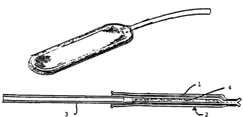
preferred chamber of the present invention (Figure 2) is a device comprising
two
bilayer membranes (1) sun-ounded by a polyester mesh (2) sonically welded
together,
with a port (3) for access to the lumen (4). Each bilayer comprises a 5 ~m
PTFE
membrane manufactured by Gore, Flagstaff, Arizona, Product No. L3 1324 and a
0.45
pm PTFE membrane manufactured by Millipore, Bedford, Massachusetts, Product
No. SF1R848E1. At one end there is a polyester (PE 90 ID 0.034" by OD 0.050")
port to permit access to the interior of the device for loading cells. The
device has an
interior lumen. Previous studies have shown that this preferred device has the
advantage (though not required for all embodiments of the present invention)
of being
able to protect
2194555
W0 96/01611 PCTIUS95IOS151
X19 -
allograft tissue from immune rejection for extended periods (Carr-Brendel et
al., J.
' Cellular Biochem. 18A, p. 223 (1994) and Johnson et al., Cell
Transplantation 3,
p. 220 (1994)).
' Other cell containing chambers which those skilled in the art may find
useful
$ in the present invention include: Agarose microcapsules (Iwata et al., J.
Biomed.
Mater. Res. 26, p. 967-977 (1992); J. Bioact. and Comp. Polymers 3, p. 356-369
(1988), and Depuy et al., J. Biomed. Mater. Res. 22, p. 1061-1070 (1988));
Hollow fibers of XM50 (Wine et al., J. Biomed. Mater. Res. 23, p. 31-44 (1989)
and Altman et al., Proc. of Third Meeting of hSAO, Supp. 5, p. 776-779 (1981);
Diabetes, 35, p. 625-633 (1986)); Alginate-polylysine (along et al., Biomat.,
Art.
Cells and Immob. Biotech. 19, p. 675-686 (1991)); Microcapsules of alginate-
polylysine (O'Shea et al., Biochim. et Biophy. Acta 804, p.133-136 (1984), Sun
et
al., App. Biochem. and Biotech. 10, p. 87-99 (1984), Chicheportiche et al.,
Diabetologia 31, p. 54-57 (1988) and Goosen et al., U.S. Patent No. 4,689,293,
IS August 25,1987; U.S. Patent No. 4,487,758, December 11, 1984; U.S. Patent
No. 4,806,355, February 21, 1989, and U.S. Patent No. 4,673,566, June I6,
1987); Chitosan-alginate (IvIcKnight et al., J. of Bioact. and Comp. Poly. 3,
p.
334-355 (1988)); Polyacrylonitrile or other ultrafiltration membranes in a U-
shaped
device (Moussy et al., Artif. Org. 13, p. 109-115 (1989), Lepeintre et al.,
Artif.
Org. 14, p. 20-27 (1990), and Jaffrin and Reach, U.S. Patent No. 4,578,191,
March 25, 1986); Hollow fibers of polyacrylonitrile (Aebischer et al., Biomat.
I2,
p. 50-56 (1991) and Lacy et al., Science 254, p. 1782-1784 (1991)); Track-
etched
polycarbonate membrane diffusion chambers (Gates and Lazarus, Lancet, Dec. 17,
p. 1257-1259 (1977)); Polymerized 2-hydroxyethyl methacrylate pHEMA
membrane devices (Ronel et al., J. Biomed., Mater., Res, 17, p. 855-864
(1983));
Microcapsules of polyacrylates (Douglas and Sefton, Biotech and Bioeng. 36, p.
653-664 (1990); Trans Am. Soc. Artif. Inter. Org. 35, p. 791-799 (1989));
Acrylic
copolymer hollow fibers (Lama et al., Proc. Natl. Acad. Sci. 88, p. I 1100-
11104
(1991)); polyol copolymer film WO 93/22427; Intravascular devices (Berguer,
U.S. Patent No. 4,309,776, January 12, 1982) and Gaskill, U.S. Patent No.
4,911,717, March 27,1990); Cationic-anionnc crosslinked membranes, e.g.
chitosan and polyaspartic or polyglutamic acid (Jarvis, U.S. Patent No.
4,803,168,
February 7, 1989); Surface-conforming bonding bridge layer of a
multifunctional
material and semipermeable polymer layer for cell encapsulation (Cochrum, U.S.
W096101611 ~ -- PCTItJS95108151
-20-
Patent No. 4,696,286, September 29,1987); Vascular perfusion devices (Chick et
al., U.S. Patent No. 5,002,661, March 26,1991); Macromer polymer
encapsulation, Desai et al. WO 93/16687; Barium-alginate cross-linked
microcapsules (Zekorn et al., Acta. Diabetol. 29, p. 99-106 (1992)), other
membrane devices (Ward et al., Fourth World Biomat. Con., Berlin, p. 152,
April
24-28, 1992)); other encapsulation devices (Aebischer, WO 94/07999; U.S.
Patent
No. 5,283,187; WO 93/00128; WO 93/00127; WO 93/00063; WO 92/19195; WO
91/10470;, WO 91/10425; WO 90/15637; WO 90/02580) and cellular implant
devices: U.S. Patent Nos. 4,241,187; 4,892,538; and 4,391,909. "Islet
Transplantation with Immunoisolation," Lanza, R. P. et al., 41 Diabetes, p.
1503
(1992) reviews various chambers used for containing cells; as do Langer and
Vacanti in "Tissue Engineering," 260 Science, p. 920 (1993). In the event that
the
particular chambers described above are not permeable enough to allow ingress
and
egress of the subcellular material to and from implanted tumor cells, one
skilled in
the art will understand that the permeability of such chambers may be altered
without changing the basic design of such chambers.
Furthermore, the applicants believe that other devices, not mentioned here,
may be used in the invention if they have the property of housing implanted
cells in
such a way as to prevent direct contact of graft cells and host immune cells,
and
allow the release of the subcellular antigenic material that stimulates the
patient
immune response. While applicants have not isolated or characterized the
subcelIular material which causes the patient immune response, they believe it
to
include immunogenic molecules (antigens) shed or secreted from the contained
tumor cells. The tumor cells shed many antigens, not just tumor associated
antigens. This is thought to recruit high numbers of macrophage and antigen
presenting cells to the site which in turn provide an enhanced immune
response.
The administration of an immunopotentiating molecule, such as a cytokine,
further
enhances the immune response at the site.
Applicant's invention provides numerous advantages over current cancer
immunotherapies. Many of the studies published to date require the sole
administration of "free" irradiated cells; i.e. cells not contained in a
chamber. The
cells are im~adiated as a safety precaution to prevent them from proliferating
and
causing additional tumors. However, they are cleared from the body within 1 to
2
weeks providing only a transient dose, and in some cases, the irradiation may
219455
W0 96101611 PCT'/U895108151
_.~ -~~21 -
interfere with production of any cytokines engineered into the cells. In
applicant's
invention it is not always necessary to irradiate the contained cells.
Although even
when irradiated cells are used they likely remain present as immunogens in the
chamber for periods of time longer than the free irradiated cells of the prior
art.
Applicant's use of a chamber offers the safety of sequestering the tumor cells
so
that, unlike the prior art, free tumor cells need not be introduced into
patients.
Moreover, in a preferred embodiment using the preferred chamber, the chamber
itself acts as an adjuvant for the subcellular antigen materials. Macrophages
are
attracted to the outer surface of the device and thus are in a position to
pick up
antigenic materials as they are shed from the tumor cells within the device.
Examples of engineered cells which may be used in accordance with the
present invention include tumor cells engineered to secret cytokines (Sobol et
al.,
WO 95/07105; Addision et al., Gene Therapy Weekly, p. 19 (November 1994));
cells engineered to express foreign antigens to increase the cellular and/or
humoraI
antitumor activity (Plantz et al., PNAS 90, p. 4645 (1993) (allogenic
histocompatibility gene); Gansbacher WO 94/18995 (allogenic tumor cell
engineered to express cytokines, adhesion molecules, constimulatory factors or
tumor associated antigens); Allione et al., Gene Therapy Weekly, p. 20
(January
1995) (mammary adenocarcinoma cells engineered to express 1L,-2, IL-4, Ilr6,
IL-
7, IL-10, TNF~, CMCSF); Hock et al., Gene Therapy Weekly, p. 22 (January
1995) (murine neuroblastoma expressing Class II MHC) (although this approach
is
thought to require direct cell-cell contact, shed MHC would be an
immunopotentiating molecule in accordance with the present invention)); and co
expression in tumor derived cells of both an immunopotentiating molecule and a
suicide gene (Frost et al., WO 92/05262).
Overall, the following examples and data presented in the figures
demonstrate effectiveness of applicant's invention in a number of different
experimental situations. When a chamber containing tumor cells is' used as a
vaccine (i.e. before tumor formation) it can be effective in preventing tumor
formation in as much as 100% of experimental animals. When implanted in the
presence of microtumor we demonstrate effectiveness in greater than 75% of the
animals tested. Finally, when combined with surgical resection of large
tumors,
implantation of devices prevented tumor regrowth in 60% of the implanted
animals.
CA 02194555 2001-06-O1
-22-
Taken together, several conclusions can be drawn from these data. First,
when reviewing all the examples, one can conclude that although cure is not
achieved
in 100% of the animals, it is nevertheless better to have a chamber than not.
At worst,
the tumors develop more slowly, at best, the animals never develop tumor; in
no case
do animals develop tumors more quickly or have larger tumors in the presence
of a
chamber than the control animals. The data further demonstrate that when using
the
chamber, tumor cells without genetic modification can be used effectively to
generate
an anti-tumor immune response. Tumor cells in a chamber are much more
effective
than free tumor cells in generating this immune response and irradiated cells
in the
chamber appear to be more effective than living cells. It is assumed that this
is due to
an enhancement of the immunogenicity of the cells due to irradiation induced
changes
in the cells.
The following examples are provided for illustration of several embodiments
of the invention and should not be interpreted as limiting the scope of the
invention.
Example 1: Rodent Adenocarcinoma Model
Cell lines used: MCA-38 (a generous gift of Dr. Augusto Ochoa, NCI) is a
murine colon carcinoma which can be maintained in vivo or in vitro. For in
vitro
maintenance, the cells were grown in RPMI (Sigma Chemical Company, St. Louis
MO) supplemented with 1 ~mIVI HEPES, 1 % non-essential amino acids, 1 % L-
glutamine, 1% sodium pyruvate, 1% pencillin/streptomycin (Sigma), 0.1% (3-
mercaptoethanol and 10% fetal bovine serum (Irvine Scientific, Irvine CA).
Cells
were routinely passaged by trypsinization twice a week.
Animals used: For most experiments, female C57/B6 mice (Harlan Sprague
Dawley) were used. Where indicated, athymic mice (Harlan Sprague Dawley) were
used. All animals were maintained according to standard procedures for care
and use
of laboratory animals.
Device: These studies utilized sonically sealed 4.5 pl or 20 pl ported devices
employing laminated membranes described above. Devices were sterilized
overnight
in 70% ethanol and then the ethanol was removed by three washes in sterile
saline
(Baxter Scientific Products, Waukegan IL).
z ~ ~~~~~
w0 96!01611 PCT/US95108151
.. a: 23 _
~Dlantation of devices: For loading, MCA-38 cells were trypsinized,
washed and pelleted by centrifugation. Except where indicated, MCA-38 cells
were
encapsulated into 4.5 ltl ported devices by loading 106 cells in 3 Itl into
the central
lumen of the device using a Hamilton syringe. The larger device was loaded
with
107 cells in 20 pl. Devices were sealed with a silicone.plug laid down using a
23
gauge needle andsyringe. The device port was completely filled with silicone
and
the port was cut in half. The remaining port was dipped briefly in 70%
ethanol.
The loaded devices were washed through three changes of saline. Devices were
placed in RPMI 1640 supplemented as described above and incubated at
37°C until
implantation.
The animals receiving implants were anaesthetized by intraperitoneal
injection of 0.2-0.3 mI of the mixture of 1 ml ketamine (Fort Dodge
Laboratories,
Fort Dodge, Iowa) and 0.75 ml xylazine (Rugby Laboratories, Rockville Center,
New York) diluted into 1 ml of sterile saline. The abdominal area was swabbed
with betadine and a ventral midline incision was made. Using a hemostat a
small
pocket subcutaneous was made on either side of the midIine incision and one
4.5 ltl
device was inserted into each pocket. Once the devices were inserted the
incision
was closed using sterile staples and the abdominal area swabbed again with
betadine. When using the 20 NT device, only one is inserted.
Tumor challenee: At the indicated times the animals were challenged with an
injection of unencapsulated MCA-38 cells. For challenges after implantation,
106
freshly trypsinized MCA-38 cells were diluted in 50 Ill of sterile saline and
injected
into the muscle of the right hind leg. In the case of rechallenge, the second
injection
of 106 cells was made into the left hind leg and, where applicable, the third
injection of 106 cells was made into the right leg. For challenge at the time
of
implant, animals were challenged with 103 free MCA-38 cells. Preliminary
studies
have shown that as few as 500 free MCA-38 cells are sufficient for tumor
formation.
HistQloev: Upon completion of each experiment, implanted devices were
recovered, fixed in glutaraldehyde, sectioned and analyzed by hematoxylin and
eosin staining for the presence of surviving tumor cells within the device
using light
microscopy.
survival of MCA-38 cells within ilmmunoisolation devices: To assess the
ability of MCA-38 to survive within the device in the absence of immune
attack,
w0 96!01611 PCT/US95I08151
104 or 106 cells were encapsulated within 4.5 ltl devices and implanted into
athymic mice. At the end of the three week implant period, the devices were
explanted, processed histologically, and analyzed for the presence of living
tissue.
MCA-38 cells did survive within the device. In all cases there was a
substantial
necrotic area in the center of the device but healthy appearing cells were
present
along the periphery. The width of the necrotic area depended upon the initial
number of cells loaded into the device (i.e. greater necrosis in devices
containing
106 cells than those containing 104 cells).
Use of devices containing MCA-38 cells as a tumor vaccine: Syngeneic
C57B6 mice were implanted with two devices each containing 106 MCA-38 cells
for three or four weeks. These animals were then challenged with an injection
of
106 free MCA-38 cells as described above. As shown in Figure 3, 0/8 animals in
two experiments developed tumors at the challenge site while all of the
control
animals developed tumors within ten days of the injection. Empty devices
implanted into mice did not protect them against a subsequent challenge with
free
MCA-38 cells.
Five of these animals received a second challenge of 106 cells 8-11 weeks
after the initial implant. In this case 4/5 of the implanted animals remained
tumor
free at both implant sites; once again all of the control animals developed
tumors at
the injection site. One experimental animal did develop a tumor at the site of
the
second injection, this animal was implanted with only one device.
Three of the animals that remained tumor free were given a third challenge
seven months after the devices were implanted. For two of the animals the
subcutaneous devices were removed before the tumor challenge was given, the
third animal was challenged with the devices remaining in place. The animal
with
the devices remained tumor free after the third challenge while both of the
animals
from which devices were removed developed tumor as did all the control animals
which had never received a device. However, while the animals which had their
devices removed did develop tumors, they did so much more slowly than the
control animals. Controls developed tumor 10 days after the challenge. One of
the
experimental animals developed tumor 25 days after the challenge and the
second
developed tumor 36 days after the challenge. Histology of the removed devices
revealed that >90% of the cells were dead and that there was extensive
calcification
of the material inside of the device. However, there was evidence of a few
CA 02194555 2001-06-O1
-25-
remaining live cells. These results suggest that the device itself does not
mediate the
anti-tumor effect since the .animals whose devices were removed did not
develop
tumors at the same rate as controls that had never been implanted with
devices. At
the same time, the device appears to be necessary to maintain the
immunological
protection, against the tumor: while tumors appear more slowly in animals
whose
devices are removed, there appears to be no long-term immunity in animals
which had
been implanted with device in the absence of those devices.
Use of devices containin~~MCA-38 cells as a tumor therapy: In another series
of experiments, animals were challenged with free MCA-38 cells at the time of
implant of devices containing MCA-38 cells. Ln this case, the animals were
challenged with 10-' free tumor cells. In one experiment we were able to
significantly
delay the time of tumor formation (Figure 4) p= 0.036. In this experiment one
animal
was tumor free at time of sacrifice at day 32. In a second experiment, all of
the
animals without devices had developed tumor by day 16 at which time only 1/10
of
the implanted animals ha<1 developed a tumor at the challenge site. These
results
suggest that the implantation of a device containing tumor cells can delay or
prevent
the growth of tumors introduced at the time of implantation.
Example 2: Canine Model
Animals Used: A dog in the Baxter animal facility (142-3) was identified
with several subcutaneous m;~sses ranging in size from pinhead to about the
size of
a quarter. Histological analysis diagnosed these masses as epithelial
inclusions
cysts. A second dog (4008) was purchased from an outside vendor. This dog had
a mammary tumor approximately 10 cm in diameter that was biopsied and
diagnosed as an intraductular mammary carcinoma.
Device: These studies utilized the sonically sealed 40 pl ported device
employing laminated membranes described above. Devices were sterilized
overnight
in 70% ethanol and then the ethanol was removed by three washes in sterile
saline
(Baxter Scientific Products, Waukegan Illinois).
Implantation of Devices: Dogs were anaesthesized by standard methods. The
area around the tumors was shaved. l:n the case of dog 142-3, the largest mass
was
surgically excised and placed into sterile saline. The mass was cut into small
WO 96101611 PCT/US95108151
-26- ~194~5~
pieces using two pairs of surgical scissors. The minced pieces were loaded
into the
immunoisolation device as follows: 80 pI of gravity settled tissue was taken
up into
a Hamilton syringe. The needle of the syringe was inserted into the port of
the
device and the contents emptied into the lumen of the device. Devices were
sealed
with a silicone plug laid down using a 23 gauge needle and syringe. The device
port
was completely filled with silicone and the port was cut in half. Using a
hemostat a
small pocket was made on either side of the site from which the tumor was
explanted and one device was inserted into each pocket (a total of two devices
were
implanted). Once the devices were inserted the incision was closed and sutured
and
the abdomenal area swabbed again with betadine.
In the case of dog 4008 -95% of the tumor mass was surgically excised
with cauterization of involved blood vessels. The incision was then sutured
and a
baseline measurement of the remaining tumor was taken. The excised tumor was
cut open and several 0.5 cm diameter pieces removed at various depths. These
pieces were further minced using two pairs of surgical scissors. The minced
pieces
were loaded into eight devices as described above. Four small ventral
subcutaneous incisions were made dorsal to the site from which the tumor had
been
excised and two devices were inserted into each incision. The incisions were
then
sutured and the abdominal area swabbed with betadine.
Monitoring of animals: The remaining masses in dog 142-3 were measured
2 to 3 times a week. Measurements were taken in two dimensions and used to
calculate total surface area of each mass. Duplicate measurements were made by
two different technicians and the values were averaged for each time point.
Similarly, the tumor remaining in dog 4008 was measured three times a week in
two dimensions.
Following excision of the largest mass from dog 142-3 and insertion of the
devices containing tissue from the excised mass, two of the four remaining
growths
showed a dramatic decrease in size (Figure 5) as determined by two independent
measurements. The other two, which were morphologically distinct, showed no
change in size. This decrease in size in the remaining masses occurred without
any
additional manipulation of the animal.
The size of the remaining tumor in dog 4008 appeared to increase initially
but this was probably due to edema resulting from surgical trauma (note
increase at
CA 02194555 2001-06-O1
-27-
day 7, Figure 6). Subsequently there has been a steady decrease in the size of
the
remaining tumor as determined by two sets of independent measurements with
some
leveling out at >30 days post surgery.
Example 3: Small Pre-Existily'umors
Cell lines used: MCA-38 is a murine colon carcinoma which can be
maintained in vivo or in vitro. For in vitro maintenance, the cells were grown
in
RPMI (Sigma Chemical Company, St. Louis MO) supplemented with 1mM HEPES,
1 % non-essential amino acids, 1 % L-glutamine, 1 '% sodium pyruvate, 1 %
penicillin /
streptomycin (Sigma ), 0.1 °io P-mercaptoethanol and 10% fetal bovine
serum (Irvine
Scientific, Irvine CA). Cells were routinely passaged by trypsinization twice
a week.
Animals used: Female C57/B6 (Harlan Sprague Dawley) were used. All
animals were maintained according to standard procedures for care and use of
laboratory animals.
Immunoisolation Device: These studies utilized sonically sealed 4.5 ~l ported
devices employing laminated membranes. Devices were sterilized overnight in
70%
ethanol and then the ethanol was removed by three washes in sterile saline
(Baxter
Scientific Products, Waukegan IL)
lantation of devices: For loading, MCA-38 cells were trypsinized, washed,
and pelleted by centrifugation. Except where indicated, 106 MCA-38 cells were
encapsulated into 4.5 ~I ported devices by loading 3 pl of the pelleted cells
into the
central lumen of the immunoisolation device using a Hamilton syringe.
Where indicated, cells were exposed to 3500 Rads before loading. Devices
were sealed with a silicone plug laid down using a 23 gauge needle and
syringe. The
device port was completely filled with silicone and the port was cut in half.
The
remaining port was dipped briefly in 70% ethanol. The loaded devices were
washed
through three changes of saline. Devices were placed in RPMI 1640 supplemented
as
described above and incubated at 37°C until implantation.
The animals receiving implants were anaesthetized by intraperitoneal injection
of 0.2-0.3 ml of the mixture of 1 ml ketannine and 0.75 ml rompum diluted into
1 ml
of sterile saline. The abdominal area was swabbed with betadine and a ventral
midline incision was made. lJsing a hemostat a small pocket was made on
R'O 96101611 PCTlUS95108151
-2g- ~'~94555
either side of the midline incision and one device was inserted into each
pocket.
Once the devices were inserted the incision was closed using sterile staples
and the
abdominal area swabbed again with betadine.
1$jgction of free irradiated cells: At the time of implant some experimental
animals were also given an injection of free irradiated tumor cells. For
irradiation
the cells were prepared as described above for loading. The cells were
suspended
at a concentration of 106 cells in 50 ml. The cells received 3500-4000 Rads
from a
cobalt -60 source. 106 cells were injected.
Tumor chal_lenee: For initiating tumors before implant, animals were
injected with 103 free MCA-38 cells 3-7 days before implantation. Injections
were
made either by intramuscular injection into the right hind leg or into the
dorsal
subcutaneous space. Preliminary studies have shown that as few as S00 free MCA
38 cells are sufficient for tumor formation. In the case of a second challenge
following implant, the second injection was made into the left hind leg.
TrParment of ore-existing tumors: Animals were injected with 103 free
tumor cells three days before implantation. At time of implant they received
two
devices containing irradiated MCA-38 cells and were also given an injection of
106
free irradiated tumor cells exterior to the devices. As shown in Figure 7,
none of
the five animals treated with irradiated cells both inside and outside of the
device
developed tumor at in the first 90 days. On day 90 two of these animals were
challenged with 106 tumor cells, one of these two animals developed a tumor
from
this challenge. All of the other animals have remained tumor free for > 150
days.
As illustrated in Figure 8, this treatment works best with the combination of
irradiated cells in the device and an injection of free irradiated cells
outside of the
device. Although some protection is afforded by administering unirradiated
tumor
cells in the device in combination with free irradiated cells, it is less
effective than
administering irradiated cells in the device in combination with free
irradiated cells.
Injection of free irradiated cells alone has no effect on tumor development.
When tumors were initiated in the dorsal subcutaneous space, implantation
of devices containing irradiated cells along with an injection of free
irradiated cells
were able to rescue 60% of the treated animals (all of the untreated animals
developed tumor at the site where the original tumors were initiated) (Figure
9).
CA 02194555 2001-06-O1
-29-
Example 4: Device Therapw after Tumor Resection
Cell lines used: MCA-38 is a murine colon carcinoma which can be
maintained in vivo or in vit,Yo. For in vitro maintenance, the cells were
grown in
RPMI (Sigma Chemical Connpairy, St. Louis MO) supplemented with 1mM HEPES,
1 % non-essential amino acids, 1 % L-glutamine, I % sodium pyruvate, 1
penicillin/streptomycin (Sigma), 0.1% P-mercaptoethanol and 10% fetal bovine
serum
(Irvine Scientific, Irvine C.A). Cells were routinely passaged by
trypsinization twice a
week.
Animals used: Female C57/B6 (Harlan Sprague Dawley) were used. All
animals were maintained according to standard procedures for care and use of
laboratory animals.
Immunoisolation Device: These studies utilized the sonically sealed 4.5 ~l
ported devices employing laminated membranes. Devices were sterilized
overnight in
70% ethanol and then the ethanol was removed by three washes in sterile saline
(Baxter Scientific Products, Waukegan IL)
Implantation of devices: For loading, MCA-38 cells were trypsinized, washed
and pelleted by centrifugation. Except where indicated, 106 MCA-38 cells were
encapsulated into 4.5 ~l ported devices by loading 3 F1 of the pelleted cells
into the
central lumen of the immunoisolation device using a Hamilton syringe. Devices
were
sealed with a silicone plug laid down using a 23 gauge needle and syringe. The
device port was completely filled with silicone and the port was cut in half.
The
remaining port was dipped briefly in 70% ethanol. 'The loaded devices were
washed
through three changes of saline. Devices were placed in RPMI 1640 supplemented
as
described above and incubated at 37°C until implantation.
The animals receiving; implants were anaesthetized by intraperitoneal
injection
of 0.2-0.3 ml of the mixture of 1 ml ketamine and 0.75 ml rompum diluted into
1 ml
of sterile saline. The abdominal area was swabbed with betadine and a ventral
midline incision was made. Using a hemostat a small pocket was made on either
side
of the midline incision and one device was inserted into each pocket. Once the
devices were inserted the incision was closed using sterile staples and the
abdominal
area swabbed again with betadine.
Tumor challenge: To initiate tumors, animals were injected with 105 free
MCA-38 cells 3-7 days before implantation. Injections were made into the
dorsal
CA 02194555 2001-06-O1
-30-
subcutaneous space. Preliminary studies have shown that as few as 500 free MCA-
38
cells are sufficient for tumor formation.
Use of immunoisolatuon devices containing MCA-38 cells as a tumor thera~y-
for treatment following turnc>r resection: The protocol for this experiment is
outlined
in Figure 10. Briefly, animals were injected with 105 cells in the dorsal
subcutaneous
space and monitored until palpable tumors were observed. On day 10 the tumors
were surgically removed from all of the animals. Half of the animals (n=5)
received
no further treatment The other half (n=5) were implanted with two devices each
containing 106 unirradiated MCA-38 cells. The devices were implanted
subcutaneously on the ventral side. Both groups of animals were monitored for
redevelopment of tumor at the original tumor site. As shown in Figure 1 l, all
of the
control animals (without implants) redeveloped tumor at the original tumor
site within
thirty days of the surgery. While two of the animals with implants also
redeveloped
tumor, three did not and have remained tumor free for >180 days. The
differences
between these groups are highly significant (p<0.05).
Example 5: Rodent Melanorr~a Model
Cell lines used: B 16 is a murine melanoma which can be maintained in vivo
or in vitro. For in vitro maintenance, the cells were grown in RPMI (Sigma
Chemical
Company, St. Louis MO) supplemented with 1 mM HEPES, 1 % nonessential amino
acids, 1 % L-glutaniine, 1 °i° sodium pyruvate, 1 %
penicillin/streptomycin (Sigma ),
0.1 % P-mercaptoethanol and 10% fetal bovine serum (Irvine Scientific, Irvine
CA).
Cells were routinely passaged by trypsinization twice a week.
Animals used: Female C57/B6 were used. All animals were maintained
according to standard procedures for care and use of laboratory animals.
Immunoisolation Device: These studies utilized the sonically sealed 4.5 ~1
employing laminated membranes. Devices were sterilized overnight in 70%
ethanol
and then the ethanol was renu>ved by three washes in sterile saline (Baxter
Scientific
Products, Waukegan IL).
Implantation of devices: For loading, B16 cells were trypsinized, washed and
pelleted by centrifugation. >=?xcept where indicated, 106 B 16 cells were
encapsulated
into 4.5 pl ported devices by loading 3 pl of the pelleted cells into the
2194~a~
w0 96/01611 PCT/US95108151
-31-
central lumen of the immunoisolation device using a Hamilton syringe. Devices
were sealed with a silicone plug laid down using a 23 gauge needle and
syringe.
The device port was completely filled with silicone and the port was cut in
halt The
remaining port was dipped briefly in 70% ethanol. The loaded devices were
washed through three changes of saline. Devices were placed in RPMI 1640
supplemented as described above and incubated at 37°C until
implantation.
The animals receiving implants were anaesthetized by intraperitoneal
injection of 0.2-0.3-ml of the mixture of 1 ml ketamine and 0.75 ml rompum
diluted
into 1 ml of sterile saline. The abdominal area was swabbed with betadine and
a
ventral midIine incision was made. Using a hemostat a small pocket was made on
either side of the midline incision and one device was inserted into each
pocket.
Once the devices were inserted the incision was closed using sterile staples
and the
abdominal area swabbed again with betadine.
ejection of free irradiated cells: At the time of implant all animais with
implanted devices were also given an injection of 106 free irradiated cultured
B I6
cells at the site of the challenge. For irradiation tile cells were prepared
as described
above for loading. The cells were suspended at a concentration of 106 cells in
50
mI. The cells received 3500-4000 Rads from a cobalt -60 source.
Tumor challenee: Four weeks after the implantation of devices, animals
were challenged with an injection of unirradiated B 16 cells. For challenge 5
x 105
freshly trypsinized B 16 cells were diluted in 50 ul of sterile saline and
injected into
the muscle of the right hind leg. Preliminary studies have shown that 104 free
B 16
cells are sufficient for tumor formation.
Uce of ~~nnnicolaton devicec con ain;n~ in vivo derived tumor ticcu c a
tumor vaccine: The protocol for this experiment is outlined in Figure 12,
briefly,
animals were treated in a first round with either devices containing
unirradiated B 16
cells or by injection of free, irradiated B 16 cells. All of these animals
developed
tumor after challenge with 5 x 10⁴ B 16 cells. The tumors from both groups of
animals were surgically excised, chopped up into approximately 1 mm2 pieces
and
loaded into 4.5 p,l devices. A third set of devices was also prepared
containing
cultured unirradiated B16 cells. These three sets of devices were implanted
into a
second set of naive animals and all three groups also received an injection of
free,
irradiated cultured B 16 cells; control animals received no treatment. The
animals
were challenged with cultured B 16 cells four weeks later. As shown in Figure
13,
CA 02194555 2001-06-O1
-32-
one of three animals immunized with devices containing tumor grown up in
animals
treated only by injection of :free irradiated cells remained tumor free for
>140 days
after challenge. Thus, the tumor cells used to immunized the naive animals
were not
cultured cells but rather they were cells subject to evolution while growing
up in the
first round of animals. These data indicate that chambers containing tumor
cells
which have been subject to evolution, such as autologus human tumor cells, are
likely
to be useful in practicing applicants' invention.
Example 6: Rodent Ovarian 'lf umor
Cell lines used: C57ov is a murine tumor which can be maintained in vivo or
in vitro. It was identified in t:he laboratory in an animal given an LV.
injection of B16
cells (5 x 10⁵). Histological Examination suggests that it was not B16
derived. For in
vitro maintenance, the cells were grown in RPMI (Sigma Chemical Company, St.
Louis MO) supplemented wiith 1mM HEPES, 1% non-essential amino acids, 1% L-
glutamine, 1% sodium pyruvate, 1% penicillin/streptomycin (Sigma), 0.1 % P-
mercaptoethanol and 10% fecal bovine serum (Irvine Scientific, Irvine CA).
Cells
were routinely passaged by trypsinization twice a week.
Animals used: Female C57/B6 were used. All animals were maintained
according to standard procedures for care and use of laboratory animals.
Immunoisolation Device: These studies utilized the sonically sealed 4.5 pl
employing laminated membranes. Devices were sterilized overnight in 70%
ethanol
and then the ethanol was removed by three washes in sterile saline (Baxter
Scientific
Products, Waukegan IL).
Implantation of devices: For loading, C57ov cells were trypsinized, washed
and pelleted by centrifugation. Except where indicated, 106 C57ov cells were
encapsulated into 4.5 ~1 pot-ted devices by loading 3 ~l of the pelleted cells
into the
central lumen of the immunoisolation device using a Hamilton syringe. Devices
were
sealed with a silicone plug laid down using a 23 gauge needle and syringe. The
device port was completely frlled with silicone and the port was cut in half.
The
remaining port was dipped briefly in 70% ethanol. The loaded devices were
washed
through three changes of saline. Devices were placed in RPMI 1640 supplemented
as
described above and incubated at 37°C until implantation.
219~~55
WO 96101611 PG°I'/US95lOS151
,. .,
=33=
The animals receiving implants were anaesthetized by intraperitoneal
injection of 0.2-0.3 ml of the mixture of Iml ketamine and 0.75 ml rompum
diluted
into 1 ml of sterile saline. The abdominal area was swabbed with betadine and
a
ventral midline incision was made. Using a hemostat a small pocket was made on
S either side of the midline incision and one device was inserted into each
pocket.
Once the devices were inserted the incision was closed using sterile staples
and the
abdominal area swabbed again with betadine.
~iection of free irradiated cells: At the time of implant all animals with
implanted devices were also given an injection of 106 free irradiated cultured
C57ov
cells at the site of the challenge. For irradiation the cells were prepared as
described
above for loading. The cells were suspended at a concentration of 106 cells in
50
~tI. The cells received 3500-4000 Rads from a cobalt -60 source.
Tumor challenee: Four weeks after the implantation of devices, animals
were challenged with an injection of unirradiated C57ov cells. For challenge 5
x
104 freshly trypsinized C57ov cells were diluted in 50 pl of sterile saline
and
injected into the muscle of the right hind leg. Preliminary studies have shown
that
103 free C57ov cells are sufficient for tumor formation.
Use of irxLm_unoisola 'on devi~ec containine C57ov as a tumor vacr;nP~
Animals were implanted with two devices each containing 106 irradiated C57ov
cells. At the time of implantation the animals also revieved an injection of
106
irradiated C57ov cells. The animals were challenged with C57ov four weeks
later.
The results are shown in figure 14, 60% of the animals have remained tumor
free
for > 30 days.
While the present invention has been described in terms of specific methods
and devices, it is understood that variations and modifications will occur to
those
skilled in the art upon consideration of the present invention.
Numerous modifications and variations in the invention as described in the
above illustrative examples are expected to occur to those skilled in the art
and
consequently only such limitations as appear in the appended claims are to be
placed
thereon. Accordingly, it is intended in the appended claims to cover all such
equivalent variations which come within the scope of the invention as claimed.