Note: Descriptions are shown in the official language in which they were submitted.
2194814
STIMULATION OF PROTECTIVE T CELLS TO rK~v~..~ AUTOIMMUNE
DISEASE
' This invention relates to methods and compositions
for preventing the development of autoimmune disease in
susceptible subjects. More particularly, the invention
relates to treatment with an agonist of CD28 to prevent
autoimmune disease development.
Background of the Invention
Insulin-dependent diabetes mellitus (IDDM) or
autoimmune diabetes is a polygenic, multifactorial,
autoimmune disease heralded by T cell infiltration of the
pancreatic islets of Langerhans (insulitis) and the
progressive T cell-mediated destruction of insulin-
producing ~ cells (Bach, 1994; Atkinson and Maclaren,
1994; Tisch and McDevitt, 1996).
Non-obese diabetic (NOD) mice are susceptible to the
development of IDDM and are an accepted model for the
development of autoimmune IDDM in humans.
CD4+ T helper cells are required for the adoptive
transfer of IDDM into recipient neonatal NOD mice or
immunodeficient NOD.Scid mice (Bendelac et al., 1987,
- Christianson et al., 1993; Rohane et al., 1995).
Cooperation between CD4+ and CD8+ T cells is required to
initiate IDDM, and islet ~ cell destruction is CD4+ T
cell-dependent (Haskins and McDuffie, 1990; Wang et al.,
1991). Current evidence suggests that the CD4+ effector
cells of IDDM in NOD mice at Thl cells which secrete IL-
2, IFN-~ and TNF-a and that the regulatory CD4+ cells are
Th2 cells which secrete IL-4, IL-5, IL-6, IL-10 and IL-13
(Rabinovitch, 1994; Liblau et al., 1995; Katz et al.,
1995).
NOD mouse T cells show proliferative
hyporesponsiveness to T cell receptor (TCR) stimulation
2 2l 94 8l4
and this hyporesponsiveness may be causal to the
development of IDDM.
It has been shown that, beginning at 3-5 weeks of
age, T cell receptor (TCR) ligation in NOD mice induces
the proliferative hyporesponsiveness of NOD thymic and
peripheral T cells, which is mediated by reduced IL-2 and
IL-4 production (Zipris et al., 1991; Rapoport et al.,
1993a; Jaramillo et al., 1994).
Decreased IL-4 production by human T cells from
patients with new onset IDDM has also been demonstrated
recently (Berman et al., 1996). Whereas addition of IL-
4, a Th2-type cytokine, potentiates Il-2 production and
completely restores NOD T cell proliferative
responsiveness, addition of IL-2, a Thl-type cytokine,
even at high concentrations, only partially restores NOD
T cell responsiveness. These findings suggest that Th2
cells may be compromised in function to a greater extent
than Thl cells in NOD mice, and raise the possibility
that Th2 cells require a higher threshold of activation
than Thl cells in these mice. Il-4 not only restores NOD
T cell responsiveness in vitro, but prevents insulitis
and IDDM when administered in vivo to prediabetic NOD
mice (Rapoport et al., 1993a) or when transgenically
expressed in pancreatic ~ cells (Mueller et al., 1996).
The proliferative hyporesponsiveness of regulatory
Th2 cells in NOD mice may favour a THl cell-mediated
environment in the pancreas of these mice, and lead to a
loss of immunological tolerance to islet ~ cell
autoantigens. This is consistent with the notion that
restoration of the balance between Thl and Th2 cell
function may prevent IDDM (Rabinovitch, 1994; Liblau et
al., 1995i Arreaza et al., 1996).
Optimal T cell activation requires signalling
through the TCR and CD28 costimulatory receptor (June et
al., 1994i Bluestone, 1995; Thompson, 1995).
Crosslinking of the TCR/CD3 complex in the absence of a
3 2194814
CD28-mediated costimulatory signal induces a
proliferative unresponsiveness that is mediated by the
inability of T cells to produce IL-2 (Jenkins et al.,
1991). CD28 costimulation prevents proliferative
- 5 unresponsiveness in Thl cells by augmenting the
production of IL-2, which in turn promotes IL-4 secretion
by T cells (Seder et al., 1994). The costimulatory
pathway of T cell activation involves the interaction of
CD28 with its ligands B7-1 and B7-2 on an antigen
presenting cell (APC), with B7-2 considered as the
primary ligand for CD28 (Linsley et al.,1990; Freeman et
al., 1993; Lenschow et al., 1993; Freeman et al., 1995).
When costimulation is blocked by either CTLA4-Ig or by
anti-B7-1 or anti-B7-2 monoclonal antibodies (mAbs),
differential effects on the incidence of various
autoimmune diseases (e.g. IDDM) and on the development of
Thl and Th2 cells are observed (Kuchroo et al., 1995;
Lenschow et al., 1995). Furthermore, in vivo studies
have demonstrated that the generation of Th2 cells is
more dependent upon the CD28-B7 pathway than the priming
of Thl cells, and suggest that the development of Th
subsets in vivo may be influenced by limited CD28-B7
costimulation (Corry et al., 1994; Lu et al., 1994).
Analyses of the development of human Th2 cells have
yielded results similar to those observed in the mouse
(King et al., 1995; Kalinski et al., 1995; Webb and
Feldman, 1995). Interactions between CD28 and its B7-2
ligand are essential for the costimulation of an IL-4-
dependent CD4+ T cell response, and IL-4 increases B7-1
and B7-2 surface expression on certain professional APCs
(eg. Langerhans cells) and B cells (Freeman et al., 1995;
Kawamura et al., 1995; Stack et al., 1994). Thus,
failure to activate NOD thymocytes and peripheral T cells
sufficiently may be due to functional and/or
differentiation defects in NOD APCs, which remain able to
optimally activate islet ~ cell autoreactive CD4+ effector
4 2194814
T cells, but not regulatory CD4+ T cells (Serreze et al.,
1988; Serreze et al., 1993). Functional defects that
compromise antigen presentation by NOD APCs, such as
deficient CD28 costimulation, may lower their ability
stimulate reguIatory Th2 cells without compromising their
ability to stimulate autoreactive effector Thl cells.
- Proliferative hyporesponsiveness of T cells has been
observed in other autoimmune diseases such as multiple
sclerosis and myasthenia gravis.
If proliferative hyporesponsiveness of T cells in
autoimmune disease could be overcome, it might be
possible by that means to prevent the development of
autoimmune diseases.
Summary of Drawings
Certain embodiments of the invention are described,
reference being made to the accompanying drawings,
whereln:
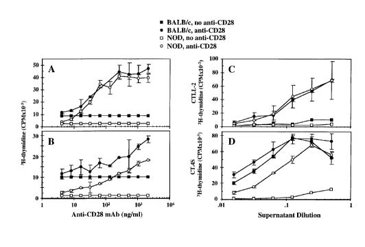
Figure lA shows thymocyte proliferation, and Figure
lB shows splenic T cell proliferation, expressed as 3H
thymidine incorporation in cpm, in the presence (circles)
or absence (squares) of various concentrations of anti-
CD28 monoclonal antibody.
Solid symbols : BALB/c mice
Open symbols : NOD mice
Figure lC shows IL-2 production and Figure lD shows
IL-4 production, expressed as 3H thymidine incorporation
into CTLL-2 and CT.4S cells respectively, by NOD (open
symbols) or BALB/c (solid symbols) thymocytes activated
by anti-CD3 in the presence (circles) or absence
(squares) of 1 ~g/ml anti-CD28 MAb.
Figure 2 shows insulitis scores in 8 week-old NOD
mice tPanel A) and 25 week-old NOD mice (Panel B) treated
with anti-CD28 antibody or hamster Ig (control).
2194814
Figure 3A shows diabetes incidence (%) at various
ages in NOD mice treated at age 2 to 4 weeks with anti-
- CD28 antibody ( ~ ) or control hamster Ig ( ~ ).
Figure 3B shows diabetes incidence (%) at various
ages in NOD mice treated at age 5 to 7 weeks with anti-
CD28 antibody ( ~ ) or control hamster Ig ( ~ ).
Figure 4, upper panel, shows IL-4 production (pg/ml)
by thymocytes, splenic T cells and islet infiItrating
cells isolated from anti-CD28 antibody-treated (solid
bars) or control (open bars) NOD mice (8 weeks or 25
weeks of age) in the presence or absence of 145-2Cll
anti-CD3~ mAb.
Figure 4, lower panel, shows IFN-r production (pg/ml)
by cells as described for Figure 4, upper panel.
Figure 5 shows proliferation (expressed as
incorporation of 3H-thymidine in cpm x 10-3) of
thymocytes, splenic T cells or islet infiltrating cells
from 8- or 25-week old, anti-CD28 antibody-treated NOD
mice, in response to anti-CD3~ antibody.
Stimulation indices (SI) were calculated as the
ratio of average cpm of anti-CD3 stimulated
cultures/average cpm of control cultures, and are shown
in parentheses. Values (mean cpm + SD) shown are
representative of three separate experiments.
Figure 6A shows pancreatic level of IL-4 and IFN-r
(ng cytokine/mg tissue) in NOD mice treated with anti-
CD28 antibody (solid bars) or control hamster Ig (open
bars).
Figure 6B shows serum levels of IgGl and IgG2a
isotype anti-GAD67 antibodies in anti-CD28 antibody-
treated NOD mice and controls.
Figure 7 shows the incidence of autoimmune diabetes
in NOC.Scid mice injected with splenic T cells from anti-
CD28 antibody-treated NOD mice (solid symbols) or control
6 ~194814
NOD mice (open symbols) at various times after injection
(transfer) of splenic T cells.
Description of the Invention
The present inventlon provides a method for
preventing the development of an autoimmune disease in a
susceptible subject by treating the subject with an
agonist of the T cell CD28 co-receptor. Autoimmune
diseases preventable by this method include IDDM,
multiple sclerosis and myasthenia gravis.
In accordance with a preferred embodiment,
development of an autoimmune disease is prevented by
treating a susceptible subject with an antibody to the
CD28 co-receptor.
Human subjects susceptible to the development of
IDDM may be identified by screening based on a subject's
HLA genetic make-up (Undlien et al., (1997)) or based on
detection of predictive serum autoantibodies such as
anti-insulin or anti-GAD antibodies (Verge et al.,
(1996)).
Treatment in accordance with the method of the
invention should be administered in the neonatal period,
from about 6 months to about 2 or 3 years of age. A
series of treatments may be required over the 6 month to
2 year period of life.
A monoclonal antibody which specifically recognises
human T cell CD28 receptor may be raised in a suitable
animal such as a mouse by conventional methods for
raising antibodies. Briefly, the mouse is injected with
human T cells and a hybridoma producing a monoclonal
antibody of the desired specificity is selected and
cloned.
Alternatively, antibodies raised against the T cell
CD28 co-receptor of a non-human mammal such as a mouse
may be used in the methods and compositions of the
7 21 94 81 4
- invention, in view of the high degree of conservation of
amino acid sequence among mammalian CD28 receptors.
Techniques are available and well known to those in
the art to prepare humanised antibodies which have a
variable region, specific for the CD28 receptor,
synthesised in a non-human mammal, combined with a human
constant region. Such humanised antibodies may be
preferable for treatment of human subjects.
Optimal T cell activation requires signalling
through both the TCR and the CD28 costimulatory receptors
of the T cell. The T cells of NOD mice have been shown
to be hyporesponsive to T cell receptor-stimulation of
proliferation.
The present inventor has shown that this
hyporesponsiveness of NOD T cells is associated with
defective CD28 receptor costimulation.
It has been shown by the inventor that treatment of
NOD mice with a CD28 agonist prevented the development of
autoimmune diabetes. Treatment of neonatal NOD mice with
an anti-CD28 antibody which gave CD28 costimulation
completely restored the proliferative responsiveness of
NOD thymocytes and peripheral T cells by augmenting their
levels of secretion of IL-2 and IL-4. The stimulated
increase in IL-4 secretion was predominant.
The antibody treatment effectively prevented the
development of destructive insulitis in NOD mice and
prevented the expected development of diabetes.
It is postulated that the antigen presenting cell
(APC)-derived costimulatory signal transduced by the CD28
receptor on NOD mouse T cells may be insufficient to
stimulate optimum T cell activation and that such CD28-
signalled activation of IL-4-producing Th2 cells is
necessary for protection from IDDM. The work of the
inventor suggests that anti-CD28 antibody prevents IDDM
in NOD mice by activating the CD28 signalling pathway in
8 ~1~48~4
NOD T cells rather than by blocking the interaction
between CD28 and its ligand, B7.
Prevention of IDDM by CD28 costimulation may be
mediated by the activation of a subset of CD4+ regulatory
T cells that confer protection against IDDM. This subset
of CD4+ regulatory T cells may be hyporesponsive in NOD
mice and may not receive a sufficient amount of the
CD28/B7 costimulatory signal required for clonal
expansion and effector function in NOD mice.
It has been proposed that precursor CD4+ Th2 cells
require a strong initial T-cell stimulation, and that the
amount of IL-4 produced is proportional to the magnitude
of the initial T cell stimulation. In the absence of
CD28 costimulation, the production of IL-4 remains below
the threshold required for the optimal development of Th2
cells (Seder and Paul, 1994; Thompson, 1995; Bluestone,
1995). It is of interest that B7-1 and B7-2 ligation of
CD28 mediate distinct outcomes in CD4+ T cells. B7-2
costimulation signals naive T cells to become IL-4-
producing T cells, and thereby directs an immune responsetowards ThO and Th2 cells (Freeman et al., 1995; Kuchroo
et al., 1995). B7-1 costimulation seems to be a more
neutral differentiative signal, and initiates the
development of both Thl and ThO/Th2 cells. Presumably,
B7-2 plays a dominant role in the production of IL-4 due
to its early expression during T cell activation (Freeman
et al., 1995; Thompson, 1995). Thus, an insufficient or
inappropriate signal resulting from a CD28/B7-2
interaction may be delivered to a subset of regulatory
CD4+ T cells in NOD mice, and this subset may not
differentiate properly into functional IL-4 producing Th2
cells.
The inventor has examined whether anti-CD28 mAb
treatment of NOD mice provides the costimulation required
for the expansion of and cytokine production by
regulatory IL-4 producing Th2-like cells. Figure 4 shows
9 21~4814
-
that anti-CD3 stimulated (in vitro) NOD thymocytes
obtained at 8 weeks, peripheral splenic T cells obtained
at 8 weeks and 25 weeks and islet infiltrating T cells
examined at 25 weeks of age produce significantly higher
levels of IL-4 compared with the same subpopulations of
cells isolated from control mice treated with a hamster
Ig. Shortly after termination of treatment with anti-CD28
mAb, thymic and splenic T cells showed a higher basal (no
stimulation) production of IL-4 compared to cells
obtained from age-matched (8 week-old) control mice.
With the exception of higher splenic T cell basal
responses in 8 week-old mice, no differences were
detected between the proliferative responses of
thymocytes, splenic T cells and islet infiltrating cells
from 8 and 25 week-old anti-CD28 treated NOD mice and
those of the age matched controls (Figure 5). The
increase in basal T cell proliferation and IL-4
production may reflect the preferential costimulation of
Th2 cells by anti-CD28 treatment in vivo. It has been
found that anti-CD28 treatment in vivo leads to an
increased production of IgGl (which reflects increased
IL-4 production by T cells) rather than IgG2a anti-GAD67
antibodies (Figure 6B). Moreover, the total number of
splenic lymphocytes was increased about l.9-fold at 8
weeks of age and l.7-fold at 25 weeks of age in anti-CD28
treated NOD mice relative to that of control treated mice
(data not shown). These findings support the idea that
anti-CD28 treatment elicits the expansion and survival of
IL-4 producing Th2 cells in NOD mice.
Anti-CD28 treatment did not significantly alter the
level of IFN-~ secretion by T cells from 8 week-old NOD
mice compared with that observed in age-matched control
mice. However, levels of IFN-r secretion by thymocytes
and splenic T cells from 25 week-old anti-CD28 treated
NOD mice were markedly reduced in comparison to-those
levels detected in control mice. These data demonstrate
lo 2194814
-
the long term down regulation of Thl cell function, which
may arise from the preferential activation of Th2 cells
induced by CD28 costimulation during the inductive phase
of the autoimmune process. The downregulation and/or
functional deviation of Thl cells towards a Th2 cell
phenotype by IL-4 is more effective than and dominant
over the inhibition of Th2 cell function by IL-12 (Perez
et al., 1995; Szabo et al., 1995; Murphy et al., 1996).
These results also agree with reports that IFN-~ secreting
Thl cells potentiate the effector phase of insulitis,
IFN-r is directly involved in ~ cell destruction (Pilstrom
et al., 1995; Rabinovitch et al., 1995; Herold et al.,
1996; Shimada et al., 1996) and the early differentiation
of naïve T cells into Th2 cells is dependent on CD28
signalling (Webb and Feldman, 1995; Lenschow et al.,
1996). It is noteworthy that in human T cells, CD28
costimulation is a critical requirement for the
development of Th2-like cells but not Thl-like cells, and
Th2 cell function remains CD28-independent after the
initial costimulation (Webb and Feldman, 1995).
Although anti-CD28 mAb treatment protects from IDDM,
this treatment still allows for the development of a non-
destructive insulitis. Therefore, this treatment does
not interfere with the migration of diabetogenic T cells
to the pancreatic islets. Rather, anti-CD28 treatment
appears to induce regulatory T cells in the pancreas to
suppress islet ~ cell destruction and progression to
overt IDDM. Evidence in support of this is derived from
assays of secretion of IL-4 and IFN-~ by infiltrating
cells from mice treated with anti-CD28 or control Ig
(Figure 4) and from analyses of the levels of expression
of these cytokines in the pancreas of anti-CD28 treated
NOD mice at 25 weeks of age (Figure 6A). The intra-
pancreatic expression of IL-4 was significantly higher in
anti-CD28 mAb treated mice, whereas the expression of
11 2194~t4
-
IFN-r remained essentially unaltered in these mice.
Committed autoreactive cells, including Thl cells, may
accumulate in pancreatic islets but the functions of IL-4
predominate to inhibit IFN-~ mediated ~ cell damage.
FACS analyses of the phenotype and surface
expression of various cell adhesion molecules in anti-
CD28 treated and control NOD mice at 8-25 weeks of age
also indicated that anti-CD28 mAb treatment did not
interfere with the migration of diabetogenic T cells to
pancreatic islets (data not shown). The levels of
surface expression of LFA-1, L-selectin and CD44 on the
surface of splenic T cells did not suffer significantly
between untreated and anti-CD28 treated NOD mice.
Similarly, the levels of surface expression of markers of
activation such as CD-69, ICAm-1 and B7-2 on B cells were
increased only slightly in anti-CD28 treated NOD mice.
The T (CD3+):B (CD19+) and CD4:CD8 T cell ratios in NOD
mice were not altered by anti-CD28 treatment.
The activation of the CD4+ Th2 cells may arise from
the ability of CD28 ligation to sustain the proliferative
response and enhance the longer term survival of T cells
by the delivery of a signal that protects from apoptosis
through the upregulation of survival factors such as Bcl-
XL .
EXAMPLES
The examples are described for the purposes of
illustration and are not intended to limit the scope of
the invention.
Methods of molecular genetics, protein and peptide
biochemistry and immunology referred to but not
explicitly described in this disclosure and examples are
reported in the scientific literature and are well known
to those skilled in the art.
Materials and Methods
12 2lq 481 4
Mice NOD/Del mouse colony was bred and maintained in
a specific pathogen-free facility. Diabetes incidence
among females in NOD colony was 40-50% at 15 weeks of age
and 80-90~ by 25 weeks. NOD-scid/scid mice generously
provided by Dr. L. Shultz (The Jackson Laboratory, Bar
Harbor, ME) were bred in the colony and used as
recipients in T cell transfer experiments. The age-and
sex-matched BALB/c mice used as controls in the in vitro
T cell proliferation experiments were also bred in the
colony.
Anti-CD28 mAb Treatment Either anti-CD28 mAb (50
~g), from supernatants from 37.51 hybridoma cells
(provided by Dr. J. Allison, University of California,
Berkeley, CA and also obtainable from ATCC, Ma.)
secreting hamster anti-murine CD28 mAbs (Gross et al.,
1992), purified by protein G affinity chromatography
(Pharmacia Biotech, Uppsala, Sweden), or control hamster
Ig (50 ~g, Bio/Can Scientific, Mississauga, ON) was
administered i.p. every other day to female NOD mice
(n=20/group, randomized from 10 different litters) from
2- to 4-weeks of age. These mice were then boosted at 5,
7 and 8 weeks of age. Other groups of NOD mice
(n=10/group, randomized from 5 different litters)
received the same treatment starting at 5 weeks of age.
Blood glucose levels (BGL) were measured weekly with a
Glucometer Encore (Miles/Bayer, Toronto, ON). Animals
with BGL >11.1 mmol/L (200 mg/dl) during two consecutive
weeks were considered diabetic.
Histopathology Analyqi-Q Mice were harvested
periodically during the course of anti-CD28 or control Ig
tréatment, and pancreatic tissue was removed, fixed with
10% buffered formalin, embedded in paraffin and sectioned
at 5 ~m intervals. The incidence and severity of
insulitis was examined by hematoxylin and eosin staining
as well as insulin staining. A minimum of 20 islets from
13 2l9 481 4
each mouse were observed, and the degree of mononuclear
cell infiltration was scored by two independent, blinded
observers using the following ranking: 0-normal, 1-peri-
insulitis (mononuclear cells surrounding islets and
ducts, but no infiltration of the islet architecture); 2-
moderate insulitis (mononuclear cells infiltrating <50%
of the islet architecture); and 3-severe insulitis (>50%
of the islet tissue infiltrated by lymphocytes and/or
loss of islet architecture). The immunohistochemical
detection of insulin was performed using a porcine anti-
insulin antibody (Dako Corp., Carpenteria, CA).
Cell Proliferation and Cyto~; ne Secretion
Splenocytes and thymocytes from NOD or control mice
were isolated as described in Rapoport et al., 1993.
Splenic T cells were isolated on T cell columns (R & D
Systems, Minneapolis, N) to a purity of 298%, as assayed
by FACS analysis of CD3 cell surface expression. Cells
(106/ml) were cultured in RPMI 1640 medium supplemented
with 10% heat-inactivated FCS, 10 mM Hepes buffer, lmM
sodium pyruvate, 2mM L-glutamine, 100 U/ml penicillin,
0.1 mg/ml streptomycin, and 0.05 mM 2-ME (all purchased
from Gibco Laboratories, Grand Island, NY) with plate- -
bound 145-2C11 anti-CD3~ mAb (1/500 dilution of ascites;
hybridoma kindly supplied by Dr. J. Bluestone, University
of Chicago, Chicago, IL) in the presence or absence of
various concentrations of the 37.51 anti-CD28 mAb. Cells
were harvested after either 48 hr (splenocytes and T
cells) or 72 hr (thymocytes), and were then assayed for
the incorporation of [3H]thymidine (1 ~Ci/well; Amersham,
Oakville, ON) added during the last 18 hr of culture.
Islet infiltrating cells were purified after
isolation of pancreatic islets from collagenase P
(Boehringer Mannheim, Laval, QC) digestion and
centrifugation of the islets on a discontinuous Ficoll
gradient. Free islets were hand-picked under a
14 2~ 9 48l 4
dissecting microscope to a purity of 295%, and purified
islets were cultured for 24 hr to allow the emigration of
lymphocytes from the islets. After culture harvest and
isolation of viable lymphocytes by density gradient
centrifugation on Lympholyte-M (Cedarlane Laboratories,
Hornby, ON), the cells were cultured for 48 hr with anti-
CD3~ as above. Culture supernatants were assayed for
their concentration of cytokines by ELISA. IL-4 levels
were interpolated from a standard curve using recombinant
mouse (rm) IL-4 captured by the BVD4-lD11 mAb and
detected by the biotinylated BVD6-24G2 mAb while IFN-
~concentrations were measured using rmIFN-r, the R4-6A2 mAb
- and biotinylated XMG1.2 mAb (all obtained from
PharMingen, Mississauga, ON). Standard curves were
linear in the range of 20-2000 pg/ml.
In some experiments, the relative levels of IL-2 and
IL-4 secreted were quantified in a bioassay using the IL-
2 dependent CTLL-2 T cell line (Gillis et al., 1977) and
IL-4 dependent CT.4S T cell line (Li et al., 1989)
(supplied by Dr. W. E. Paul, Laboratory of Immunology,
National Institute of Allergy and Infectious Diseases,
Bethesda, MD). Two fold serial dilutions of test
supernatants were added to CTLL-2 cells (1.5 x 10⁴) and
Ct.4S cells (5 x 10³), which were cultured for 24 h and 48
h, respectively, in flat-bottomed 96 well-plates. Cell
proliferation was assessed by addition of [3H]-thymidine
for 8 h prior to termination of culture, and [3H]thymidine
incorporation was determined as above.
Intrar~n~-eatic Cyto~ine Analysis Intrapancreatic
IL-4 and IFN-~ concentrations in tissue samples were
quantified, as described in Chensue et al., 1992; and
Lukacs et al,. 1994. Briefly, pancreata were isolated
and snap frozen in liquid nitrogen. Upon analysis, the
samples were homogenized and sonicated in protease
inhibitor buffered cocktail followed by filtration
2194814
through 1.2 ~m filters (Gelman Sciences, Ann Arbor, MI).
The filtrates were analyzed for IL-4 and IFN-r
concentrations by ELISA, and the ELISA results were
normalized relative to the total amount of protein per
pancreas and recorded as ng/mg tissue.
GAD Antibody ELISAs The presence of anti-GAD
antibodies in collected sera was determined by ELISA as
previously described (Elliott et al., 1994). Briefly,
sera samples were added at appropriate dilutions to
plates coated with murine GAD67 (10 ~g/ml). Using AP-
conjugated goat anti-mouse isotype (IgG1 or IgG2a)
antibodies with p-nitrophenylphosphate disodium in
diethylamine buffer (substrate) the optical density was
read at 405 ~m to determine the relative amount of the
individual anti-GAD isotype. All sera were titrated at
1:20, 1:40, 1:80, and 1:160 dilutions for anti-GAD67
antibodies. Since no significant differences were found
between the IgG1 and IgG2a ratio at the 1:20 dilution
between treated and untreated mice, all sera were tested
for the specific isotypes (IgG1 and IgG2a) at the 1:20
dilutions.
Adoptive Cell Tran~fer Female NOD.Scid mice
(n=5/group) 6 to 8 weeks of age were each injected i.p.
with splenic T cells (107) from pre-diabetic female NOD
mice previously treated with anti-CD28 mAb or control Ig.
The recipients were followed for a maximum of 12 weeks
after transfer and blood glucose levels (BGL) were
monitored weekly.
Flow Cytometry Splenic T cells and thymocytes (105)
were suspended in 0.1% BSA and PBS/0.001% NaN3, and were
then incubated for 30 min at 4~C with various FITC- or PE-
conjugated mAbs against different murine lymphocyte
subpopulations and functional markers, including CD3E,
CD4, CD8, CD19, CD25, CD69, CD44, L-selectin, CD40, LFA-
1, B7-1 and B7-2 (PharMingen). Isotype matched (Ig)
16 2I q 48l 4
' -
antibodies were used as negative controls. Cell
fluorescence was analyzed using a FACScan and Lysis II
software (both from Becton-Dickinson, San Jose, CA).
Example 1 - Restoration of NOD T cell proliferative
responsiveness by CD28 costimulation
Thymocytes and splenic T cells from 8 week-old NOD
and control BALB/c mice were activated by plate-bound
anti-CD3 in the absence or presence of varying dilutions
(2 ng/ml - 2 ~g/ml) of soluble anti-CD28 mAb. Cell
proliferation was determined by [3H]thymidine
incorporation. The results are shown in Figures lA and
lB. The results of triplicate cultures are expressed as
the mean values + SD, and are representative of three
different experiments.
Figure lA shows that CD28 costimulation provided by
anti-CD28 markedly enhanced the anti-CD3-induced
proliferative responses of NOD and BALB/c thymocytes,
yielding 19.5- and 5.6-fold increases (at the highest
concentration of anti-CD28) in these responses,
respectively. Similar results were observed when an
anti-TCRa~ mAb was substituted for anti-CD3 (data not
shown). However, when quiescent NOD and BALB/c
thymocytes were stimulated by anti-CD28 in the absence of
anti-CD3 (or anti-TCRa~), a low level of proliferation
was observed which was equivalent to the basal
proliferative response detected in the absence of any
stimulus (data not shown).
Anti-CD28 mAb also significantly enhanced the NOD,
and to a lesser extent the BALB/c, anti-CD3-induced
splenic T cell proliferative response (Figure lB). NOD
and BALB/c splenic T cells were less responsive to CD28
costimulation (in terms of fold increases) than
thymocytes from these mice, consistent with the notion
that primed and naive T cells have different requirements
for costimulation. Whereas primed splenic T cells
17 2 1 948 1 4
require only TCR engagement to proliferate and produce
IL-2, naive thymocytes require at least one additional
costimulatory signal for optimal proliferation.
NOD and BALB/c thymocytes obtained from 8 week old
mice were activated by plate bound anti-CD3 in the
absence or presence of 1 ~g/ml soluble anti-CD28 mAb
(optimal concentration). Culture supernatants were
removed, diluted and assayed for their IL-2 and IL-4
content by stimulation of proliferation of the CTLL-2 and
CT.4S T cell lines, respectively. The results are shown
in Figure lC and lD. In Fig. lC, the CTLL-2 cpm values of
[3H]thymidine incorporation for anti-CD3 activated NOD and
BALC/c T cells represented by the highest supernatant
dilution were 9,064 + 1,246 and 3,715 + 940,
respectively. The results of triplicate cultures are
expressed as the mean values + SD, and are representative
of four different experiments.
Figure lC demonstrates that anti-CD3 plus anti-CD28
costimulation significantly increased IL-2 production by
both NOD (21.6-fold) and BALB/c (5.5-fold increase) but
not BALB/c thymocytes (Figure lD). This may be
attributable to the higher basal level of IL-4 production
by BALB/c T cells than NOD T cells. CD28 costimulation
augmented the proliferative responsiveness, as well as
IL-2 and IL-4 production, of NOD thymocytes to levels
comparable to those of BALB/c thymocytes. This may occur
by a CD28-mediated pathway that significantly enhances
the differentiation and ability of NOD thymocytes to
produce IL-4, which can subsequently stimulate T cell
proliferation in an autocrine and/or paracrine fashion.
The finding that IL-4 restores the proliferative
responsiveness of NOD thymocytes by increasing their
level of IL-2 production agrees closely with the reported
role for IL-4 in the stimulation of IL-2 production by
mouse T cells in response to plate-bound anti-CD3.
18 2194~14
~,
The data above suggest that the induction of NOD T
cell responsiveness is dependent largely on the ability
of IL-4 to increase IL-2 production and stimulate NOD T
cell proliferation. These results also suggest that both
NOD Thl and Th2 cell proliferative responsiveness can be
restored by CD28-mediated costimulation via a mechanism
that is partially, if not primarily, dependent on the
enhancement of IL-2 and IL-4 production, respectively.
Example 2 - P e~e-,tion of In~ulitis by anti-CD28 Antibody
8 week-old and 25 week-old NOD mice (n 2 5 in each
group) were injected with either anti-CD28 mAb or control
hamster Ig.
Following hematoxylin and eosin staining of
pancreata, a minimum of 20 islets from each NOD mouse
were observed and the degree of mononuclear cell
infiltration was graded independently by two observers as
follows: 0-normal; l-peri-insulitis (mononuclear cells
surrounding islets and ducts but not infiltrating the
architecture); 2-moderate insulitis (mononuclear cells
infiltrating <50% of the islet architecture); 3-severe
insulitis (>50% of the islet tissue infiltrated by
lymphocytes and/or loss of islet architecture).
Scores are shown in graphical form in Figure 2.
Anti-CD28 treatment of NOD mice during the inductive
phase (2-4 weeks of age) of development of IDDM prevented
destructive insulitis. At 8 weeks of age, these anti-
CD28 treated NOD mice had 70% of healthy islets
(insulitis score=0) as seen in Figure 2A.
At 25 weeks, in these anti-CD28 treated NOD mice
(Figure 2B), the percentage of islets displaying severe
insulitis (insulitis score=3) was considerably lower
(19%) than that observed in control treated mice (46%),
and anti-CD28 treated animals still possessed 22% of
normal healthy islets (insulitis score=0) while normal
islets were not present in the control animals. In
19 ~lq4814
contrast, when anti-CD28 treatment was initiated after
the onset of insulitis at S weeks of age, significantly
less protection from insulitis was found (data not
shown).
Example 3 - E-eve~.tion of autoi~une diabete~ in NOC mice
Twenty female NOD prediabetic mice (randomized from
five different litters) were injected three times weekly
from 2 to 4 weeks of age with 50 ug of either the 37.51
anti-CD28 mAb or control hamster Ig, and then boosted at
6, 7 and 8 weeks of age. Another group of ten females
(randomized from three different litters) were similarly
treated from 5 to 7 weeks of age. Mice were screened
weekly for the presence of hyperglycemia (BGL >11.1
mmol/L) starting at 8 weeks of age. Diabetes was
diagnosed when mice were hyperglycemic for two
consecutive readings. The results are shown in Figure 3.
Treatment of pre-diabetic NOD mice with anti-CD28
antibody at 2 to 4 weeks of age completely prevented the
development of IDDM. At 28 weeks of age, 16 of 20
control mice had developed IDDM whereas none of 20
treated mice had developed IDDM (Figure 3A). If anti-
CD28 antibody treatment was delayed until after 5 weeks
of age, significantly less protection against IDDM was
obtained (Figure 3B).
Anti-CD28 antibody treatment was unable to prevent
cyclophosphamide-induced IDDM in NOD mice, regardless of
whether cyclophosphamide was injected before or after
anti-CD28 antibody administration (data not shown).
This results indicates that cyclophosphamide-
sensitive regulatory T cells must be present and
stimulated by anti-CD28 mAb in order to prevent IDDM by
antibody treatment. Thus, CD28 costimulation represents
a form of immunostimulation of NOD T cells which
effectively protects against IDDM, particularly when
anti-CD28 treatment is administered during the inductive
phase of the disease.
2 1 948 1 4
,
Example 4 - Induction of IL-4 production in vivo by
anti-CD28 antibody treatment
Thymocytes, splenic T cells and islet infiltrating
cells (106/ml~ were pooled from at least 3 age-matched NOD
mice at various times after treatment at 2-4 weeks with
anti-CD28 mAb or control Ig, and were then stimulated
with the 14.5-2C11 anti-CD3~ mAb (plate bound, 1/500
ascites dilution). After either 72 hr (thymocytes) or 48
hr (T cells and islet infiltrating cells) of culture, the
concentration of IL-4 and IFN-~ in cell supernatants from
triplicate cultures were determinèd by ELISA. The
results are shown in Figure 4. Values shown are the mean
+ SEM of three separate experiments.
~5 Example 5 - Lack of ~nhAnc4ment of anti-CD3-stimulated
T cell proliferation by treatment with
anti-CD28 antibody
Thymocytes, splenic T cells and islet infiltrating
cells (2xlO5/well) from 8 and 25 week-old NOD mice (n23)
injected at 2 to 4 weeks with either anti-CD28 mAb or
control hamster Ig were cultured in triplicate wells in
the presence or absence of the plate-bound 145-2C11 anti-
CD3~ mAb (1/1000 ascites) for 48 hr (T cells,
infiltrating cells) or 72 hr (thymocytes). Cell
proliferation was determined by [3H]thymidine
incorporation. Results are shown in Figure 5.
Example 6 - Pancreatic IL-4 and IFN-~ ~nhancement by
anti-CD28 antibody treatment
NOD mice were treated with either anti-CD28 mAb
(n=7) or control hamster Ig (n=5) at 2-4 weeks. Mice
were sacrified at 25 weeks of age, and intrapancreatic
IL-4 and IFN-~ concentrations were determined by ELISA.
Results are shown in Figure 6A. Values were expressed as
mean ng cytokine/mg tissue. Comparison between means was
performed by Student's t test, and a p2vla~ue8olf4<0.05 was
chosen as the level of significance (**p<0.001).
Serum samples were assayed for anti-GAD antibodies
as described above. Results are shown in Figure 6B.
Example 7 - Delay of IDDM onset by T cell tran-~fer
Splenic T cells (107) from 25 week-old, pre-diabetic
female NOD mice previously untreated or treated at 2-4
weeks with anti-CD28 mAb were injected into 6-8 week-old
female NOD.Scid mice (n=5/group). The recipient NOD.Scid
mice were followed for a maximum of 12 weeks after
injection, and BGL were monitored weekly. Results are
shown in Figure 7.
When splenic T cells from non-diabetic NOD mice (25
weeks of age) were transferred into NOD.Scid recipients,
the transfer of IDDM was either prevented or
significantly delayed if recipient mice received T cells
from anti-CD28 treated donors rather than T cells from
control Ig treated mice (Figure 7j. All (5/5) of the
mice injected with T cells from control Ig treated mice
became diabetic between 35-40 days after transfer, while
only 2/5 of the mice injected with T cells from anti-CD28
treated animals developed diabetes by 90 days post
transfer.
~ 2l94814
Arreaza et al., 1996, Clin. Immunother., v. 4, pp. 251-
260;
Atkinson and Maclaren, 1994, New Engl. J. Med., v. 331,
pp. 1428-I436;
Bach, 1994, Endocrine Rev., v. 15, pp. 516-542;
Bendelac et al., 1987, J. Exp. Med., v. 166, pp. 823-832i
Berman et al., 1996, J. Immunol., v. 157, pp. 4691-4696;
Bluestone, 1995, Immunity, v. 2, pp. 555-559;
Christianson et al., 1993, Diabetes, v. 42, pp. 44-55;
Corry et al., 1994, J. Immunol., v. 153, pp. 4142-4148;
Elliott et al., 1994, Diabetes, v. 43, pp. 1494-1499;
Freeman et al., 1993, J. Exp. Med., v. 178, pp. 2185-
2192;
Freeman et al., 1995, Immunity, v. 2, pp. 523-532;
Gillis et al., 1977, Nature, v. 268, pp. 154-156;
Gross et al., 1992, J. Immunol., v. 149, pp. 380-387;
Haskins and McDuffie, 1990, Science, v. 249, pp. 1433-
1436;
Herold et al., 1996, J. Immunol., v. 156, pp. 3521-3527;
Jaramillo et al., 1994, Life Sciences, v. 55, pp. 1163-
1177;
Jenkins et al., 1991, Adv. Exp. Med. Biol., v. 292, pp.
167-176;
Kalinski et al., 1995, J. Immunol., v. 154, pp. 3753-
3760;
Katz et al., 1995, Science, v. 268, pp. 1185-1188;
Kawamura et al., 1995, Eur. J. Immunol., v. 25, pp. 1913-
1917;
King et al., 1995, Eur. J. Immunol., v. 25, pp. 587-595;
Kuchroo et al., 1995, Cell, v. 80, pp. 707-718;
Lenschow et al., 1993, Proc. Natl. Acad. Sci USA, V. 90,
pp. 11054-11058;
Lenschow et al., 1995, J. Exp. Med., v. 181, pp. 1145-
1155;
23 21948l4
Lenschow et al., 1996, Immunity, v. 5, pp. 285-293;
Li et al., 1989, J. Immunol., v. 142, pp. 800-807;
Liblau et al., 1995, Immunology Today, v. 16, pp. 34-38;
Linsley et al., 1990, Proc. Natl . Acad. Sci USA, v. 87,
pp. 5031-5035;
Lu et al., 1994, J. Exp. Med., v. 180, pp. 693-698;
Mueller et al., 1996, J. Exp. Med., v. 184, pp. 1093-
1099;
Murphy et al., 1996, J. Exp. Med., v. 183, pp. 901-913;
Perez et al., 1995, Int. Immunol., v. 7, pp. 869-875;
Pilstrom et al., 1995, Cytokine, v. 7, pp. 806-814;
Rapoport et al., 1993a, J. Exp. Med., v. 178, pp. 87-99;
Rabinovitch, 1994, Diabetes, v. 43, pp. 613-621;
Rabinovitch, 1995, J. Immunol., v. 154, pp. 4874-4882;
Rohane et al., 1995, Diabetes, v. 44, pp. 550-554;
Seder and Paul, 1994, Annu. Rev. Immunol., v. 12, pp.
635-673;
Seder et al., 1994, J. Exp. Med., v. 179, pp. 299-304;
Serreze et al., 1988, J. Immunol., v. 150, pp. 2534-2540;
Serreze et al., 1993, J. Autoimmun., v. 6, pp. 291-300;
Shimada et al., 1996, Diabetes, v. 45, pp. 71-78;
Stack et al., 1994, J. Immunol., v. 152, pp. 5723-5733;
Szabo et al., 1995, Immunity, v. 2, pp. 665-675;
Thompson, 1995, Cell, v. 81, pp. 979-982;
Tisch and McDevitt, 1996, Proc. Natl. Acad Sci. USA, v.
88, pp. 527-532;
Undlien et al., 1997, Diabetes, v. 46, pp. 143-149;
Verge et al., 1996, Diabetes, v. 45, pp. 926-933;
Wang et al., 1991, Proc. Natl. Acad. Sci. USA, v. 88, pp.
527-532;
Webb and Feldman, 1995, Blood, v. 86, pp. 3479-3486; and
Zipris et al., 1991, J. Immunol., v. 146, pp. 3763-3771~.