Note: Descriptions are shown in the official language in which they were submitted.
21~48~'~
DISTENSIBLE ANNULOPLASTY RING FOR SURGICAL REMODELLING
OF AN ATRIOVENTRICULAR VALVE AND NONSURGICAL METHOD
FOR POST-IMPLANTATION DISTENSION THEREOF
TO ACCOMMODATE PATIENT GROWTH
Field of the Invention
The present invention relates generally to medical
devices, and more particularly to annuloplasty ring useable
for surgical correction of certain disorders of the atrio-
ventricular (i.e., mitral and tricuspid) valves of the
human heart.
Background of the Invention
In many patients who suffer from disfunction of the
mitral and/of tricuspid valves) of the heart, surgical
repair of the valve (i.e. "valvuloplasty") is a desirable
alternative to valve replacement. One specific group of
patients who are typically candidates for such surgery are
children who suffer from congenital atrioventricular septal
defect (AVSD) .
Remodelling of the valve annulus (i.e. "annuloplasty")
is central to many reconstructive valvuloplasty procedures.
Such remodelling of the valve annulus may be accomplished
by implantation of a prosthetic ring (i.e. "annuloplasty
ring") to stabilize the annulus and to correct or prevent
valvular insufficiency which may result from defect or
disfunction of the valve annulus.
The prior art has included numerous annuloplasty
rings, such as those described in United States Patents
Nos: 4,042,979 (Angell); 4,290,151 (Massana); 4,898,446
(Reed); 4,602,911 (Ahmadi et al.); 5,061,277 (Carpentier et
al.); and 5,201,880 (Wright et al.), as well as
International Patent Publication WO 91/17721 and Foreign
Patent Publication SU197710.
One problem associated with the annuloplasty rings of
the prior art is that, when such annuloplasty rings are
implanted into children (such as pediatric patients with
219486'
2
AVSD) the subsequent growth of the patient may render the
annuloplasty ring too small for its intended function.
Thus, follow-up surgery my be necessary to replace the
originally implanted annuloplasty ring with a larger ring
suitable for the then-current size of the patient.
Although some of the annuloplasty rings of the pr-for
art have incorporated means for adjusting the size of the
ring at the time of implantation, applicant is aware of no
prior art annuloplasty ring which is constructed and
equipped for post-implantation size adjustment, in situ, to
accommodate changes in annular size due to growth of the
patient.
Summary of the Invention
The present invention provides a distensible
annuloplasty ring which may be expanded, in situ, by way of
a transvascularly and/or transeptally positionable valve
expansion apparatus.
In accordance with a presently preferred embodiment of
the invention, the annuloplasty ring may be made up of a
plurality of separate segments or leaves which are slidably
or movably secured to one another to form a ring having the
desired configuration of the mitral or tricuspid valve
annulus. When dilatory or outward pressure is exerted
against the inner surface of the ring, as may be
accomplished by way of a radially expandable member (e. g.,
a balloon or expandable wire cage) introduced within the
annulus of the remodeled valve, such pressure will cause
the segments or leaves to slide or distend relative to one
another. Such sliding or distention of the segments or
leaves will expand the ring to a larger annular size.
It is preferable that the individual segments or
leaves which form the ring incorporate locator lugs and
notches, or other suitable registry apparatus or frictional
locator apparatus, for controlling the amount of distension
CA 02194867 2002-02-05
3
which results from eawh appl=_cation of dilatory pressure,
and for preventing the segments or leaves from
inadvertently sl_ippin~_I or mo~,ring relative to one another.
The ring ma~% k:~e ~~overted by a stretchable or
distensible sheat::)n to prevent: blood f=rom entering into
and/or stagr;atincx in ~:he spaces between the articulating
surfaces of the =i.ndividua:l segments or leaves. Also, a
stretchable or d_istensible suture ring, formed of :needle-
penetrable mater_i.al such as I:)acron mesh, i;~ mounted on
the ring to faci=l.i.tate sut,~zr_~ng--in-place of the ring at
the time of impla.nt.ation.
In accordancve wi.:h an alternative embodiment of the
invention, the annuloplasty ring may be formed of a
nonelastic polymE>r or othex, distensible material which
will remain distended after t:,he application of outward
dilatory pressurc:~ has been terminated.
Still f=urthc>r in accordance with the invention,
there is provided a method f<7r performing remodelling
annuloplasty of an atx-iove_nt=-ic,~zlar val ve, with a
subsequent t.rans:Lt.~rlinal and/or t_ranseptal procedure for
enlargement of the a annu:lop_~asty ring to accommodate
growth of the pat::.ient.
According tc_~ one aspect of the invention, there is
provided a distensible annulc:~plasty ring for implantation
in a heart valve annulus, compr=i.sing:
a plurality of b=~ocompatib=1_e ring segments defining
a periphery of tine ring;
a first cooperating structure formed on at least one
segment; and
a second coc:aperat:ing structure formed on another
segment, the fir;~t and se~om:a cooperating structures
engaging to previ:~r~t. contraction of the ring and allow
distension thereof= .
CA 02194867 2002-02-05
?a
According r_c~ another a.s~:;ect: of the invention, there
is provided a di~;tens=ibi.e annuloplasty rind having a
plurality of biooom,pat=.ib-~e ring segments defining <~
periphery of the zing hav-r:g a first. size, wherein a
first cooperating struct~.;re i s formed on at: least one
segment, and a :~Ec~~nd cooper~<ting structure is formed on
another segment, t:-~e firjt: arid second cooperating
i0 structures engagi.na to prevent vontraction of the ring
and allow di.sten"i~~n t=herec: f , aCad Followed by the use of
a dilation apparatus ~~nsertec; into the ring to distend
the annuloplasty ring to a ss.ze larger than the first
size, as a mean: to support t::e annulus of a heart ..
Further objE~cts and advantages of t:he invention wil,~
become apparent t o those ~~~,il.leca in the art , upon reading
of the following Det<~;yled De~;cription of the Preferred
Embodiments and consideration of the accompanying
drawings.
Brief Description of the Drawings
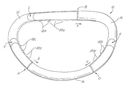
Figure 1 i:~ a part=ia1- perspective view of a first
embodiment of the adjustable an~nuloplast~y ring of t:he
present inventiom..
Figure 2 is a partial- cut--~~way perspective view of a
second embodiment cf the ad just:~~ble annuloplasty rung~ of
the present inver~.tion.
2~.~4~f'~
4
Figure 3 is a reduced perspective view of a third
embodiment of the adjustable annuloplasty ring of the
present invention.
Figure 3a is an enlarged, cut away perspective view,
of a portion of the annuloplasty ring of Figure 3.
Figure 4 is a cut-away illustration of a human heart
having an adjustable annuloplasty ring of the present
invention implanted at the mitral position, and showing the
manner in which a dilation apparatus (e. g., a balloon
l0 catheter or expandable cage) may be advanced through a
catheter, positioned transeptally, and utilized to effect
in situ enlargement of the adjustable annuloplasty ring in
accordance with the method of the present invention.
Detailed Description of the Preferred Embodiment
The following detailed description and the
accompanying drawings are intended to describe and show
certain presently preferred embodiments of the invention
only, and are not intended to limit the spirit or scope of
the invention in any way.
With reference to the drawings, Figures 1-3 show
alternative ways of constructing the adjustable ring
members 10, 10a and lOb of the present invention. The ring
members 10, 10a and lOb shown in Figures 1-3 have a
generally "D-shaped" configuration which corresponds to the
normal anatomical shape of the mitral valve annulus. It
will be appreciated that, if these ring members 10, 10a and
lOb were intended for use in remodelling of the tricuspid
valve, they would have the generally round configuration of
the normal anatomical shape of the tricuspid valve annulus.
The ring member 10 shown in figure 1 comprises first
12, second 14 and third 16 tubular segments. Each segment
12, 14, 16 is joined to the two other segments to form a
substantially unitary ring structure. The first segment 12
is tubular in configuration, having open ends A and B into
~1~48fi7
which the corresponding ends of the second and third
segments 14, 16 are inserted. The second segment 14 has a
blunt tipped or closed first end C and an open tubular
second end D. The third segment 16 has blunt tipped or
5 closed first and second ends E and F, respectively.
The first end C of second segment 14 is inserted into
the open second end B of the first segment 12. A series of
raised lugs or teeth 18a protrude from one side of the
portion of the second segment 14 which inserts into the
second end B of the first segment 12. A corresponding
series of apertures or detents 20a is formed in the side
wall of the first segment 12. The individual teeth 18a
snap into and fractionally engage the individual detents
20a, as shown.
Similarly, the first end E of the third segment 16 is
inserted into the open second end D of the second segment
14. A series of raised lugs or teeth 18b protrude from one
side of the portion of the third segment 16 which inserts
into the second end D of the second segment 14. A
corresponding series of apertures or detents 20b is formed
in the side wall of the second segment 14. The individual
teeth 18b snap into and fractionally engage the individual
detents 20b, as shown.
Also, the second end F of the third segment 16 is
inserted into the open first end A of the first segment 12.
A series of raised lugs or teeth 18c protrude from one side
of the portion of the third segment 16 which inserts into
the f first end A of the f first segment 12 . A corresponding
series of apertures or detents 20c is formed in the
sidewall of the first segment 12. The individual teeth 18c
snap into and fractionally engage the individual detents
20c, as shown.
The individual teeth 18 are conf figured and constructed
such that, when sufficient dilatory pressure is applied to
the inner surface of the ring 10, the segments 12, 14, 16
. 219~:~~'~
6
will spread apart and the teeth 18 will be caused to move
out of the detents 20 within which they are positioned and
will slidably advance and snap into the next available
detents in the series, thereby effecting one incremental
increase in the annular size of the ring. Further
application of additional dilatory pressure will cause the
teeth 18 to move to the next available detents 20 in the
series, thereby effecting a second incremental increase in
size, and so on.
Figure 2 shows an alternative ring 10a comprising
first and second semi-annular tubular segments 30, 32 which
are joined together in end to end fashion, as shown, to
form the desired annular configuration of the ring 10a.
Rack bars 34, 36 insert into the opposing open ends of the
first and second tubular segments 30, 32. Teeth 18
protrude laterally from the portions of each rack bar 34,
36 which insert into the juxtaposed ends of the first and
second semi-annular tubular segments 30, 32 ,as shown.
Corresponding apertures or detents 20 are formed in the
side walls of the tubular members 30, 32. The individual
teeth 18 snap into and fractionally engage the individual
detents 20, as shown. As described here above with respect
to the embodiment shown in Figure 1, the application of
dilatory pressure against the inner surface of the ring 10a
wyo cause the semi-annular tubular segments 30, 32 to move
apart and the individual teeth 18 will advance to, and seat
within, the next available detents 20, thereby causing the
size of the ring 10a to increase by a predetermined
incremental amount.
It will be appreciated that the components which make
up the ring member 10 need not necessarily be of tubular
configuration as shown in the embodiments of Figures 1 and
2. Indeed, as shown in Figure 3, the ring member 10b may
comprise of a plurality of non-tubular arcuate leaves 40,
42, 44, 46 assembled in overlapping relation to one another
CA 02194867 2001-05-03
7
and contained within a distensible outer sheath 50, as shown.
In any embodiment of the invention, a suture ring 52,
formed of material such as Dacron Mesh TM, is mounted about the
periphery of the ring member 10, is mounted about the periphery
of the ring member 10, 10a, lOb to facilitate suturing-in-place
of the ring member 10, 10a, lOb to surrounding anatomical
tissue.
Figure 4 shows a schematic illustration of the human
heart having an adjustable annuloplasty ring 10 of the present
invention implanted at the mitral position therein. The
anatomical structures and major blood vessels of the heart are
labeled, on Figure 4, in accordance with the following legend:
PV . . . . . . Pulmonary Veins
PA . . . . . . Pulmonary Artery
RPA . . . . . . Right Pulmonary Artery
LPA . . . . . . Left Pulmonary Artery
SVC . . . . . . Superior Vena Cava
IVC . . . . . . Inferior Vena Cava
AO . . . . . . Aorta
RA . . . . . . Right Atrium
RV . . . . . . Right Ventricle
LA . . . . . . Left Atrium
LV . . . . . . Left Ventricle
IS . . . . . . Interatrial Septum
AV . . . . . . Aortic Valve Position
MV . . . . . . Mural Valve Position
TrV . . . . . . Tricuspid Valve
PuV . . . . . . Pulmonic Valve
As shown in Figure 4, one method by which the size of the
annuloplasty ring 10 may be adjusted is through introduction of
a guide catheter 50, via catheterization of the superior vena
cava such that the distal end of the catheter is passed through
the interatrial septum IS, using known septal penetration
technique, and into the left atrium LA. A balloon dilation
catheter 52, such as a valvuloplasty plasty catheter of the
type commercially available, is then advanced through the lumen
of the guide
21~486'~
8
catheter 50, and positioned such that the balloon 60 of the
balloon catheter 52 is within the annulus of the mitral
valve MV. Thereafter, the balloon 60 is inflated, as
shown, to cause the adjustable annuloplasty ring 10 to
expand to a larger annular configuration.
In embodiments, such as those described and shown
hereabove in Figures 1-3, it will be appreciated that the
balloon 60 may be expanded to a specific diameter which
will evoke a single incremental increase (i.e., from one
notch to the next) of the mechanical expansion-controlling
system of teeth and notches formed in the annuloplasty ring
10.
Similarly, when the annuloplasty ring 10 is implanted
at the tricuspid valve TrV it will be desirable to advance
the guide catheter 50 through the superior vena cava SVC to
a point where the distal end of the guide catheter 50 is
positioned within the right atrium RA of the heart.
Thereafter, the balloon dilation catheter 52 may be
advanced to a point where the distal portion of the balloon
catheter 52 extends through the tricuspid valve TrV.
Thereafter, the balloon 60 will be dilated so as to expand
an annuloplasty ring of the present invention (not shown)
when implanted within the tricuspid valve TrV.
It will be appreciated by those skilled in the art
that various modifications additions and deletions may be
made to the above-described embodiments, without departing
from the intended spirit and scope of the invention.
Accordingly, it is intended that all such modifications
additions and deletions be included within the scope of the
following claims.