Note: Descriptions are shown in the official language in which they were submitted.
21 9~308
WO96/02212 r~
--1--
TRANSLUMINALLY IMPLANTABLE INFLATABLE PROSTHETIC CARDIOVASKULAR VALVE
Field of the Invention
The present invention pertains generally to medical
equipment, and more particularly a catheter-introducible
prosthetic valve which may be implanted into a ~l;~n
heart or elsewhere in the cardiovascular system to
augment or replace a malfunctioning endogenous valve.
Backgrouna of the Invention
The prior art has included numerous surgically
implantable prosthetic valves which may be utilized to
replace malfunctioning heart valves, such as the aortic
valve and the mitral valve. Some of the prosthetic heart
valves of the prior art are " - '-n;cal" valves of non-
biological origin. Others are "biological" valves
wherein all or a portion of the valve consists of
harvested r~r~-l;An (e.g., porcine) tissue which has been
preserved by way of a chemical f ixation process.
Although surgically implantable prosthetic heart
valves have become widely used in clinical practice,
their implantation involves a major cardiothoracic
surgical procedure wherein the patient must be placed on
full cardioplllr -ry bypass for a significant period of
time. As a result, patients who have severe
complications of their valvular disease or who are
otherwise severely ill or elderly may be unable to
undergo the rigors of such major cardiothoracic surgical
procedure and are, thus, unable to
2 1 95~
WO96102Z12 l~ll-_ .3
-2-
receive the benefits of a surgically implanted prosthetic
cardiovascular valve.
A number of prior investigators have proposed
various "coll ~r~ i hl e" cardiovascular valves and other
cardiovascular apparatus (e.g., embolus traps) which may
be collapsed and inserted into the mammalian vasculature
through the lumen of a tubular catheter or introducer.
Examples of collapsible cardiovascular valves and related
apparatus are found in United States Patent Nos.
3,671,979; 4,056,854; 4,592,340; 4,727,873; 4,817,600;
4,960,424; 4,994,077; 5,163,953; and 5,207,695, as well
as the following foreign patents and/or patent
publications WO91/17720; DT 2700-531 and WO93/01768.
Although various collapsible, catheter deployable,
heart valves and/or other cardiovascular apparatus may
have been proposed in the prior art, there remains a need
for further r~fin~--nt and dev~ L of such devices so
as to arrive at a clinically useful prosthetic
cardiovascular valve which may be implanted, through the
lumen of a cardiovascular catheter, without the need for
major cardiothoracic surgery.
~ ry of the Invention
sroadly stated, the present invention comprises an
inflatable prosthetic cardiovascular valve which, when in
a deflated state, is sufficiently collapsible to be
passed through the lumen of a tubular cardiovascular
catheter ~nd which, when subsequently inflated, will
assume a fully functional operative cardiovascular valve
configuration.
In accordance with a first integral or annular
: ~--S;r-nt of the invention, there is provided a
collapsible prosthetic cardiovascular valve comprising an
annular inflatable toroidal valve body and one or more
2~95308
W096/022l2
-3-
occluder members (e.g., pliable leaflets) affixed
thereto.
A plurality of legs or strut members may extend from one
side of the toroidal valve body to facilitate attachment
of, and/or to maintain operative positioning of, the
valve leaflets.
In accordance with a second split or linear
omho~;r~ L of the invention, there is provided a
collapsible prosthetic cardiovascular valve comprising an
inflatable valve body having a split or separation formed
therein and one or more occluder members (e.g., pliable
leaflets) affixed thereto. When deflated, the valve body
may be separated at its split or separation and extended
into an elongate linear deflated configuration. When
inflated, the valve body assumes a function annular or
circular configuration. The inflatable valve body may be
inherently biased to assume said annular or circular
configuration upon inflation thereof, or may be provided
with one or more tether lines or other guide me_bers
useable to guide or pull the valve body into the desired
annular configuration as inflation of the valve body is
accomplished.
Although the prosthetic cardiovascular valves of the
present invention may in~L~uL~Le various numbers of
individual valve leaflets, a preferred omho~;r~~t of the
valve inc~L~L~Les three (3) valve leaflets, each having
three (3) inboard edges which meet along a tre-foil
margin within the annular central passageway of the
inflatable valve body.
Although the collapsible cardiovascular valves of
the present invention may be inflated by various means,
one preferred embodiment of the invention employs a
detachable inflation tube which is initially connected to
the valve, and which may be subsequently severed from the
valve and removed following inflation thereof.
W096/022l2 2~q 5 ~0~ s~ l~
-4-
The inflatable cardiovascular valves of the present
invention may be inflatea with any suitable inflation
fluid. In some ~mho~;r ~S, the valve may be initially
inflated with material(s) which will react or otherwise
undergo gelation or solidification within the valve body,
thereby resulting in a gel-filled or solid-filled valve.
The collapsible cardiovascular valves of the present
invention may be specifically sized and configured for
implantation at various sites or locations within the
cardiovascular anatomy. In particular, collapsible
valves of the present invention may be sized or
configured to replace or augment any natural heart valve,
including the mitral and aortic valves of the human
heart. Similarly, collapsible cardiovascular valves of
the present invention may be sized and configured for
implantation in veins of the extremities to replace or
augment absent or malfunctioning venous valves. In
instances when the valves of the present invention are
utilized to replace or augment the aortic valve of the
heart, the positioning and location of the prosthetic
valve of the present invention will be such that the
prosthetic valve does not interfere with blood flow into
the coronary circulation.
Further in accordance with the invention, there are
provided apparatus and methods for percutaneous
transluminal insertion and utilization of the
collapsible/inflatable cardiovascular valves of the
above-described character.
Further objects and advantages of the invention will
become apparent to those skilled in the art upon reading
and understanding of the following detailed description
and the accompanying drawings.
srief DescriPtion of the Dr winqs
WO96/02212 2 1 95~08 1~I/U~
~ -5-
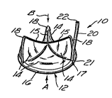
Figure 1 is a perspective view of a catheter-
introducible cardiovascular valve of the present
invention having an inflatable toroidal support
structure.
Figure la is a perspective view of the inflatable
toroidal support structure portion of the catheter-
introducible cardiovascular valve of Figure 1.
Figure lb is a cut away plan view of the portion of
the inflatable toroidal support structure shown in Figure
la.
Figure 2 is a perspective view of an alternative
"split" embodiment of a catheter-introducible prosthetic
valve of the present invention.
Figure 3 is a cross-sectional diagram of a human
heart having an inflatable prosthetic valve of the
present invention implanted adjacent the endogenous
aortic valve.
Figure 3a is an enlarged view of region AV of Figure
3.
Figures 4a-4c provide a step-by-step diagram of a
method by which the prosthetic valve of Figure 1 may be
inserted, inflated and implanted in the body of a human
being or other mammal.
Figures 5a-5d are a step-by-step diagram of a method
by which the prosthetic cardiovascular valve of Figure 3
may be inserted, inflated and implanted in the body of a
human being or other mammal.
Detailed DescriPtion of Preferred ~o~ir t~
The detailed description set forth below in
connection with the ~ppPnAPd drawings is intended merely
as a description of the presently preferred embodiments
of the invention, and is not intended to represent the
only form in which the present invention may be
constructed or utilized. The description sets forth the
W096102212 2 1 9 5 3 0 8 rcl~.
-6-
functions and sequence of steps for construction and
implementation of the invention in connection with the
illustrated embodiments. It is to be understood,
however, that the same or equivalent functions and
sequences may be accomplished by different omhoS;~-nts
that are also intended to be Pnr~rp~so~ within the
spirit and scope of the invention.
i. Construction of Inflatable Cardiovascular
Valves of the Pre~ent Invention
With reference to Figures 1, la and lb, there i8
shown a first P~ho~; r-nt of a prosthetic cardiovascular
valve 10 of the present invention comprising an
inflatable support body 12 having one or more pliable
valve leaflets 14 mounted thereon. The leaflets 14 are
configured and c~ ed 80 as to move, in response to
hemodynamic v ~ of the blood, between a) an "open"
configuration wherein blood may flow through the valve in
a first direction A and, b) a "closed" configuration
whereby blood is prevented from back flowing through the
valve in a second direction B.
In the ~;r ~ shown in Figures 1, la and lb, the
inflatable stent or support body 12 of the valve 10
comprises an inflatable annular ring or toroid 16 having
a plurality of inflatable legs or struts 18 extending
therefrom. An inflation tube 20 is initially attached to
an inflation port 17 on the inflatable body 12 of the
valve 10 to permit infusion of inflation fluid into the
inner cavity 24 of the inflatable support body 12. Such
inflation tube 20 preferably comprises a pliable,
elongate tube having a hollow lumen 22 extending
longitudinally therethrough.
The distal end of the inflation tube 20 may be
~ot~h~h1e from the inflatable body 12 of valve 10 such
that, after the valve 10 has been fully inflated, the
WO96102212 2 I q 5 3 ~ 8 . ~l/L~ .5
-7-
inflation tube 20 may be volitionally detached from the
valve lO and subsequently extracted and removed.
In embodiments wherein the inflation tube 20 is
detachable from the valve lO, a sealing element 21 such
as a check valve or sphincter like elastic ring may be
~icr~c~ within or adjacent the inflation port 17 or
other location where the inflation tube 20 separates or
~icconn~rts from the valve 10, so as to result in closure
of the inflation port 17 or residual portion of tube
lumen 22, thereby preventing leakage or seepage of the
inflation material or fluid from the inner cavity 24 of
the valve 10. The valve leaflets 14 may be formed of any
material(s) suitable for performing the leaflet function,
including thin membranes or sheets of pliable synthetic
or biological material capable of flexing back and forth
between the above-described "open" and "closed"
configurations in response to hemodynamic movement of the
blood against the leaflets 14, yet sufficiently resistant
to extension that leaflet(s) perform their intended
function.
As such, the leaflets 14 are preferably constructed
and configured to mimic the function of the leaflets or
cusps of a healthy endogenous cardiovascular valve.
Synthetic leaflets 14 may be formed of elastomeric
materials such as polyurethane, silicone, rubbers, etc.
suitably modified to provide limited extensibility.
Biological leaflets 14 may be formed of chemically fixed
l; An valvular tissue or other biological tissue
(e.g., pericardium) which is sufficiently thin and
pliable to perform the desired valving function of the
leaflets 14.
Although the leaflets may vary in number and
configuration, the presently preferred embodiments shown
in the drawings utilize three (3) separate leaflets 14
having ~LLe~ondingly configured inboard edges 15 which,
WO96/02212 2 ~ ~ 5 ~ ~ 8 r ~"~ ~ .3
-8-
when in their closed position, interact or meet with one
another in a tre-foil margin as shown.
The valve leaflets 14 may be mounted on or attached
to the inflatable support body 12 by any suitable means,
including suturing or adhesive. For example, in some
Pmho~ i r- -nts the material of which the leaflets 14 are
formed may be wrapped around portions of the inflatable
body 12 and subsequently sutured in place to hold such
biological leaflet material on the inflatable support
body 12. In other embodiments wherein biological or
synthetic material is employed, it may be desirable to
apply quantities of adhesive to affix the leaflets 14 to
the inflatable support body 12. Also, in some
PmhQ~ nts, the leaflets 14 may be formed of synthetic
material integral with the inflatable support body 12.
The presently preferred embodiments of the invention
include an integral or annular Pmho~;r-nt (Figs. 1 and
la) as well as a split or linear Pmho~;r nt (Figs 2 and
2a).
In the integral or annular PmhoA;r?nt of the valve
10 (Figs. l and la) the inflatable valve support body 12
is in the form of a continuous ring or annulus which,
when deflated, may be compressed or compacted into a
sufficiently small space to pass through a catheter lumen
of approximately 4-7mm in diameter. Thereafter, the
valve support body 12 may be inflated to cause the valve
to assume the operative annular or ringli~e structure
shown in Figure l.
In the alternative linear or "split" embodiment of
the valve lOa shown in Figure 2, a division or closed
split 23 is formed vertically through one of the strut
members 18a, as shown. In such Pmho~i ~, the valve lOa
may be deflated to a flaccid state and subsequently
extended into an elongate outstretched configuration
whereby a first end lla of the split 23 is situated at
WO96/02212 2 1 9 5 3 ~ 8 r ~ s~ .s
g
one longitudinal end of the valve 10a and the opposite or
second end llb of the split 23 is situated at the
opposite end of the valve 10a, as illustrated in Figure
5b.
Optionally, in the "split" embodiment shown in
Figure 2, one or more pull line(s) or tether(s) 40 may be
attached to the valve 10a to facilitate movement of the
valve 10a from its deflated linearly extended and compact
configuration (Fig. 5b) to its generally circular
inflated operative configuration (Fig. 5d). The
tether(s) 40 may initially extend separately from and
alongside the inflation tube 20a or may be contained
within one or more tether guides or passageways
associated with or formed within the inflation tube 20a.
Specifically, in the embodiment shown in Figure 2, a
separate tether guide lumen 44 extends longitn~inA1ly
through the inflation tube 22a, which is incuL~uoLated
with the first end member of split support strut 18a, and
into that first end strut member 18a where it terminates
distally in a tether entry apertures formed in the face
of the split portion lla, opposite the points on
corr~cpon~inr~ split portion llb where the distal ends of
tethers 40 is rrnn~~t~d to the second portion llb of the
split support strut 18a. From this connection, tethers
pass into tether entry ap~LLuLes formed in the
corresponding split portion llb and extend proximally
through tether guide lumen 44 of inflation tube 20a. The
proximal ends of the tethers 40 emerge out of and/or
apart from the proximal end of the inflation tube 20a so
as to be grasped and retracted by the operator. As such,
retraction of the tethers 40 in the proximal direction
(arrow C) will guide or pull the second split portion llb
of the split support strut 18a into juxtaposition with
the first split portion lla thereof.
WO96102212 2 1 ~ 5 3 Q 8
--10--
The connection or attachment of the distal ends of
tethers 40 to the second split portion llb of the split
valve support body 12a may be a releasable connection
whereby the distal ends of tethers 40 may be released and
pulled awfiy from the valve lOa simultaneously or separate
from detachment of the inflation tube 22a from the valve
body 12a. The releasable or tearable connection of the
distal ends of the tethers 40 to the second portion llb
of the split support strut 18a may be specifically
constructed such that the amount of tension re~uired to
tear or separate the tethers 40 from the valve lOa can
only be exerted when the valve lOa is properly anchored
in its desired position within the mammalian body.
The valve 10, lOa of the present invention may be
implanted at many different desired locations in the
mammalian cardiovascular system. For example, the valves
10, lOa may be implanted adjacent to or in replacement of
a malfunctioning heart valve, as shown in the
illustration of the human heart shown in Figure 3. The
anatomical ~Lu~uLe and major blood vessels of the heart
are labeled
on Figure 3 in accordance with the following legend:
PV . . . . . . Pulmonary Veins
PA . . . . . . Pulmonary Artery
RPA . . . . . Right Pulmonary Artery
LPA . . . . . Left Pulmonary Artery
SVC . . . . . Superior Vena Cava
IVC . . . . . Inferior Vena Cava
A0 . . . . . . Aorta
RA . . . . . . Right Atrium
RV . . . . . . Right Ventricle
LA . . . . . . Left Atrium
LV . . . . . . Left Ventricle
AV . . . . . . Aortic Valve Position
MV . . . . . . Mitral Valve Position
TrV. . . . . . Tricuspid Valve
PuV. . . . . . Pulmonic Valve
~,l ss3as
WO 96/02212
--11--
In ~mho~ir-nts where the valve 10, lOa is implanted
in the aortic position on the outflow side of the
endogenous aortic valve, (see Figure 3) it will be
appreciated that the valve 10, lOa must be carefully
positioned so as not to impede or block bloodflow into
the coronary ostia. Additionally, it will be appreciated
that the valves 10, lOa of the present invention may be
useful in various peripheral, extracardiac locations,
such as in the veins of the lower extremities as
replacements and/or augmentations for absent or
malfunctioning endogenous venous valves.
ii. Inflation of the Cardi~vasçular Valves
of the Pr~nt Tnvention
The inflatable cardiovascular valve 10, and in
particular the inflatable support body 12 of the valve 10
may be inflated by any suitable inflation fluid or
substance.
In some ~hod;~~lLs and applications, it may be
desirable to inflate the support body 12 with a gas or
liquid (e.g., carbon dioxide or saline solution) which
may subsequently be extracted or removed from the valve
body 12 if it is subsequently desired to deflate the
valve 10.
In other embodiments and applications, it may be
desirable to inflate the valve lo with a fluid which
subsequently gels or solidifies within the inflation
space 24 of the valve support body 12, thereby minimizing
any lik~lih~od that the inflation substance would
inadvertently leak or seep from the implanted valve 10.
For example, one or two ~- ~nt elastomer-forming
chemical reactants may be initially instilled into the
inflation space 24 of the support body 12 through
inflation tube 20 while in a liquid state, and
2 1 953~
W096/02212
-12-
subsequently allowed to gel or solidify within the
inflation space 24 of the valve support body 12 so as to
result in a gelatinous or solidified (e.g., elastomeric
or rigid) filling material within the inflation space 24
of the inflated valve 10.
In embodiments wherein the valve 10 is inflated with
flowable liquid or gaseous inflation fluid(s), it may be
possible to subsequently deflate and remove the valve,
while in : ~:'; Ls wherein gelling or solidifying
inflation materials are employed, subsequent deflation
and removal of the valve may be rendered non-feasible.
Accordingly, the selection of the type of inflation
material to be employed may depend on whether it is
desirable to subsequently deflate the valve.
Alternatively, a two stage inflation technique may
be utilized whereby an initial temporary liquid or
gaseous inflation fluid is initially utilized to inflate
the valve, but is subsequently replaced by a more
permanent solidifying or gelling inflation substance.
This two-staged inflation technique would permit the
valve to be deflated and/or manipulated as needed during
the implantation and affixation processes, but would
subsequently allow the valve to becomQ permanently
inflated and configured after the proper positioning and
affixation of the valve has been achieved. In accordance
with this aspect of the invention, a bioacceptable or
biologically inert temporary inflation fluid (e.g., 0.9
percent NaCl solution) may be initially passed into the
valve 10 to effect inflation thereof. After the valve 10
has been appropriately positioned and affixed in its
desired operative location, an escape opening may be
formed in the body of the valve by puncture thereof, or
by other suitable means, and a more permanent inflation
substance, such as a material which will subsequently gel
or solidify, may be passed into the valve lo, thereby
2 1 95308
WO96102212
-13-
displacing the temporary inflation fluid out of the
escape aperture, and allowing the valve to become filled
with a more permanent non-escaping inflation material.
iii. Insertion and Positionina of the Inflatable
Valves of the Pre3ent Invention
The inflatable cardiovascular valve lO of the
present invention may be inserted, deployed and implanted
at their intended anatomical locations, by any suitable
means.
a) A ~referred method of im~lantinq an annular
inflatable valve of the first emho~ir t
Figures 4a-4c provide a stepwise illustration of a
presently preferred method for percutaneous transluminal
catheter introduction of the "integral" or annular first
embodiment of the inflatable valve lO of the present
invention.
As shown, a tubular cardiovascular guiding catheter
30 having a hollow lumen 32 extending longitn~;nAlly
therethrough is initially inserted, by a standard
percutaneous introduction technique, into a blood vessel
(e.g., the femoral artery). The catheter 30 is advanced
through the vasculature until the distal tip of the
catheter 30 is positioned adjacent the intended site for
implantation of the valve lO.
The valve lO is initially deployed in its deflated
compact configuration and is mounted or attached on an
introducer member 34, such as a bendable cardiovascular
guidewire or elongate tubular member having an annular
slot near the distal end to hold or a~, 'Ate the
deflated valve lO therein. The elongate inflation tube
20 attached to the deflated valve lO is deployed within
or alongside the introducer member 34. The introducer
member 34 having the deflated valve lO mounted thereon is
=
2 1 95308
WO96/02212 ~ 3
-14-
then inserted into and advanced through the lumen 32 of
the pre-positioned guide catheter 30.
After the distal end of the introducer member 34
having the deflated valve lO mounted thereon has emerged
from the distal luminal opening of the guide catheter 30,
a quantity of inflation fluid is injected or infused
through the inflation tube 20 and into the inflatable
body 12 of the valve 10. The inflating valve lO is, by
virtue of its inflation or by other mechanical or
manipulative means, separated from the introducer member
34, thereby allowing the introducer member 34 to be
proximally retracted and removed as shown in Figure 4b.
After the valve 10 has been fully inflated, the
inflation tube 20 may be detached, proximally retracted
and removed, as shown in Figure 4c.
The inflated, operatively configured valve 10 (Fig.
4c) may subsequently be held in its desired position
within the heart or blood vessel by way of engagement
members (e.g., pins, hooks, etc. ...) protruding from the
valve lo or by application of one or more physiologically
compatible adhesives (e.g., polyurethane adhesive).
In other ~mho~ir-~ts and applications, surfaces of
valve 10 which contact the receptive body may be provided
with, coated, or infused with a suitable biologic element
(e.g., a biologically compatible tissue graft or cellular
matter obtained from an autologous or otherwise
genetically compatible source) such that the
aforementioned surfaces on valve 10 will become
biologically assimilated within the local tissue and
thereby fix the valve 10 as an implant.
After the in~lated valve 10 has been adhesively,
mechanically or frictionally engaged in its desired
implanted location, the catheter 30 may be removed,
thereby leaving the inflated, operatively configured
.
~ i 9~3 08
W096/02212 ~ .3
-15-
valve 10 in its desired implanted position within the
heart or vasculature.
b.) A ~referred method of ; lantina a
linear inflatable valve of the ~econd ~
The linear inflatable valve lOa of the 6econd
embodiment of the present invention may be implanted by
the transluminal catheter implantation technique
illustrated in Figures 5a-5d.
As shown, an elongate guide catheter 30a having a
hollow lumen 32a extending longitudinally therethrough is
initially inserted into the vasculature by a known
percutaneous insertion technique and is subsequently
advanced to a position whereat the distal tip of the
catheter 30a is positioned adjacent the intended site of
implantation of the valve lOa.
The deflated, linearly extended, valve lOa is
initially mounted on or in an introducer member 34a, such
as a pliable cardiovascular guidewire or elongate tube
having a linear slot for receiving and/or A~, -'sting
all or a portion of the deflated valve lOa.
The inflation tube 20a and optional ~-n;r~llAtion
tether 40 may be initially extended alongside the
introducer 34a.
The introducer 34a having the deflated, linearly
extended, valve lOa positioned thereon is then inserted
into the lumen 32a of the pre-positioned guide catheter
30a and advanced therethrough until the distal end of the
introducer 34a having the deflated valve lOa mounted
thereon has emerged from the distal opening of the guide
catheter lumen 32a.
Inflation fluid is then passed through inflation
tube 20a and into the valve lOa. The act of inflation of
the valve lOa, and/or other physical manipulation means,
is employed to separate the valve lOa from the introducer
W096l02212 2 1 9 5 3 ~
-16-
34a, thereby permitting the introducer 34a to be
proximally extracted and removed as shown in Figure 5b.
As the valve 5b is being inflated, the manipulation
tethers 40 may be pulled or otherwise manipulated by the
operator 50 as to draw the distal or second end lla of
the linearly extended valve into juxtaposition with the
proximal or first end llb thereof. After having been
placed in juxtaposition, the opposing surfaces of split
23 are held, affixed or locked together in the closure
process, thereby creating the desired annular
configuration of the inflated valve.
After the valve lOa has been fully inflated, the
inflation tube 20a and the optional manipulation tethers
40 may be detached, proximally withdrawn and removed as
shown in Figure 5d.
The inflated valve lOa may be affixed in its desired
position by way of mechanical fixation apparatus (e.g.,
pins, hooks, etc. ...) or adhesive as described above
with respect to the valve 10 constructed in accordance
with the first I mh~ L.
After the inflated valve lOa has been adequately
affixed in its intended operative location, the guide
catheter 30a may be withdrawn and removed, thereby
leaving the inflated operatively configured valve lOa at
its desired site of implantation within the heart or
vasculature.
Although the invention has been described herein
with specific reference to presently preferred
~ho~; r~nts thereof, it will be appreciated by those
skilled in the art that various additions, modifications,
deletions and alterations may be made to such preferred
embodiments without departing from the spirit and scope
of the invention. Accordingly, it is intended that all
reasonably foreseeable additions, deletions, alterations
2 1 953~
WO96102212
~ -17-
and modifications be included within the scope of the
invention as defined in the following claims.