Note: Descriptions are shown in the official language in which they were submitted.
WO96/03117 rc.,~
21 9568
_ JKATION OF RTor.~Tr~T.T.Y AGTIVE .TCGTTT.TC~ INTO BIOACTIVE
GLASSES
This i9 a continuation-in-part of U.S. Application
Serial No. 08/406,047, filed March 17, 1995, pending, which is
5 a r~nt;rll~tion-in-part of U.S. Application Serial No.
08/281,055, filed July 27, lg94, pending.
Field of the Invention
The present invention relates to the incorporation of
biologically active molecules into the matrix of glass, in
10 particular bioactive glass, using a sol-gel-derived process of
production.
sackground of the Invention
Musculoskeletal injuries have a substantial impact on
the health and quality of life of millions of Americans.
15 Delayed healing of and non-unions of fractures represent a
c~ntinn~nc orthopaedic challenge. The conventional way of
treating these problems is to use bone plates or screws in
combination with autologous bone grafting.
As a natural composite material, ~nt~gon~llc bone graft
20 has been shown to have both osteoconductive and osteoinductive
properties. In addition, it i8 a sterile, non-; -, ;c and
non-toxic material, which has the ability to be fully incorpo-
rated into the fracture site. Notwithstanding the long
duration for their activity to develop, autogenous bone grafts
25 are the gold standard by which synthetic composites are
compared. Given that there is also a limited supply and
harvest site morbidity of autogenous bone graft material, there
WO96103117 r~
2lss6~a
is significant motivation to develop synthetic composites. To
date, no synthetic bone graft substitutes have fully achieved
the properties of autogenous bone graft.
FnhAn~ing the rate and probability of fracture healing
5 and the promotion of bone formation and healing of delayed and
non-union fractures are of great clinical significance.
INI~/AAOS sponsored workshop. Bone Formation and sone
Regeneration. Tampa, F~: American Academy of Ort~opaed~c
Surgeons, 1993~. The large population of patientE with delayed
lO unions and non-unions of bone, the large direct medical costs,
and the societal costs related to their long term disability,
highlight the need for effective and improved methods of
treatment.
Advances in materials science and the identification
15 of osteogenic and osteoinductive growth factors have invited
the investigation of newer alternatives for autogenous bone
grafting. Osteogenesis, which is the process of bone forma-
tion, involves both osteoconduction and osteoinduction.
Osteoconduction is the process in which differ~ntistpd bone-
forming cells produce a bone matrix upon an existing substrate.Materials that promote this process are con3idered
osteAr~n~ ive. Osteoinduction is the process by which
undifferentiated mesenchymal precursor cells are transformed
into differentiated bone forming cells. Factors or materials
25 that promote this process are considered to be osteoinductive.
Growth factors delivered by biologically active con-
trolled release carriers have the potential for improved
fracture heallng and lower morbidity, thereby resulting in
improved patient care and a decrease in the overall costs
30 associated with fracture care. Similarly, the delivery of
antibiotics by such carriers, either alone or in addition to
growth factors, will help reduce the incidence of infections,
which can further contribute to delays in healing. In
fractures involving, for example, the spire, the incorporation
35 of anti-inflammatory agents and analgesic~ will help control
inflammation, which can also delay the healing process, and
contribute to patient comfort during the healing process.
WO96103117 r~ ,r1
21 9~680
- 3 -
Additionally, the controlled release of such materials
regardless of the bioactivity of the carrier would represent a
distinct advantage over current delivery methoùs and assist
fixation of implants.
The ideal synthetic graft would be a scaffolding
material that would stimulate bone tissue to grow in place of
the scaffold as it degrades. ~Damien et al., J. Applied
Biomater. ~l99l) 2:187-20.) Synthetic materials intended as
bone graft substitutes should have r chRn;~Al and other
lO properties similar to those of bone, and should be biocompa-
tible with the surrounding tissues. In order to provide a
union across the fracture site they must serve not only as
scaffolding materials but also, similarly to native bone, have
a stimulatory effect on bone tissue regeneration.
The currently used synthetic bone graft materials are
considered ost~onnn~ tive in that they elicit the formation of
the bone matrix at their surfaces. Furthermore, they lead to
a contiguous interface with bone or are replaced by bone
tissue. Such properties suggest a chemical interaction between
zo these bioactive materials and the bone environment. Cells
existing in the bone matrix environment exhibit a beneficial
response to these materials.
The materials studied most for use as synthetic grafts
have been calcium phosphate ceramics and bioactive glasses.
25 Calcium phosphate ceramics ~CPCs) are very similar in composi-
tion to the mineral phase of bone. sioactive glass are capable
of forming a hydroxyapatite layer on their surface that mimics
the mineral phase of bone.
The most commonly used calcium phosphate ceramics
include: hydroxyapatite ~HA), in either dense or porous forms,
and ~-tricalcium phosphate ~-TCP). HydL~y~tite is of
limited effectiveness as a grafting material. When HA
particulate material in porous and dense form was evaluated as
a grafting material in the alveolar ridge it was found that
fibrous ~nnRrcl.lRtinn formed in perosseous 6ites. Migration of
the particles was also found to be a problem. ~Ducheyne P., J.
siomed. Mater. ~es. ~l987) 21(A2 Suppl):2l9.) Further, HA
wos6lo3ll~ F~ .,i0l
21 956~
cannot be used as a scaffolding material since its rate of
degradation is slow. [Cornell et al., Clin. Orthop. ~1992~
297; and Radin et al., ~. Biomed Mater. Res. ~19g3) 27:35-4s.)
~-TCP, on the other hand, is a biodegradable material
5 which i9 osteoconductive. However, its degradation rate has
been found to be too fast to serve as an effective aynthetic
graft material in load-bearing situations. (Damien et al.,
supra. ) Thus, clinical evaluations and applications of the HA
and ~-TCP materials, either dense or porous, have demonstrated
that both materials are limited by a lack of controlled rate of
reactivity.
Bioactive glasses were first found to bond to living
bone by Dr. ~arry Hench in the late 1960's. Since that time,
more than ten ~roups around the world have shown that glasses
~t~n~inin~ sio" CaO, P2O , ~azO and other smaller amounts of
oxides in various compositions bond to bone. (Ducheyne P., J.
i3iomed Mater~ Res. ~1987) 21(A2 8uppl~:219; Hench, ~.L., Ann.
N. Y, Acad. 5ci. (1988) 523:54; ~n~t~r.cfit~n et al., ~. Biomed
~ater. Res. (1991~ 25:1019-1030; Andersson et al., ~. Non-Cryst
Solids ~1991) 129:145-151; Boone et al., J. Biomed Mater. Res.
(1989) 23(A2 suppl~:183; Ducheyne et al., Clin. Orthop. Rel.
Res. (1992~ 76:102-114; Hench, L.~., ~. ~iomed Mater. Res.
(1989~ 23:685-703; Xokubo, T., ~iomaterials (1991) 12(2):155;
and R~wlings, R.D., ~. Mater. Sci. Letters (1992) 11:1340-
1343.)
Bioactive glass-ceramics undergo surface corrosion
reactions when exposed to body fluids. These corrosion
reactions form a silica-rich surface layer. This layer serves
as a nucleation site for the deposition of calcium ph~srh~te,
30 which evolves into a thick l-yd~v~y~atite layer. When in
contact with bone forming cells, this layer will form the basis
of the chemical bond between the glass and the bone matrix.
(Ducheyne, supra; Hench (1988), supra; and Hench, (1989),
supra.~ Dr. ~ench~s 45S5 bioactive glass has been the most
extensiYely studied of the bioactive glass-ceramics. Its
composition by weight ~ is: 4S~ SiOz, 24.5~ CaO, 6~ P~05 and
24.5% Na~O.
WO9G~03117 P~Il~ .'b~
21 95~0
In U.S. Pat. No. 5,204,106 (incorporated herein by
reference), 45S5 glass in particulate form in a narrow size
range was described as being an effective bone graft substitute
in the alveolar ridge model and as being well incorporated into
5 the surrounding bone. The glass granules were described as
causing the upregulation of osteoprogenitor cells to
osteoblasts that actively lay down bone tissue. ~Schepers et
al., ~. Cral Rehabil. (1991) 18:439-452.)
The following parameters are important for bone-
10 bioactive synthetic grafts: controlled resorption andreactivity, immersion induced transformation of the synthetic
materials' surface into a biologically-equivalent
hydLo~ydpatite-like mineral, relatively large surface area, and
porosity (to create a network for osteoblastic activity).
15 Bioactive glass can pot~nt;Ally be tailored to fit these
parameters. In addition, the following requirements are
important for a successful delivery system for biologically
active molecules: 1) controlled release of the molecules; 2)
delivery of adequate amounts of the molecules; 3) rapid growth
20 of bone tissue into the carrier; 4) biocompatibility,
osteoconductivity, and osteoinductivity of the implant
material; and 5) resorption of the carrier once bone tissue has
completely formed. (Lucas et al., ~. B-omed Mater. Res. (1989)
23(A1 Suppl):23.) No delivery system currently available meets
25 all of these criteria. (Damien et al., supra; and Cornell and
Lane, Cli~. Orth. Rel. Res. (1992) 277:297-311.) Certainly, no
delivery system results in controlled delivery.
Attempts have been made to try to improve calcium
phosphate ceramics by using them as delivery vehicles for bone
30 growth factors. To date, there has been no success in
incorporating growth factors into calcium phosphate ceramics in
a way that will lead to a sustained release of the added growth
factor. Mostly, one achieves a "burst" release, which is a
rapid initial release of most of the material over a short
35 period of time. (Campbell et al., ~rans. Orthop. Res. Soc.,
40:775, 1994.)
WO9G/03lt7 r ~u. ~ ~ I
21 9568rJ
Carriers made of ~-TCP, or nonsoluble collagen, have
been moderately successful when combined with bone morphogenet-
ic protein in attaining good acceleration of bone tissue heal-
ing. (Damen et al., ~. Dental Res. (1989~ 68:1355-1359)
5 ~owever, these systems have not been able to produce a measur-
able, controlled release of growth factor for time spans
approaching those needed for bone tissue regeneration to span
large bone filling defects. In one study, large amounts of
growth factors, i.e. greater than 50 milligrams, were required
to fill defects greater than three (3) ~Pnt;--tP~s. (Johnson
et al.l C7in. Orthop. (1992) 277:229237.)
In most of the systems studied with osteoconductive
materials used as carriers, the method of incorporation has
been that of simple immersion of the material into a growth
15 factor solution. The growth factor is then adsorbed either
onto the material surface or into the pore structure, but is
then quickly released upon immersion in an aqueous solution in
a bur~t effect. (Campbell et al., ~rans. Ortho~. Res. Soc.,
40:775, 1994.)
Published application WO 92/07554 reports a material
which can be implanted in living tissue which has a
~iodegradation rate matching the rate at which the tissue
regenerates. It is reported that the material may include an
active substance providing an extended therapeutical effect.
25 The material includes a calcium rh~rh~t~, biodegradable oxide
or polyoxide, and an active substance having amine groupings
such as netilmicin and/or g~nt~m; cin in sulphate form.
Published application Wo 53~05323 reports a
composition for st; l~t;ng bone growth comprising at least one
of FGF, TGF-~, IGF-II, PDGF, and their biologically actLve
mutants and fL _ c, or bone extracts with corresponding
activityl or bone extracts with BMP activity, and a suitable
application material.
United Kingdom Patent Application ~B 2255907 A reports
35 a delivery system for biologically active growth and
morphogenetic factors comprising a solid adsorbent selectcd for
its specific affinity for the factor and the factor adsorbed
WO96/03ll7 21 q5680 PCT~S95/09401
-- 7
thereon. In one embodiment, porous hydroxyapatite is specified
as the solid adsorbent.
U.5. Pat. No. 4,869,906 describes a resorbable porous
tricalcium phosphate in which the pores are sealed with a
filler mixture of antibiotic and a filler.
U.S. Pat. Nos. 5,108,436 and 5,207,710 describe
stress-bearing prostheses having a porous region in~ ~;n~tinn
with an osteogenic factor extract or a purified osteogenic
inductive protein, optionally in ~ ';n~tinn with a TGF-
~
l0 cofactor, in a pharmaceutically acceptable carrier. Thecarrier is either a collagen composition or a ceramic. The
osteogenic factor extract is dispersed in the porous region.
Other procedures for , ;n;ng the stress-bearing member with
the osteoconductive material inrln~;nr~ coating, saturation,
15 applying vacuum force to get the material into the pores, and
air-drying or freeze-drying the material onto the member. It
is further described that the pharmaceutically acceptable
carriers preferably include a matrix that is capable of
providing a structure for developing bone and cartilage. Some
20 preferred pharmaceutically acceptable carriers listed include
collagen, hydL~y~dtite, tricalcium phosphate, and bioactive
ylass. However, there is no description of a preparation
rnnt~in;ng bioactive glass as a pharmaceutically acceptable
carrier.
U.S. Pat. No. 4,772,203 describes implants having a
core and a matrix, with the matrix being at least partially
resorbable. The resorbable matrix is one or both of bioactive
and nst~ng~n~ci5-inducing. Tricalcium phosphate,
hydroxylapatite [sic], and bioactive glass are listed as such
30 matrixes. It is further stated that if a resorbable matrix is
employed, it is further possible to embed antibiotics in the
latter.
U.S. Pat. No. 4,976,736 describes biomaterials useful
for orthopedic and dental applications having a base portion of
35 calcium carbonate and a surface layer of a synthetic phosphate
such as hydroxyapatite. One advantage asserted for hydroxyapa-
tite is absorbency. It is further described that antibiotics
WO9C/~311~ PCT~Sg51~9~1
21 956~ ~
or growth iactors can be introduced into the pore cavities of
the implant or attached, respectively. Alternatively, the
antibiotic or growth factor can be intermixed with a preferably
biodegradable polymer and injected or vacuum infiltrated into
the porosity of the phn~ph~ surfaced material
Gombotz et al., ~. App. i3ioma~., (1994) 5:141-150
describe the incorporation of transforming growth factor-~ into
a composite implant made from poly~lactic-co-glycolic acid) and
demineralized bone matrix. It i5 reported that the implants
exhibited an inflammatory response with little miner~l;7~tion
or bone formation. Similar results were reported in Meikle et
al., BiomaterIals~ ~1994) 15(7):513-521 with poly DL-lactide-
co-glycolide discs having bone matrix extract incorporated
therein.
U.S. 2at. No. 4,563,350 describes a composition
suitable for inductive bone implants comprising a purified form
of osteogenic factor in admixture with a carrier having a
percentage of non-fibrillar collagen. The factor is added to
the collagen either in solution or gelatin form and stirred in
20 dilute mineral acid ~or 1-2 hours at approximately 4OC. The
material is then dialyzed and lyophilized.
~ apanese Laid-Open Patent Publication No. 5253286
describes a bone restoring material comprising Ca-r~n~;n;ng
glass powder and or cryatallized glass powder, an aqueous
solution composed mainly of phosphate, and a medical 5nh5t~nr~
in release-controlled form. The medical substance is described
as being in particulate form and can be coated with materials
capable of oppressing the releasing of the substance
temporarily.
As can be seen from the foregoing, a carrier
providing for the controlled release o~ biologically active
molecules is needed. Such materials which are additionally
osteoconductive and~or osteoinductive are also needed.
Bioactive glasses are osteoconductive but are usually
formed by ~mh;n;ng the different oxides in a platinum crucible
and melting the mixture at a t ,-ratnre of 1300-1400~C. This
is the melt-derived, or conventional, method o~ obtaining
W096/031l7 r~"~ ~.c
21 9~6~0
g
bioactive glasses. Such temperatures, however, would destroy
the function of most biologically active molecules during
preparation.
Another method which can be used to synthesize bioac-
5 tive glass is that of sol-gel processing. Sol-gel synthesis of
glasses is achieved by combining a metal alkoxide precursor,
such as tetraethylorthosilane ~TEOS, Si(oCaHs)4 in the case of
silica), with water and an acid catalyst to produce a
hydrolysis reaction with consequent polymerization of the metal
10 alkoxide species and production of a gel. This gel will
consist mostly of the metal oxide when dried and will attain
the consistency of glass.
Several investigators have reported the incorporation
of proteins into a sol-gel-type glass produced using silicon
15 alkoxide precursors and water with a maintenance of function.
Braun et al., ~. of ~on-Crystalline Solids, ~1992) 147 and
143:739-743; Yamanaka et al., Chemistry of ~aterials, ~1992)
4(3):495-497; Ellerby et al., Science, (1992) 255:1113-1115;
and Avnir et al., EncaDsulation of Oraanic Molecules and
20 Enzvmes, Ch. 27, pp385-404, American Chemical Society ~1992i.
Methods for synthesizing low temperature, low alcohol, low
proton-concentration sol-gels for enzyme incorporation are
described. The incorporated proteins ~in~;n~d their
functionality. However, the focus of such ~L~C~d~l~S was the
immobilization of the protein within the sol-gel in a manner
which retains the protein of interest within the gel. When the
sol-gel material functions as a sensor, very small molecules,
such as glucose, can pass through the pores for assay. The
incorporation within the sol-gel provides for repeated use of
30 the protein. Release of the protein from the sol-gel was not
desired and would actually be counter to maintenance of long-
term activity.
In U.S. Pat. No. 5,074,916, alkali-free bioactive sol-
gel compositions based on SiO2, CaO, and PzOs are described.
35 Compositions ranges are 44-86, 4-46, and 3-15 weight percent,
respectively. Calcium nitrate was used as the calcium source,
and tetraethoxysilane ~TEOS) was used as the silicon ~1 kn~i
WO'~6103117 P~T/US95~9401
21~5~
- 10 -
source. However, the process described utilizes temperatures
around 600-300~C, and an acid to water volume ratio of l/6.
Such a process is totally inc~mp~tihle with the incorporation
of biological molecules.
Su3rary o~ the Invention
The present invention is directed to controlled-
release carriers. In the carriers according to the invention,
biologically active molecules are incorporated within the
matrix of a silica-based glass. We have found that a
lC derivation of the sol-gel techniriue facilitates such
incorporation without negatively affecting subse~u~l,L activity
of the molecules. In the case of pure silica glass, the
release of the biological molecules from the carrier i9
effected primarily by diffusion through the pore structure. In
the instance the glass contains oxides in addition to silicon,
the release of biological molecules is e~fected by diffusion
and reaction when immersed in fluids such as, for example, body
fluids.
The sol-gel derived technigue allows extensive control
20 of the glass ultrastructure and, thus, further control over the
timing and guantity of release of the biologically active
molecules, such as drugs or growth factors. Such carriers can
be both osteoconductive and osteoinductive through the forma-
tion of a calcium phnCph~e surface layer (i.e. bioactive~ and
the release of protein factors that attract and stimulate
mesenchymal cells to differentiate into bone forming cells on
the carrier surface, as well as increase the proliferation of
osteoblasts in the local area. The net effect can be the
acceleration of bone tissue regeneration and reduction in the
;nn;~rnre of infection in the area adjacent to the sol-
gel/biolng;c~lly active molecule carrier composite, making the
same particularly attractive as implants. The bioactive
composite materials have a synergistic effect in promoting bone
formation and, as such, can serve as an acceptable substitute
for autogenous bone graft material.
WOg~l03117 Icl~u~ ,a.l''l
~ 21 q56~0
11 -
In one aspect, the present invention relates to a
carrier for controlled release of biologically active molecules
over time comprising silica-based glass having biologically
active molecules incorporated within the matrix of the glass.
In another aspect, the present invention relates to
a method for preparing silica-based glass having biologically
active molecules incorporated in the matrix comprising reacting
a silicon metal alkoxide with water and methanol in a molar
ratio of from about 6:1 to about 20:1 water/alkoxide, adjusting
the pH to a value between 1 and 4.5, adding the biologically
active molecule, allowing the mixture to gel and age at
temperatures from about 0~C up to about 40~C, and then drying
the aged gel at temperatures from about 15~C to about 40~C.
In another aspect, the present invention relates to
15 a method for preparing silica-based glass having biologically
active molecules incorporated in the matrix comprising reacting
a silicon metal alkoxide and other ~l kn~ .c with water and
methanol, adjusting the pH to a value between 1 and 4.5, adding
the biologically active molecule, allowing the mixture to gel
20 and age at temperatures from about 0~C up to about 40~C, and
then drying the aged gel at temperatures from about 15~C to
about 40~C.
In another aspect, the present invention relates to
a method for preparing silica-based glass having biologically
25 active molecules in.u~uL~Led in the matrix comprising reacting
a silicon metal alkoxide with water, adjusting the pH to a
value between 1 and 4.5, adding a calcium salt and, optionally,
a phosphorous pPn~n~i~, adding the biologically active
molecule, allowing the mixture to gel and age at temperatures
irom about 0~C up to about 40~C, and then drying the aged gel
at temperatures from about 15~C to about 40~C.
In another aspect, the present invention relates to
a method for preparing pure silica glass having biological
molecules incorporated in the matrix comprising reacting a
silicon metal alkoxide with water and methanol in a molar ratio
of about 10:1 water/alkoxide, a methanol~alkoxide molar ra~io
WO~103117 1~
219568~ ~
- 12 -
of about l:l, adjusting the pH to a value between l 5 and 3,
adding the biologically active molecule, allowing the mixture
to gel and age at temperatures ~rom about 0~C to about 40~C,
and then drying the aged gel at temperatures from about l5~C to
about 40~C.
In another aspect, the present invention relates to
a method for delivering biological molecules to a bony defect
comprising implanting a material comprising a controlled-
release carrier of silica-based glass having biological
lO molecules incorporated within the matrix of the glass in the
bony defect.
In another aspect, the present invention relates to
a method for delivering antibiotics in situ comprising
contacting a sample with silica-based glass having antibiotics
incorporated within the matrix of the glass.
In another aspect, the present invention relates to
a method for preparing a controlled-release carrier comprising
silica-based glass having a porous matrlx and biologically
active molecules incorporated in said matrix comprising
combining a silicon alkoxide and calcium alkoxide and mixing
under an argon atmosphere for up to about 15 minutes without
any water, alcohol, or acid, being added. The biologically
active molecules are then added to the mixture in acld and the
mixture is allowed to gel and age at temperatures from about
0~C to about ~0~C, and then dried at temperatures ~rom about
15~C to about 40~C until a weight loss of from about 50 percent
to about 80 percent is achieved.
I n another aspect, the present invention relates to
a pre-treated carrier comprising silica-based glass having
30 biologically active molecules incorporated within the matrix of
the glass. The carrier has been treated by immersion in a
solution c~nt~ning ions typical for interstitial fluid for a
period of up to about seven days prior to use.
In another aspect, the present invention relates to
35 an improved implant for filling a bony defect. ~he improved
implant comprises a coating of a silica-based glass having
WO9610311~ 21 95680 P~ 5~ l
- 13 -
biologically active molecules incorporated within the matrix of
the glass.
In another aspect, the present invention relates to
a composition for varying release rates of biologically active
5 molecules comprising granules of carriers for controlled
release of biologically active molecules over time comprising
silica-based glass having biologically active molecules
incorporated within the matrix of the glass. To effect the
varying release rate, granules of different sizes in the range
from about 500 ~m to about 5 mm are ;n~lu~oA,
In another aspect, the present invention relates to
a composition comprising different populations of granules of
carriers for controlled release of different biologically
active molecules over time. The composition comprises silica-
15 based glass having biologically active molecules incorporated
within the matrix of the glass, each population having a
different biologically active molecule incorporated therein.
srief Description of the Figure~
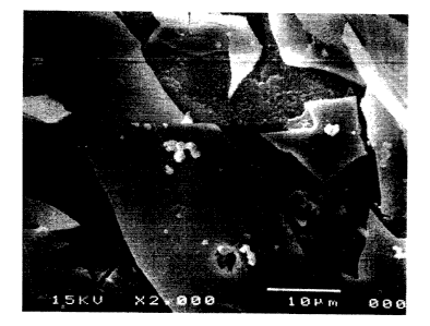
Figure l depicts a scanning electron micrograph of
silica-based glass immersed in simulated physiological
solution.
Figure 2 depicts energy dispersive x-ray analysis of
a nodule detected on silica-based glass immersed in simulated
25 physiological solution.
Figure 3 depicts the release of vancomycin, over time,
from granules and discs of pure silica glass immersed in a
~; ~lat~ physiological solution.
Figure 4 depicts the effect of concentration on
30 vancomycin release versus time.
Figure 5 depicts a comparison of the zones of bacteria
inhibition of vancomycin dissolved in simulated physiological
solution and vancomycin released from pure silica glass.
Figure 6 depicts the zone of bacteria inhibition size
35 versus immersion time and concentration of vancomycin released
from pure silica glass.
~VOg~i~3117 P~ 15~CI
21 9568~ --
- 14 -
Figures 7 a and b depict the relationship between
trypsin inhibiror concentration and release through 7 weeks and
4 weeks, respectively.
Figure 8 depicts the effect of incorporated content,
s 0.5 vs. l.o ~g, on the cumulative release of active TGF-~l from
granules dried to a 50~ weight 1CGS~
Figure 9 depicts the effect of the degree of drying,
50 vs. 70~ weight 105e, from granules loaded with l.o ~g TGF-
~1 .
10Figure 10 depicts the effect of SA/V, granules vs.
disks, loaded wlth 1 ~g TGF-~1 and dried to 50~ weight 108s.
Figure 11 depicts release of active TGF-~1, per time
period and cumulative, from disks loaded with .5~g and dried to
57~ weight loss (n=3).
15Figure 12 depicts an absorption isotherm of silica-
based glass rnn~;n;~g other oxides.
Figure 13 depicts FTI~ spectra of silica-based glass
c~ntR;n;ng other oxides before (lower spectrum~ and after
(upper spectrum~ immersion in SPS.
20Figure 14 depicte the release of trypsin inhibitor
from sol-gels c~ntR;n;ng Ca and F.
Figure 15 is an FTIR spectrum of a Ca-P sol-gel
containing trypsin inhibitor before and after immersion in a
tris buffered electrolyte solution.
Detailed Description
Utili~ing the method according to the present
invention, proteins and other biologically active molecules can
be incorporated into silica-based glass carriers in a way that
leads to sustained release of the added molecules and does not
destroy their function. Such a controlled release delivery
system can be used in implant materials, for example, to fill
in bony defects, including defects larger than three
centimeters without requiring an excessive quantity of growth
factors. Such a controlled release delivery system also finds
use in other applications with site-specific targeting needs
such as, for example, chemotherapy. The carriers can be
W096/0311~ 2 I q 5 6 8 0 ~ " l
synthesized under sterile conditions or can be sterilized
subsequently using conventional sterilization methods
Controlled-release carriers according to the invention
comprising antibiotics can be used in tissue culture for
5 preventing rnntAminRt;nn, particular that which develops upon
consumption of antibiotic added with medium, by contacting the
carrier with the culture through, for example, immersion.
Controlled release carriers according to the invention
comprising growth factors, in particular bone growth factors,
can be used to test the effect of the cnntinnRl, controlled
release of different factors on bone cells in vitro. It is
also contemplated that such carriers can be used for the
development of immortal bone cell lines in vitro.
Sol-gel derived processing can be done at low
temperatures -- i.e. approximately 40~C or below -- and low p~.
Both of these conditions can be important for m~;ntA;ning the
functionality of biologically active molecules incorporated
into the sol-gel matrix.
The advantages of sol-gel derived processing include
20 the following: l) a sol, which is a 5~Rr~nR;n~ of c0ll9i~Al
size particles, is in liquid form before it gels; 2) the whole
reaction can be done at room temperature; and 3) the
microporosity of sol-gel glasses can be controlled by, for
example, varying water content, timing of proton addition,
25 proton concentration, aging time, and drying time. The pore
sizes achievable with sol-gel processing in general are in the
nRn, -t~r range. During the liquid phase of the reaction,
proteins and other biologically active molecules can be added
to the liquid sol before it gels. These molecules then become
30 encased in the solid matrix. Because of the controllable
microporosity, a subsequent controlled release of molecule is
achieved.
As used herein, ~controlled-release carrier~ refers
to carriers for biologically active molecules, as defined
35 below, which provide for the release of the biologically active
molecules over time when immersed in solutions rnntA;n;ng, for
example, ions typical for interstitial fluid. An example of
~'0~6/031l7
21 9568Q
- 16 -
such a solution is simulated physiologic solution ~SPS), used
in some of the examples below. SPS is made by dissolving
reagent grade NaC1, K~1, NaHCO3, K2HPO;, CaCl2, MgCl" and MgSO~
in a 0.05 M Tris[hydroxymethly]~~; n~ n~ hydrochloride
(tris) buffered solution (pH 7.3 at 37~C) resulting in ionic
crncPntr~t;~n~ similar to plasma: Wa~ 2~M, K~=5mM, Ca~Z=2.5mM,
Mg~'=1.5mM, HCO,-=27mM, HPO4~2=lmM, and 0.5mM so~-7. Another
example is tissue culture medium.
As used herein, "bioactive" refers to a bone bioactive
10 material having a calcium phosphate rich layer present, or
which develops during appropriate in ~itro or in vivo
conditions. As observed by Pereira et al., ~. of ~iome~. ~at.
Res., 1199~) 28:693-698 (incorporated herein by reference),
pure silica gel having a porous hydrated layer i8 able to
induce a carbonated hydroxyapatite layer when soaked in a
simulated body fluid r~nt~;n;ng calcium and phosphate ions.
Pure silica hydrogels produced using TEOS and drying
temperatures of around 400~C were immersed in simulated body
fluids having different magnesium, calcium, and phosphate ions.
It was reported that apatite nucleation induction periods were
decreased with the addition of small amounts of calcium and
phosphate ions to the fluids, as well as increase in p~. Li et
al., J. Ap~l. ~omater., (1993) 4:221-229 and Li et al., ~.
Amer. Ceram. scc., (1993~ 75:2094-2097 Iboth incorporated
25 herein by reference~.
As used herein, "silica-based7 refers to the inclusion
of a silicon oxide in the composition of the glass. other
oxides may also be present.
As used herein, ~biologically active molecules" are
30 defined as those organic molecules having an effect in a
biological system, whether such system is in vitro, in vivo, or
in 8i tu. Biologically active molecules include, but are not
limited to, the following categories: growth factors,
preferably bone growth factors, cytokinea, antibiotics, anti-
inflammatory a~ents, analgesics, and other drugs. The term~type~ as used hereinafter in reference to biologically active
molecules re~ers to biologically active molecules of the
~'096/03117 2 1 95~80 P~
- 17 -
previously listed categories, as well as specific compounds,
i.e. vancomycin, T~F-~, etc. These specific c~ , In~q can be
in the same or different categories.
The term ~matrix" includes the solid framework of the
5 bioactive glass structure itself, as well as the pores. The
phrase ~incorporated within said matrix" denotes that the
molecules are inc~L~L~ted throughout the gla~s network.
The term "bony defect" refers to regions necessitating
repair including, but not limited to, fractures, areas of wear
10 and tear, holes resulting from removal of screws and pins,
replacements, periodontal applications, and deterioration of
bone due to old age or disease.
The term "implant" refers to a material for filling
bony defects as described above. The implant preferably
15 comprises a silica-based glass further comprising calcium. The
implant can be in the form of granules, discs, blocks, or
monoliths, and can comprise the controlled release carrier or
simply be coated with the carrier. The implant can also
comprise porous materials for use in bone surgery such as
20 porous hydL~y~dLite or, as described in W0 94/04657, porous
bioactive glass. The term also includes prosthetic devices
which, according to the invention, can have a coating, or
partial covering, of glass or bioactive glass having
biologically active molecules incorporated within the matrix.
25 Examples of such prosthetic devices include, but are not
limited to, hip and joint prostheses.
The implant can comprise a "cocktail" providing for
a combination of materials and/or release rates. The cocktail
can include a population of granules of different sizes, all
r~ntnining the same type of biologically active molecules.
Alternatively, granules r~nt~ining different types of
biologically active molecules can be combined. The granules in
such a cocktail can ~e the same size or different sizes,
thereby providing for the release of different molecules at
35 different rates. For example, a cocktail including
antibiotics, anti-inflammatory agents, and growth factors can
be prepared.
W0~6/03117 r~
21 956~ ~
It i8 also contemplated that two or more types of
biologically active molecules can be contained in each implant
material as defined herein. This can be effected by
simultaneous addition of the molecules into the solution.
5 Alternatively, implants cnnt~ining one or more biologically
active molecules can be prepared and then these implants can,
themselves, be coated with, or incorporated within, a solution
co~t~;n;ng one or more different types of biologic~lly active
molecules, and/or at different ~un~ellLL~tions.
The term ~'antibiotic" includes bactericidal,
fungicidal, and infection-preventing drugs which are
substantially water-soluble such as, for example, gentamicirl,
vancomycin, penicillin, and c~ph~lospnrins.
The term "growth factors" includes growth factors
identified as having osteogenic or osteoinductive properties.
Included among the many factors identified with the control of
bone formation are platelet derived growth factors ~PDGF), the
transforming growth factors (TGF-~), insulin-like growth
factors ~IGFsl, fibroblast growth factors (FGFs), and the bone
morphogenetic proteins (BMPs) . These growth factors are
present at the site of fracture healing in vivo and are
produced at the time of injury through platelet lysis (PDGF and
TGF-~) and by the resorption of bone matrix ~TGF-~ ar.d sMPs).
The individual factors will be discussed in more detail below.
~5 The term "contacting~ includes, but is not limited to,
contacting the carrier with the sample for which release of the
biologically active molecules is targeted through, for example,
immersion, implantation, and ~ ing.
The identification of osteogenic and osteornn~nrt;ve
30 growth factors has spawned the search for new graft substances
obtained through genetic engineering concepts. The controlled
delivery of these recombinant molecules, however, is important.
Growth factors with known effect on bone tissue must be
delivered at the site in sufficient doses to stimulate healing.
35 Glasses synth~; 7~d following a room temperature sol-gel-
derived ~Luccd~ are outstanding candidate materials for the
controlled release of such osteoinductive molecules. The
WO96/03117 pcT~rs9-clug4~l
~ 21 ~-~68~
- 19 --
processing of the glass allows one to control the ultrastruc-
ture of the glass such that the timing and quantity of release
are tailored to fit the specific therapeutic needs. In addi-
tion, these glasses can be osteoconductive, thereby providing
5 a substrate for bone tissue development.
The effects of the growth factors when exogenously
applied to in vitro and in vivo experimental models of bone
fnrr-tinn have demonstrated their biological properties.
~Cornell et al., supra; and Mohan et al., supra.)
Consequently, any material which affords the sustained delivery
of such factors is beneficial. Although most of the previous
studies clearly demonstrate the osteogenic and osteoinductive
effects of these proteins, the precise biological properties of
these growth factors with respect to the degree of bone
formation is greatly influenced by the following: the environ-
mental conditions of the experimental model, the timing, method
and dose of growth factor delivery, the hormonal milieau and
the synergy between the various growth factors. Thus, the
present invention provides a method to elucidate the effects of
these growth factors.
From a developmental point of view, the formation of
bone occurs in a series of discrete steps. Initially there is
a proliferative phase followed by cellular differentiation and
deposition of a collagenous matrix which in itself influences
25 subsequent expression of bone proteins. (NIri/AAOS sponsored
workshop, supra.~ Some workers view collagenous matrix
synthesis as a series of temporal events in which there is an
initial collagenous phase followed by a rise in ~lk~l;n~
phosphatase activity and the expression of osteonectin, bone
30 sialoprotein and osteocalcin. OSt~npnnt ~ n expression and
synthesis has been further dissected temporally in terms of
sulfation, phosphorylation and molecular size. Aside from the
proteins listed above, other studies have shown that at least
two forms of chondroitin sulfate proteoglycan are also synthe-
3~ sized by the osteoblast. These parameters can all be measuredby methods well known in the art. Some growth factors are
detailed below.
W0~6/(13ll7 2 1 9 ~ 6 ~ ~ , Ul,~
- 20 -
Insulin-like growth factor (IGF~ I and II are made by
bone cells as well as by other ti~sues throughout the body.
They are found in bone matrix and have presumably been secreted
by bone cells. ~Canalis et al., Calcified ~issue Int. ~1993)
53:S90-S~3; and Canalis et al., J. Bone Miner. ~es. ~1993)
8:S237.) In ~itro, IGFs have been shown to increase bone
collagen and matrix synthesis, to increase osteoblast-precur30r
replication and decrease bone collagen degradation. ~Xock et
al., Bndocrinolog,v (1988) 122(1~:254); and Mccarthy et al.,
Endocrinology (1989) 124(11:3~1.)
Growth hormone is thought to act through IGF in
stimulating bone growth, but it has also been shown to have
local effects on mesenchymal cell proliferation and
differentiation. ~Downes et al., ~. Mater. Sci.:Mater. Med.,
~1991) 2:176-180; and Silbermann, M., ~iomaterials, (1990)
11:47-49.) ~uman growth hormone has two molecular weight
species, one of 20,0~0 and the dominant species of 22,000.
Platelet derived yrowth factor ~PDGF), a polypeptide
of approximately 30kD in molecular weight, exists as a dimer
composed of two A subunits or two B subunits or as a
heterodimer of an A and a B subunit, creating three separate
forms of PDGF. These subunits are the products of two separate
genes. ~hile all three forms are found in bone matrix, only
PDGF AA is made and secreted by bone cells in vitro. P~GF s3
25 has been found to be the most active of the three ~orms.
(Mohan et al, 6upra.)
PDGF has been sho~n to have bone resorbing activity
in vitro; a number oi inve8tigators have reporSed increased
bone resorption in response to administration of physiological
30 doses of PDGF. ~Tashjian et al.~ Endrocrinology ~1582)
111:118-124.~ Additionally, PDGF has been shown to increase
osteoprogenitor cell replication.
Transforming growth factor-beta (TGF-~) is a family
of molecules which may have bone prcmoting properties for
fractures. TGF-~ is a h~ '; riC peptide with a molecular
weight of 25 kD. The most abundant sources of this molecule
are platelets and bone. This multifunctional peptide has a
WO96103117 re~ ,.C.~I
~ 2 1 q5680
- 21 -
broad range of cellular activities, including control of the
proliferation and expression of the differentiated phenotype of
several cell types specific to bone, among them mesenchymal
precursor cells, chondrocytes, osteoblasts, and osteoclasts.
(Beck et al., J Bone ~iner. Re5. (1991) 6(9~:961; Joyce et al.,
Orthop Clin. North Am. (1990) 21~1):199; and Joyce et al., J.
Cell Biol. (1990) 11016):2195.) Although it exists in several
distinct forms, two of these, TGP-~1 and 2, have been isolated
from bone in approximately a 4:1 ratio. ~n vivo studies based
10 on both i t~;ctochemical staining and in sit~ hybri~i7-tt;nn
have demonstrated the synthesis of TGF-~ by both thul.dLo~yLes
and osteoblasts and the accumulation of TGF-~ in models of
endochondral ossification. (Joyce et al., Orthop Clin. North
Am. (1990) 21tl):199; and Joyce et al., tJ. Cell Biol. (1990)
15 110(6):2195.) In a study in which TGF-~ 1 or 2 was introduced
by daily injection into the subperiosteal region of newborn rat
femurs, (Joyce et al., J. Cell Biol. (1990) 110(6):2195)
demonstrated that me~llch~, 1 precursor cells in the periosteum
were st;r~lated by TGF-~ to proliferate and differentiate in
20 much the same manner as that which is observed in embryological
bone formation and early fracture healing. After the tt~qA~inn
of injections, t3n~,.B.",.l.~l ossification also occurred,
resulting in the rt~pl~t t of cartilage with bone.
The implantation of a bone morphogenetic protein (BMP)
25 solution leads to a series of dev~lt~ ~tl processes including
chemotaxis, proliferation, and differentiation, which result in
the transient formation of cartilage and its replacement by
living bone tissue complete with hematopoietic marrow. (Urist,
M.R., Science ~1965) 150:893-899.) Several newly discovered
factors, BMP-l through 7, and osteoinductive factor (OIP) have
been implicated in the BMP process. sMP-2 through 7 are all
members of the TGF-~ superfamily of molecules and are closely
related to two factors Vgl and DPP which are involved in a
variety of developmental processes during '_yugt-llesis. Both
35 sMP-2A and BMP-7 have been expressed as recombinant proteins
both of which have been shown to clearly induce the entire
cartilage and bone formation process seen with bone-derived BMP
WO9h~311~ /u~,./.~SI~I
2 1 ~
- 22 -
solutions. ~Wozney, J.M., Prog. Growth Factor ~es. ~g89)
1~4):267.) At the present time~ two BMPs: BMP-aA t~.erhart et
al ., Clin. Orthop. (19g3) 31~; Wozney et al., Science ~1988)
242(488~):1ri28; and Yasko et al., ~. ~one ~oint Surg. ~Am>
5 ~Aug. 1992) 74(7):1111 and ~. Fone ~oint. Surg. cAm~ ~1992)
74t5):659) and BMP-7 ~Sampath et al., ~. Biol. Che~. ~1992)
267(28):20352) ~also known as OP-l) have been demonstrated to
increase bone formation at extraosseous sites, and to enhance
fracture healing. ~Gerhart et al., Cl~n. Orthop. ~19931 317.)
Purified BMP has been utilized in femoral and tibial non-unions
in uncontrolled clinical trials. t~ohnson et al., Clin.
orthop. (1988) 230:257-265; Johnson et al., Clin. or~hop.
(1988J 236:249-257; and Johnson et al., Clin. Orthop. (1990)
234.)
Current state of knowledge suggests that the local
growth factors most likely to increase fracture healing
significantly are PDGF, TGF-~ and BMP-2.
Maintenance of function of growth factors after
incorporation within the silica-based glasses can be tested
20 using the aforementioned technlques for determining bone
differentiation. The --;nt~n~n~e of function of antibiotics
can be ascertained using standard disc susceptibility tests
such as are described in ~ntlhi~tics in l.nhoratorv M~iC;n~
3rd P~;tion, V. ~orian, ed., chapter 2, Williams and Wilkins,
Baltimore, Md., 1991 (incorporated herein by reference).
Function of incorporated anti-;nfl t~y agents and
analgesics can be ascertained by, for example, testing for
inhibition of prostagl~n~;n synthesis in cell culture.
30 Sol-gel-derived glass ~ynthesls
Pure silica and calcium ~n~in;n~ glasses have been
synthesized with biologically active molecules incorporated
therein. Briefly, a silicon alkoxide ~e~uL~v~, preferably
tetramethylorthosilane (TMOS), in pure solution is combined
with ~ n;7ed water and stirred by magnetic or ultrasonic
means. The water to TMOS molar ratio affects porosity and
specific surface area of the gels, which, in turn, affect
WO~6J03117 r~ J~,r,~
21 '~680
- 23 -
bioactivity. As both increase, 50 can bioactivity. To
increase both, water is provided in amounts exceeding
stoichiometric, or in an H2O/TMOS molar ratio ranging from
about 6:1 to about 20:1. In a preferred : '~'; ' the molar
5 ratio of H2O/TMOS is 10:1. Alcohol, preferably methanol, can
be added at an alcohol/TMOS molar ratio of from about 0:1 to
about 1:1. Acetic acid (0.1 N) or HCl ~0.1 N) can be used as
a catalyst for the hydrolysis reaction, and i8 added to
r-;rtA;n the desired pH, as disclosed below.
Calcium methoxyethoxide (20~ solution in
methoxyethanol, Gelest Inc., Tullytown, PA) can be used as a
calcium alkoxide source. Calcium methoxyethoxide ~CME) is
added in an amount sufficient to result in a final percentage
of up to about 40 ~ by weight calcium oxide upon drying of the
15 gel. Triethyl phosphate can be used as a phosphorous p~ntn~
source. Triethyl phosphate ~TEP~ can be added to achieve a
final concentration of phosphorous pentoxide, P2O5, up to about
10 ~ by weight upon drying. Weight percentages throughout are
calculated based upon the reactions going to completion and
20 complete drying. The water, TMOS, and acid are mixed using
sonication in an ice bath, or magnetic stirring, or a
combination of both. When a calcium alkoxide is present, the
TMOS, calcium alkoxide and additional Alkn~ c, if any, are
preferably mixed under non-a~ueous conditions under an argon
25 atmosphere using either magnetic stirring or sonication for up
to about one hour.
Alternatively, a calcium salt can be used in place of
a calcium alkoxide. It has been found that the use of a
calcium salt can extend the time to gelation, thereby affording
30 a longer time for incorporation of the biologically active
molecules and, concomitantly, a ~ ; - c distribution of the
biologically active molecules. An extended time to gelation is
also helpful for the coating of implant materials with the
carrier. In a preferred c~o~; -t, the calcium salt is CaCl2.
35 The calcium salt is added in an amount su~ficient to result in
a final percentage of up to about 40 ~ by weight calcium oxide
upon drying of the gel.
WO 96~0311~ i.,,J,l~S 1-1
~1 95~0
Since the biologically active rolec~ c to be
incorporated retain their biological activities after treatment
in moderate to highly acidic conditions, an amount of acid
necessary to maintain acidity in a range of pH from about 1-
4.5, preferably about 1.5-3, prior to, or during, incorporation
of biologically active molecules is used.
The biologically active molecules to be incorporated
are added at concentrations resulting in final ~Qn~ntrAtions
ranging from about 0.0001 to about 10 ~ by weight of the glass.
lC Glasses with compositions of silicon in the range of
60-100% (by weight) with the L~ ;n~r as other oxides can be
prepared. The liquid sol can be cast into a poly~yL-nc
container. The sol is aged and allowed to gel in a sealed
c~ntA-n~r. Aging can take from about one (1) day to about four
(4) weeks. Drying can be performed for a time of from about l
to about 14 days.
In order for sol-gel derived gla85 to be an effective
carrier for biologically active molecules, the process should
be carried out at a low t~ _ ~nre (about 2-40~C) and, in the
20 case of pure silica glas~, the acidity of the sol should be
between pH l and 4.5. ~emperature, sol pH, ~ calcium content,
water to TMOS molar ratio and other factors affect the gelling
time of the sol. However, when incorporating biologlcally
active molecules, the gelling time of the sol should allow
25 enough time in the liquid state to enable the addition of the
solution of biologically active molecules for incorporation,
cast, and homogeneously mix the sol. Gelation occurs when
enough cross-links have formed such that the network spans the
length of the ~ntA~n~r~ Gross observation reveals little or
30 no movement of the cast materlal upon inversion.
A lower pH increases the gelling time. A higher
calcium content decreases gelling time. A higher gelling time
is desirable in order to see more of the sol-gel reactions
going to completion, thus ending with a final material with
less porosity and smaller pore size. ~ess porosity also means
a more mechanically strong material with longer times of
protein release. However, there are instances when greater
WO9G103117 r~ ,5/~
2 ~ 9568~
- 25 -
porosity may be desirable, for example, achieving a more rapid
release of molecules, or a more rapid degradation of the
carrier. Larger pore sizes facilitate the release of larger
molecules through diffusion.
A lower tC.. ~L~tUl~ also increases gelling times. To
achieve lower temperatures, the reaction is then carried out in
an ice-cooled water bath. A higher water content will
decrease gelling time for most metal ~lk~ri~c, although the
porosity may stay high due to increased water evaporation from
10 the material. Conditions are selected such that gelation
optimally occurs within a period ranging from at least about 30
minutes to about 48 hours for incorporating biologically active
molecules. Gelation can be performed at temperatures ranging
from about 0~C to about 40~C.
Aging of the sol-gel occurs after casting and is
performed by keeping the casting container sealed. Sol-gel
reactions continue nni, ~~ ~ during this period. Aging can be
performed at temperatures ranging from 0~C to about 40~C.
~onger aging times (of up to 1 month) result in a more
20 mechanically strong material, which undergoes less cracking
than materials that have been aged for le6ser time periods.
Aging at a lower temperature, such as 4~C, also extends the
gelling time.
Drying temperature and time can also affect the final
25 material characteristics. A fast rate of drying can produce
cracks in the final material. The final material loses about
50-80% of its weight between casting and final drying due to
evaporation of water, and alcohols produced as by-products of
the reaction. Drying is performed at temperatures ranging from
30 about 15~C to about 40~C by unsealing the casting ~nt~;n~r,
and can be performed at ~t _L'h~riC pressure, or pressures
lower than atmospheric.
As is evident from Figures 3, 7, 8, and 14, the
release kinetics of the biologically active molecules in the
35 early stages of immersion, i.e. from about one day to seven
days, is higher than those in the later stages. At about seven
days after immersion, a major change in the slope of the curveq
W0961~3117 P~-t~,~,C ~
21 9~680
- 26 -
is observed, representing a major change in rate of release.
The early higher release is not a 1'burst" effect as pre~iously
reported by several authors ~cited above). This higher early
release i9 advantageous when a dual treatment regimen i5
imposed -- an acute treatment at a high dose, followed by a
"chronic" lower dose. In cases when a steady state release is
desired right from the onset of the medical treatment, i.e., a
release without major changes in rate, the sol-gel carriers can
be treated by immersion at the time of pro~ tinn such that the
intitial higher release phase has taken place before actual use
in the patient.
Exampl~ 1
Synthe~is of 80l-gel~Yancomycin Composite
A sol-gel derived silica-based matrix-vancomycin
lS composite was synthesized employing a room-temperature, low
acidity, low alcohol concentration procedure. ~ancomycln waR
selected as the drug to be released due to its proven efficacy
against gram positive cocci, especially staphylococci, which is
a major cause of osteomyelitis. vancomycin is a water soluble
(up to lCo mg/ml~ tricyclic glyceropeptide of approximately
3,300 molecular weight.
The ~material was prepared as follows: l9.6 ml
tetramethylorthosilicate (TMOS, Aldrich, St. ~ouis1 MO,
U.S.A.), 14.2 ml water, 5.2 methanol and o.Ol ml of lN HCl was
sonicated in a glass beaker in an ice bath for 30 minutes.
Then, 4 ml of the 801 was cast into 23 mm diameter polystyrene
vials (Sarstedt, Princeton, N~) and l ml of lO mg/ml vancomycin
HC~ (Lederle, Carolina, Puerto Rico) was added to the 8018 in
the vials and the samples were mixed. The same amount of
30 water, i.e. l ml, was added to control samples. The total
H20/TMOS ratio was lO:l. The methanol~TMOS ratio was l:l. The
amount of incorporated vancomycin to sample weight was about
l~. The vials were sealed with airtight caps, gelled, aged,
and dried at room temperature. Time to gelation varied from lS
to 25 hours. Addition of the vancomycin solutions did not
change significantly the time to gelation.
W09~/0311~ 21 9 5 6 8 ~ r~
.
- 27 -
After aging for 2 weeks in the sealed ~n~;nonS, the
sols were exposed to air for drying. During drying,
evaporation of liquid from the gel pore network reeulted in
weight loss and shrinking of the gels. The weight loss
c~n~;nu~ up to 2 weeks. Drying was considered to be complete
when the weight loss reached 75-78~. The significant weight
108s and shrinking did not produce visible cracks. The
resulting products were transparent monoliths in a shape of 11
mm diameter and 8 mm high cylinders weighing 1.1 gram. The
10 density of the dried gel material was equal to 1.5 g/cm3.
Since 10 mg vancomycin was incorporated into each of the discs,
the vancomycin content in the material was 0.91~. There is no
reason to expect that other water-601uble antibiotics will
behave any differently.
15 Exa~ple 2
V ,~in Release Study
For the in vitro vancomycin elution study, a part of
the monoliths was crushed, ground, and sieved to obtain either
small granules in a size range from about 500-700 ~m, or large
20 granules of about 5 x 5 x 2 mm. The rest of the monoliths were
tested as discs.
The synthesized vancomycin composite was immersed into
a simulated physiological solution (SPS) with ion content
similar to that of plasma as ~; ~rl os~d previously. To
25 determine the effect of the sample surface area to volume
(SA/V~ ratio, the material used for the immersion experiments
was shaped as follows: small granules of 500-700 ~m (SA/V
approximately 10 mm1), large granules 5 x 5 x 2 mm (SA/V = 1.5
mm~l), discs 11 mm diameter x 4 mm (SA/V . 0.85 mm~~), and half-
30 cylinders 5.5 mm x 4 mm ~SA/V = 1.2 mm-l).
All the samples were immersed at the same vancomycin
content in sample/solution ratios equal to 1 mg vancomycin per
1 ml. The immersed samples were incubated at 37~C for time
periods ranging from about 1 hour to about 3 weeks. The
solutions were totally ~Y~h~nge~ at the following time periods:
1 hour and 1, 3, 7, 14, and 21 days.
~109610:}117 P~ I/U,. ''~
2 ~ 9568~ --
- 28 -
The released vancomycin concentrations were measured
using an automated Fluorescent Polarizing T -Rcay system
lTDxR system, Abbott Diagnostics, Irving T~J. The results of
the vancomycin release assay are presented in Figure 3 and
summarized in Table I below. In Pigure 3, open circles
represent the small granules. Open triangle9 represent the
large granules. Open squares represent the 11 mm diameter
discs. Open inverted triangles represent the 5.5 mm dlameter
discs.
TABLE I
~r
~elease Time Released/Incorporated
Sample (days) Vancomycin
Small Granules 6 100
~arge Granules 21 55
Disc (SA~V = 1.2 mm~l) 21 48
As indicated by the foregoing data, the v~ in
release rate was affected by the material shape, i.e. the
material surface area to volume ratio. Specifically, the
vancomycin release from the small granules was very rapid and
most o~ the incorporated ~ in was released during the
first day of immersion. In contrast, the large granules (SA/V
= 1.5 mm~l) and discs (SA/V = 1.1 or 0.8 mm~l) showed a
continuous vancomycin release, which started at one hour,
gradually increased to a maximum, then slowly decreased,
tailing-off up to 3 weeks later. The maximum vancomycin
release was measured during the period of immersion hetween
three days and one week.
These findings indicate that the SA/V ratio can affect
the release of materials. Comhination of the materials of
varying shape, i.e. varying SA/V ratio, can provide a con-
trolled vancomycin release which starts upon immersion andcn~tinn~s for up to one month.
Exa~ple 3
Effect of V - I~.in concelltration
W096/03117 ~ ~Jv~
~ 2 1 956~0
~9
The sol-gel derived silica based matrix-vancomycin
composites with varying vancomycin content were synthesized.
The sols were prepared as disclosed above in Example l. Then,
1.2 ml of the sol were cast into 23 mm diameter polystyrene
5 vials. The cast sols were divided into two groups and 0.3 ml
of solutions with different vancomycin concentrations were
added to the cast sols of both groups in order to keep the same
H20~TMOS molar ratio of 10:1. The amounts of the incorporated
vancomycin were 10 and 20 mg for groups 1 and 2, respectively.
10 The percentage of va~ cin to sample weight was equal to 2.8
and 5.5~, respectively. The sols were gelled, aged, and dried
to about 75~ weight loss.
Ultrastructure parameters of the sols such as specific
surface area (SSA), average pore size (PS), and pore volume
(PV) of the dried sols were determined using the monolayer gas
absorption technique (multipoint B.E.T., Quantachrome). The
measured values were as follows:
SSA, m2/g545
PS, nml.8
PV, cc/gO.45
The obtained sol-gel derived discs, 11 mm diameter x 2 mm, with
SA/V ratio equal to 1 mm~l, were subjected to vancomycin release
study as disclosed above in ~xample 2. The discs were immersed
into 5 ml SPS. The vancomycin content in sample (total weight
25 of vancomycin) to solution volume ratios (Wv~V) were 2 and 4
for groups 1 and 2, respectively. The concentrations of
released vancomycin were measured as described above in Example
2. The results of the study are presented in Figure 4. In
Figure 4, solid bars represent vancomycin at lo mg
incorporation. Hatched bars represent vancomycin at 20 mg
incorporation.
The data show that the amount of released vancomycin
increased with the amount of incorporated drug. Thus, the
released amount appears to be a function of the incorporated
35 quantity (at conditions otherwise equal). However, the drug
release profile over time appears to be similar for different
concentration. Specifically, the drug release started right
~0~6/03117 r~u~
21 ~5~0
after immersion, reached a maximum by 3 days, then gradually
decreased.
Exa~ple 4
In vltro sacterla Inhibition T-st
The SPS solutions with varying contents of vancomycin,
released from the sol-gel derived silica-based matrix from the
experiments described in Examples 2 and 3, were tested for
susceptibility of Staphylococcus aureus bacteria to the
released drug. The standard disc susceptibility test technique
lO was applied (See ~orian, supra.). The sample SPS solutions
with vancomycin released during immersion were tested and
compared with standard solutions of vancomycin in SPS with
concentrations ranging from lOO to lOrOOO ~g/ml. Concen-
trations of the sample SPS solutions with vancomycin released
lS during immersion were measured using the Fluorescent Polarizing
T ~c~y described previously. Single, twenty ~l aliquots
of each solution ~either standard or sample) were deposited
onto l/~2 inch filter paper discs (#74Q-E, Schleicher ~ Schnell,
Keene, NH). The drug solution impregnated discs were then
dried and stored in a desiccator at 4~C. A blood agar plate
inoculated with Staphylococcus aureus (ATCC 25923) was obtained
from the Microbiolooy ~aboratory, Hospital of the University of
Pennsylvania. A l.5 x lO~ CFU/ml suspension of bacteria in
0.45~ saline was created to match a McFarland E~uivalence
25 Turbidity Standard 0.5 (~emel, Lienexa, ~A), Mueller-Hinton
agar plates, 15 x lOO mm (~odel 01-620, Remel, Lienexa, KA)
were inoculated with lO ~l of the Staphylococcus aureus
suspension by streaking with a sterile swab soaked in the
suspension over the entire agar surface to ensure an even
distribution of inoculum (standard inoculation pLvcedu~ A
vancomycin impregnated disc was placed in the center of each
agar plate. The agar plates were then incubated in a
humidified air environment in a single-chamber, water-jacked
incubator ~Model 3159, Forma Scientific, Marrietta, OH) at
37~C, for 24 hours. Zones of bacteria inhibition were measured
~09~/~3117 r.llL~
~ 2 ~ ~568(~
- 31 -
using a caliper with a precision of 0.1 mm. The data are
presented in Figures 5 and 6.
The measured zone of inhibition sizes plotted against
vancomycin concentration in a logarithmic scale of vancomycin
5 released from sol-gel, as disclosed in Example 3, are presented
in Figure 5. In Figure 5, open circles represent vancomycin
dissolved in SPS. Closed circles represent vancomycin released
from the sol-gel carrier.
The discs, impregnated with 30 ~g of vancomycin,
10 either dissolved in SPS or released from the silica-based
matrix, exhibited a zone size greater than 12 mm. According to
the Zone Diameter Interpretive Standards (Lorian, supra, Tab.
2.1.), a zone of that size indicates that bacteria are
susceptible to the material, and the equivalent minimum
inhibitory concentration breakpoint is less than 4 ~g/ml. The
concentration-zone of inhibition relationship for the sample
solutions of vancomycin released from the silica-based matrix
showed a close fit to that of the standard solutions.
Figure 6 shows zone of bacteria inhibition sizes
20 versus immersion time and concentration of vancomycin
incorporated into the sol-gel derived silica matrix. Open bars
represent vancomycin at 1 mg. Solid bars represent vancomycin
at lo mg. Cross-hatched bars represent vancomycin at 20 mg.
The data demonstrate that vancomycin released from the sol-gel
25 matrix was effective to inhibit the bacteria growth up to three
(3) weeks (at 20 mg). The measured zone of inhibition sizes
appear to increase with concentration of incorporated
vancomycin, reflecting larger quantities of released
vancomycin.
The foregoing experiments demonstrated the following:
incorporation of ~n~ ~cin into the silica-based matrix using
the sol-gel technology provides a controlled drug release over
time, starting upon immersion (and thus implantation) and
continuing for at least 3 weeks; and the employed room tempera-
ture, low acidity, low alcohol concentration sol-gel procedure
did not alter the vancomycin properties since vancomycin
released from the sol-gel derived material is as effective in
F~, IIV~ IC 1
~'096/03117
21 956~ ~
- 32 -
inhi.biting bacteria as vancomycin sol~tions that were not
obtained from a sol-gel carrier.
WO~61031l~ P ~
2 1 95680
Example 5
Synthesis of Sol-Gel/Trypsin Inhibitor Composite
Sol-gel derived glass discs with trypsin-inhibitor
incorporated inside their matrix have been successfully
synthesized. Trypsin Inhibitor ~SIGMA) is a protein with
molecular weight of 21 kD. The sol-gel/protein composite
contains 1-10 mg Trypsin Inhibitor ~TI) per 150-200 mg disc.
Protein elution was measurable in samples with 2 mg or greater
of protein per disc.
The pluCd~u~ used to synthesize 1 gram ~by dry
weight) of the sol-gel derived glass was as follows: 2.48 ml
of TMOS ~Aldrich, St. ~ouis,MO~ was combined with 2 ml DI water
and 0.68 ml of methanol in a 30 ml beaker and mixed for 5
minutes using magnetic stirring. This resulted in an H~O/TMOS
15 molar ratio of 10:1 and a methanol/TMOS molar ratio of 1:1.
Then, 0.01 ml of lN HCl was added to catalyze the sol-gel
reaction. This results in a clear one phase solution which is
stirred for 15 minutes. The sol was cast in 0.8 ml volumes
into polystyrene containers and the trypsin inhibitor solution
20 was added in a volume of 0.2 ml of 0.1 N acetic acid solution
with protein concentration in the range of 1-10 mg/ml. The
solution was mixed with vortexing and the r~nt~inPrs were
capped. Gelling occurred within 1-4 days after casting. The
capped sol was allowed to gel and age for times ranging between
1 day to 2 weeks ~pPn~;n5 on the desired porosity at room
temperature. After aging, the sol-gel was allowed to dry at
either room temperature or 37~C by uncapping the casting
cnnt~;nPr. Any liquid produced was decanted off the solid. A
higher drying temperature increased the porosity and rate of
shrinkage. After drying, the resulting solid had lost 60-70~
of its original weight due to evaporation of water and alcohol
(methanol is a by-product of the sol-gel reactions).
The resulting solid material was a porous three-
~; nc;nn~l network~polymer of silica which releases
incorporated biomolecules in a controlled release fashion. The
various processing parameters are depicted in Table II. Sample
designations are of the Eormula SxxxCxxPxx (date cast), where
WO~/03117 ~ .,''C5tO1
2195~8~ ~
- 34 -
~Sxxx~ is the calculated 56 silica, RCxx" is the calculated %
calcium oxide, and "Pxx~ is the calculated % phosphorous
pentoxide. The date cast is presented as the "last two digits
of the year.month.day." The trypsin inhibitor content and form
5 of the sample is indicated in the basic Eormula TI=X-[sample
shape] where "X'~ i8 the amount of trypsin inhibltor in mg per
sample. The effect of pE~ on gelling time is apparent from
Table II.
TAsL II
I'romslnR rcnum~ l/nltf
S2mple S100 S100 510D S100 S100
~ubolutory Ddlm~6On) ~94.4.18~(94.4.18~~94.4.18) (94.4.18)194.4.18)
Tl-nwuntoftrypsbl 11-2- n=3- Tl-S- 7i--7~- li=lD-
lnbibitor in mS lu-nules]i4numlcslIf-nmrJ~ r~n~nulesljg~udes
Wa~erAMOS f~do 10 10 10 10 i0
Medtumbl~iOS Rrdb
Add Ct4dyst ml Jnd N0.1 mJ I0.1 ml t 0.1 ml I0.1 ml 1 0.1 mi I
(amourd Imd co~ntndimtl l~t RCIN lla N HCI 1~1 PtCI N RCI
Y~Silicnckr4tcd 100 loo 100 100 i00
Y. Calclum O~ide 0 0 0 D 0
cdcul~d
% Phasphcuous Pcnto~ide o o o o o
catculated
pll ~s c st 2.3 2.4 2.8 3 3.2
t3ellin~ Time houni 168 144 120 72 24
Volttme catt ml
Wci8hlascns~ mg 10D3.31009.2 1007 9972 Y60.g
Tottl Pwtein nt s4mple ~ mLt 2 3 5 7.5 10
A~m3 rlmc dnys 7 7 7 7 7
3 0 Dryin~ rlme ~ dys 5 5 5 5 5
Dryin~ Tcmperatme C 20 20 t0 20 2D
~degrees cenb~de~
U'et4ht nRcr drYine m4 3B4 8 3~02 382.5 391.4 363
'6 Wci4bl kss ~ Y. 61.6 613 62 60.8 62.1
35 Example 6
Trypsln knhlbitor Release
The initial release kinetic studies were carried out
by immersing the sol-gel/protein composite ~lO0 mg ~ol-gel/l mg
protein for each sample) in deionized ~ater inside containers
WO96/03117 r~ s.,s.~. I-l
~ ~ 21 956~
- 35 -
which were siliconized ln order to reduce protein binding. The
protein content in water was measured at different time points.
The water was replaced fresh after each time period. The
collected fluid that had been in contact with the 801-
5 gel/protein composite was analyzed for protein content using acolloidal gold/spectrophotometric method (Integrated Separation
Systems, Natick, MA, Stoscheck et al., Anal. ~iochem., (1987)
160:301-305, incorporated herein by reference) with sensitivity
down to 0.5 ~g/ml. The results are depicted in Table III
10 below.
The numbers in the table represent protein release in
~g of trypsin inhibitor after immersion in DI water. Results
for each time point are provided together with Il~tive
protein release.
T~8rS III
Pro ein Rel - -~ ed pg
I~mer~ion T~me: 3 day~ I veek 2 voo~ 3 ~eek~ ~ vee~
Sample nn~ n~ ~
5100(94.4.18~TI2 75 38 20 16
cumulative release: 75 111 133 149
S100~94.4.18)TI3 115 56 33 27
cumulative release: 115 171 204 231
5100~94.4.18~T}5 7s 82 48 30 33
cumulative release: 75 157 205 235 268
5100~94.4.18)TI7.5 142 97 70 68 78
cumulative releaae:142 239 309 377 g55
S100~94.4.1B~TI10175125 80 55 67
cumulative release: 175 300 380 435 502
For key see Table I.
The protein release kinetics of the samples and
30 results listed in Table II are depicted in Figure 7b. Protein
release was measured for a period of four weeks. "T12N (open
sc,uares) represents TI=2 from the table. "T13" (open circles)
represents TI=3 from the table. "TI5N (filled squares)
represents TI=5 and T17.5 (filled circles) represents TI=7.5.
"TI 10" ~filled s~uare within open square) represents TI=lo
wo g6/03ll7 r~ ~u~
2~ q568(}
- 36 -
sample S100 (~4.4.18~. All samples were in the form of
granules ha~ing a diameter less than about 2 mm.
As can be seen from Figure 7a, trypsin inhibitor was
continually released from all samples for a period of at least
seven (7~ weeks.
Example 7
Bioactivity of Pure Silica Glass
Sol-gel derived glass with a composition of 100%
silica and water/TMOS molar ratio of 15:1 was synthesized and
its bioacti~ity tested in vitro in SPS by measuring changes in
calcium-ion ccncentration. A 5 gram sample was made by
_ ' ining 12.38 ml of TMQS with 8.87 ml of DI water and
St~n~r~ting for 5 minutes in an ice cooled bath. To this
mixture, 8.87 ml of 0.1 N Acetic Acid was added and the mixture
sonicated for an additional 15 minutes. Then, 4.43 ml sodium
phosphate (.01 M, pH 7~ buffer was added and the mixture
sonicated for one minute. The liquid sol was cast as 3 ml
samples. The pH of the sol as cast was 4.5. The gelling time
was approximately 2 hours. Aging of the samples was done at
room temperature, for 1 day. Samples were dried for 3 days at
37~C and weighed about 500 mg.
Samples in the form of discs approximately 1 cm in
diameter and 4 mm high (1.76 cm~ SA~ were then immersed into
sps (17.6 mll for a sample Eurface area to immersion solution
~olume ratio of 0.1 cm~1. Samples were immersed for two weeks
with constant stirring at 37~C. SPS co~centration of calcium
normally averages 100 ppm. After 2 weeks of immersion of
samples, the average concentration of calcium in the retrieved
SPS averaged 25 ppm. This indicates that calcium was consumed
30 by the glass from solution, most likely by forming a calcium
ph~sph~t~ layer on its surface.
Example 8
Bioactivity of Silica-Based Glass ~nt~ni~ Other Oxides
Sol-gel samples with a composition of 65~ SiO2, 30
CaO and 5~ P20~, by weight, were made by combining 1.61 ml TMOS,
~VO96/03117 rc~ o. 1~
2 1 q~)680
5.04 ml 20$ calcium methoxyethoxide solution in methoxyethanol,
and 0.12 ml triethyl phosphate, and magnetically stirring for
5 minutes at 4~C. To this mixture, 1 ml 0.1 N HCl was added
to mimick the conditions for incorporation of proteins, and
stirred for an additional minute, for a water/TMOS molar ratio
of 5.13. Four, 1 ml samples were cast and the 301 gelled in
about 5 minutes. These samples were aged for 3 days, and then
dried for 4 days at room temperature. Samples after drying
weighed about 600 mg.
Sol-gel samples were then immersed into 12 ml of SPS
to test for calcium ~hnCph~te surface layer formation. Samples
were retrieved after being immersed 5 days in SPS with constant
stirring at 37~C. The samples were viewed using scanning
electron microscopy ~SEM) and surface analysis was performed
15 using energy dispersive x-ray analysis (EDXA). The surface of
the samples cnn~;n~ nodules 1-3 ~m in diameter (Figure 1)
that, when analyzed with EDXA (Figure 2), cnnt~;n~d high
proportions of calcium and phosphorous. This ; n~ t~p the
formation of calcium phosphate nucleation sites as a precursor
to calcium phosphate layer formation.
Example 9
synthesis of Sol-Gel/TGF-~ Composite
Recombinant human transforming growth factor beta
(TGF-~1) was incorporated into pure silica sol-gel glass. The
25 sol-gel was synthesized by , ining 5 ml TMOS with 5.4 ml of
water, and methanol, for a water/TMOS/methanol molar ratio of
9:1:1, and m-gnPtic~lly stirring for 5 minutes at room
temperature. 10 ~L of lN HCl was added to the mixture and the
sol was stirred for 30 minutes. Then, 0.9 ml of the sol was
30 cast into a polystyrene container and 0.1 ml of TGF-~ solution
with l$ bovine serum albumin ~BSA) to prevent non-specific
binding of the growth factor to the casting ~nnt~;n~r was
added. Different quantities of TGF-~ were added to each sample
cast ranging from 0.5 ~g to 2 ~g of TGF-~ for each solution
35 added with 1$ BSA. Samples were aged for 3 days at 37~C and
WO 96~03117 r~,l~.,..,.5~,, 1~1
21 9~6~ ~
- 38 -
dried at 37~C until they had lost approximately 50% of their
"as cast" weight.
Example lO
TGF-~l Releaso
Sol-gel derived silica glass was 9ynthesized by mixing
and stirring TMOS, DI water, and lN HCl in a molar ratio of
l:lO:Q.OOl. Then, 0.9 ml of the sol was cast into 15 ml
diameter polystyrene vials and O.l ml of a solution ~nnt~;ning
either 0.5 or l ~g of TGF-~l was added to the sol samples.
lO TGF-~l (Celtrix Lab., Inc.1 was prepared according to
in6tructions supplied. Briefly, aliquots from a 2.347 ~g/~l
TGF-~l stock solution were rPR"~rP"~d in 10 mM XCl following
lyophilization in 1~ bovine serum albumin (BSA) (Sigma Chemical
Co.). The resulting TGF-~l solutions were stored in l.5 ml
15 microcentrifuge tubes (USA Scientific) at -70~C until use. The
pH of the sols at the time of casting was measured to be 1.7.
The vials were sealed and the sols were allowed to gel (lS
hours) and age (24 hours) in an incubator at 37~C. Then, the
gels were dried to either 50 or 70~ weight 10~8 resulting in
20 transparent glass disks about lO mm in diameter. Part of the
samples of each group was crushed to produce granules in a size
range from about 500 to about lOOO ~m. A total of ~ sets of
silica glass/TGF-~l composites were prepared as follows: l ~g
dose: disk6 and particles dried to 50~ weight los~, and
Z5 particles dried to 70~ weight loss; 0.5 ~g dose: particles
dried to 50~ weight loss. ~n additional six silica glass disks
cn"t~ining 0.5~g TGF-~l and two controls (without TGF-~ll were
prepared and dried to 57~ weight loss. The disks had uniform
dimensions with an average diameter of 10.17 mm and an average
30 height of 4.93 mm.
The release of TGF-~l trom the sol-gel derived silica
glass particles and disks was measured by immersion in l ml of
sterile phosphate buffered saline (P~S) ~n~1n;ng 1% BSA.
Prior to immersion all the samples were sterilized by UV
irr~ tinn. The BSA prevents the non-specific binding of TGF-
WO 96/1)3117 r~ ,,,..'U5 ICI
21 95~80
- 39 -
~1 to the immersion reservoir. Concentration was determined
using an enzyme linked immunosorbent assay (EBISA).
The amount of active TGF-~1 released from the sol-gel
materials was assessed using the MvlLu mink lung epithelial
cell inhibition assay. This assay determines TGF-51 activity
based on its inhibition of MvlBu cell proliferation as
measured by [3H]-thymidine incorporation. ~ennings et al.,
"Comparison of the biological activities of TGF beta 1 and TGF-
beta 2: Differential acti~ity in endothelial cellsn, J. Cell
10 Physiol. 137:1~7-172 lg88. Confluent MvlBu cells ~ATCC) were
lifted from tissue culture flasks using Cell Dissociation
Solution (Specialty Media) and plated in Corning 24-well
polystyrene tissue culture dishes. Cells were plated at a
density 4.0xlO~ cells/well in 1 ml of Dulbecco's Modified
15 Eagle's Medium (DMEM) sùpplemented with 1% fetal bovine serum
(FBS) (Hyclone) and 50 ~g each of penicillin and streptomycin
(Sigma Cell Culture). Dishes were ;nn~h~t~d at 37~C and 5~ C0~
for 24 hours to allow the cells to adhere to the bottom of the
wells.
Following ;n~hatinnl the wells were aspirated and
treated with media containing TGF-~1 of known picomolar (pM)
concentrations as well as lyophilized 1% BSA in lO mM HCl to
serve as the control. The concentrations ranged from 0.1 pM to
lO.o pM and were added in triplicate. Aliquots from sample
solutions, i.e. cnnt~;n;nr, TGF-~1 released from silica glass
upon immersion, were diluted into the same range of
concentrations and also added to the cells. The dishes were
;nrnh~ted for additional 24 hours.
After treatment with TGF-~1 standard and sample
solutions, the wells were aspirated and the cells labeled for
2 hours with 1 ~Ci/ml of [3H]-thymidine (NEN Research Product~)
in 1 ml of tissue culture medium. At the conclusion of the
incubation period, the relative levels of radioactivity
incorporated into cellular DNA were assessed. Each well was
35 washed with 1 ml of PBS, pH 7.4, followed by 1o minutes of
treatment with trichloroacetic acid (TCA) to precipitate all
unincorporated [3H]-thymidine. Following TCA precipitation, the
W096103117 2 ~ 95680 r~
- 40 -
cells were wa3hed twice with PBS and solubilized with 2~ sodium
dodecyl sulfate by shaking at room temperature for 2 hours.
Radioisotope incorporation into each sample was determined by
liquid scintillation counting of a 200 ~l aliquots in 5 ml of
ICN Ecolume s~ntill~r;on fluid using a Beckman ~Sl800 ~iquid
Scintillation Counter. Duplicate counts were performed for
each sample.
The effect of various parameters such as the
concentration of incorporated TGF-~l, the degree of drying, and
the surface area to volume ratio, on the cumulative release of
biologically active TGF-~l vs. time is depicted in Figures 8,
9, and lO. A sustained release of biologically active TGP-~l
over a 7 day period, with maximum release occurring at 3 days,
was observed for the various group samples. The amount of the
15 released TGF-~l depended on the processing parameters.
Specifically, the released amount increased with the
concentration incorporated and decreased with the degree of
drying. The release also depended on the material shape, i.e.
SA/V ratio. ~ith an increase in the incorporated content from
0.5 to l ~g the amount released from granules increased from
3.4 to 10.5 ng after 7 days of immersion (Fig. 6). The
cumulative release also increased significantly with a
reduction in a degree of drying from 70 to 50% (Fig. 9~.
Moreover, the release from small granules was 3 times greater
~5 than that from disks due to a significant increase in the SA/V
ratio from l.l to lO mm~1 (Fig. lO). Thus, among the
experimental groups the largest released amount of lO.5 ng,
equal to 1% of the incorporated amount, was observed in the
case of samples loaded with l.0 ~g TGF-~l, dried to 50% weight
loss, and crushed to granules in a size range from 500 to lO00
~m. The measurements, conducted in triplicate, confirmed a
sustained release of biologically active TGF-~l over 7 days of
immersion (Fig. 111.
The ~ol-gel technology used to synthesize glass/TGF-~l
composites does not alter the biological functionality of T~F-
~l. The carriers showed a sustained release of therapeutic
WO96~3117 r~llu~,r'tSI~I
~ 21 95680
- 41 -
quantities of an osteoinductive growth factor in this
biologically active form.
Example 11
Synthesis and Characterization of Ca and P ~ntl~n~n5 Glass-s
A sol-gel derived silica-based glass cnnt~;n;ng Ca and
P was synthesized by mixing the three ~lkn~;d~ TMOS, CME, and
TEP under an argon ~ 5ph~re and stirring the mixture for 5
minutes using a magnetic stirrer. A glass having a final
composition of about 70% SiO2, 25$ CaO, and 5$ P~Os (percent dry
10 weight) was prepared by mixing 3,47 ml TMOS, 8.4 ml CME, and
0.24 ml TEP. Then, 1.1 ml of the sol was cast per 15 mm
diameter polystyrene vial and 0.38 ml of 0.1 N acetic acid was
added to each of the sols to mimick the conditions for
incorporation of proteins. The gels were sealed, aged for
three days at room temperature, and dried at 37OC to S0~ weight
loss.
The microstructure of the Ca-P cnnt~;ning silica-based
glass prepared was characterized using surface area analysis
(Autosorb-l, Qn~ntachrome). Prior to the analysis, the samples
20 were outgassed at 30~C. The material pore structure can be
characterized by the shape of absorption isotherms, i.e. plots
representing changes in the absorbed gas (Nz) volume vs.
relative pressure P/Pc~ The isotherm for the Ca-P cnnt~;ning
silica-based glass is depicted in Figure 12. The shape of the
isotherm is characteristic of a mesoporous material, as defined
in the Manual on Usin~ a Surface ~n~lvzer Autosorbl,
Quantachrome Corp., pp. II-4-46, 1992, incorporated herein by
reference. ~SC~U1~5 materials are defined therein as
materials having an intermediate pore size, or pores in the
30 size range of greater than 20 angstroms and less than 500
angstroms. SSA, PV, and mean pore size were determined to be
331 m~/g, 0.97 cc/g, and 58.4 angstroms, respectively.
The ability of the synthesized Ca-P ~nnt~;n;ng glass
to form a surface HA layer was assayed after immersion in SPS
35 for one week. The samples were analyzed prior to and after
immersion using ETIR. The FTIR spectra of the samples before
W'096/03117 r~
21 956~0
- 42 -
and after one week of immersion are presented in Figure 13.
The absorption bands in the spectrum before immersion (bottom
spectrum) are characteristic of silica-gel. After immersion,
a doublet of bands appeared at 603 and 580 cm~1 ~upper
spectrum). These bands indicate the formation of HA on the
glass surface, thereby indicating bioactive behavior.
Example 12
Synthesis of Ca and P containing Sol-Gel TI r _ -ite
lC Sol-gel deri~ed glass c~nt~;ning Si, Ca, and P was
synthesi2ed by _ ';n;ng T~OS (3.47 ml), CME ~8.40 ml) and TEP
(0.24 ml), to produce a composition of 70~ sio2, 25~ CaO and 5~
P2Os, under an argon atmosphere and mixing ~or 15 minutes. For
each sample, 0.75 ml of the alkoxide mixture was cast in a
15 polystyrene c~n~;n~r and then mixed by vortexing with 0.25 ml
of 0~l N Acetic acid/protein solution (TI concentration= 2, 3,
4, 5 mg). The water/TMOS ratio was l0:l and the gelling of all
the samplee occurred in under l minute. Aging took place over
3 days in sealed containers at room temperature. The samples
20 were dried to 50~ of their as-cast weight Qt 37~C by nn~rping
their containers. After drying, the samples were crushed to
produce granules in a size range o~ about l00-1000 ~m in
diameter.
Exa~ple 13
25 Tryp~in Inhibitor Rel-ase
ProteLn release studies were performed by immersing
500 mg of the granules in l ml of 50 mM tris buffer solution
(pH 7.3 at 37~C). Solutions were replaced completely after
each time point varying from l hour to seven weeks. Samples
were immersed in l ml plain tris solution at 37~C with constant
shaking. For each time point measured (l hr, 2 hrs, 4 hrs, 24
hrs, 48 hrs, 72 hrs, 96 hrs, l week, 2wks, 3wks, and 4 wks) the
solution was ~Yoh~ng~ for fresh tris solution and the protein
concentrations were measured using a colloidal gold assay.
35 The protein concentrations in the solutions were measured using
a gold colloidal assay (Integrated Separation Systems, ~atick
W096/03117 21 9 5 6 8 0 r~l,u~x~
.
- 43 -
MA). Cumulative protein release from the Ca-P rnnt~in;ng glass
is represented in Figure 14.
A comparison with Figure 7 reveals that a sustained
release over long immersion time periods was observed for both
types of glass compositions. In both cases, a somewhat rapid
release was observed up to immersion times between 4 and 7
days, after which a more gradual release was observed. The
released amount depends on the TI concentration incorporated,
i.e. with a greater TI content in the glass matrix, the amount
10 released was greater. A 10% release from the silica glass
matrix was measured after 6 weeks of immersion, while a 5%
release from the Ca-P cnnt~;n;ng glass was obtained after 4
weeks of immersion. The addition of Ca and P to the sol-gel
derived silica glass did not significantly affect the TI
release profile and the released amount.
Exa~ple 14
Bioactivlty Studies Tn~ n~ Sol-Gel~ with Trypsin Inhibitor
Samples were 5ynth~c;7ed in a similar way as described
in Example 12 above, except that the proportion of ~1kn~;~o5
20 was changed to achieve three different compositions:
(1) 70% SiO2, 25% CaO and 5% P20s
~2) 37% SiO2, 10% CaO and 3% P20s
(3) 94% sio2, 5% CaO and 1% P20s
Composition (1) was synthesized both with and without 4 mg of
25 TI, the two other compositions were synth~ci7~ without TI.
The samples were immersed as 25 mg of granules into 25 ml of
SPS solution (a tris buffered solution with electrolyte
concentrations similar to plasma) at 37~C with constant
shaking. At the end of the immersion period (either 1, 3, or
7 days) the SPS solution was pipetted out and then analyzed for
Ca and PO~ using atomic absorption spectrometry and
colorimetry. The granules after immersion were analyzed after
immersion using FTIR for the presence of P-o bend peaks at
around 600 cm~l.
The FTIR spectra of glass of composition (1) prior to
and after immersion in SPS for one week are represented in
Figure 15. The spectrum of the sample prior to immersion
~V961~3117 P~ ,5~.1~1
2 1 95b80
- 44 -
(lower spectrum) shows absorption bands of silica and proteins
in the lower ~below 1200 cm~1) and higher (above 1200 cm~1)
energy regions, respectively. A doublet of bands, located at
562 and 603 cm~1, appeared in the spectrum after lmmersion
~upper spectrumi. The doublet, characteristic of the P-O
bending mode of vibration, in~ tPc formation of a
hydroxyapatite (HA) layer on the surface of sol-gel derived
glass. Formation of the HA layer was also detected on the
glass of composition ~l~ without TI. Compositions ~2) and (3)
10 also showed formation of the HA layer.
Example l5
Synthesis Using Calciu~ Salt
A bioactive, sol-gel derived glas~ having a final
composition of about 70% SiO2, 20S CaO, and 5~ P2Os ~by weight)
15 was 5ynrh~.ei7ed as follows: 9.3 ml TMOS was combined with 5.8
ml deionized water ~DI) and stirred in an icy ultrasonic bath
for l0 minutes. Then, 15 ~l of l N HCl was added, and the
stirring rnnttm-~ until the mixture became clear. The pH
measured 3Ø The mixture was then switched to magnetic
20 stirring. While the mixture was ~nnt;n-ln-lcly being stirred,
3.08 g of CaCl2 dissolved in 5 ml 3I water and 2 ml of TEP were
added dropwise to the mixture. The total H2O~TMOS ratio was
about l0; the acid/H2O volume ratio was about 0.0015. The sol
was cast into polystyrene vials 17 mm in diameter, 1 ml per
2s vial, ~ealed, and allowed to gel at room tempera~ure. Time to
gelation was about one hour. The gels were then aged for l0
days and dried to a weight los8 of about 70S of as-cast welght,
both occurring at room temperature. The resultant glass discs
were 8 mm in diameter, 4 mm high, and were transparent,
ln~icating ~ ~e1lOus distribution of the alloying oxides. No
cracks were observed in the obtained monoliths.
W05~6/~31~ r~
21 95680
- 45 -
Exa~ple 16
Synthesis of Ca and P C~nt~;n;n~ Sol-Gel~Biologically Active
Molecule Composite ~sing Calciu~ Salt
A bioactive sol-gel derived glass is prepared as in
5 Example 15, with the addition of biologically active molecules
either before or after the sol is cast. The biologically
active molecules are added, for instance, as disclosed in
Examples 5, 10, or 11 above.
The foregoing examples are meant to illustrate the
invention and not to limit it in any way. Those skilled in the
art will recognize that modifications can be made which are
within the spirit and scope of the invention as set forth in
the ~pp~n~d claims.
All references cited herein are incorporated herein
15 by reference.