Note: Descriptions are shown in the official language in which they were submitted.
2195983
W097/00649 PCT/SG95/00009
- 1 -
AT~rT~m~Al~Rn ATM FOR M~nT~r, ~k~ ~.IIIKr~.;
FTT~T.n OF ~ ~ b~v ~ nN
This invention relates to an articulated arm for
~n~nCt; ng medical pLv~eduLe8 and relates particularly,
though not OE clu8ively, to a robotic system which is suitable
for use in medical applirat;~n~, whether it be 8urgical,
therapeutic or diagnostic. The articulated arm may be used
to provide a firm support for a range of surgical or
diagnostic tools.
0 BAI.~ K(III- -I TO ~I~T.' ,L~V~
C~ r;~ r-rh;n~ and robotic technology have
been used very successfully in many industries, especially
where repetitive (hence laborious), accurate or hazardous
tasks are to be handled with speed. The positional
repeatability, certainty, accuracy and precision of a robot
has recently found application in positioning surgical tools
in the operating theatre. In particular, m;n;~-lly invasive
procedures have great potential to exploit robotic
technology.
It is known in the art that non-invasive surgeries,
such as E5WL ~extracorporeal shockwave lithrotripsy) and high
intensity ultrasound, for the fra~ ;on of kidney stones,
or otherwise, have employed computer assisted positioning
systems to place the target, which is in the patient, in line
with the firing line of the treatment tool which is outside
the patient.
However, most m;n;r-lly invasive surgery (MIS)
procedures require the use of a tool or a number of
macroscopic scale tools called endoscopes, which can be rigid
or fl OEible, enter through small natural, or incised,
openings on the patient, to treat the diseased portions of an
organ ; nt~rn~l to the body. ~n~cor~ for MIS yloeedures
usually carry optic fibres for the delivery of cool light for
viewing the operating site. Such views can be seen through
an eyepiece, or shown on a TV monitor (called video surgery),
W097100649 1 9S9~3 F~
-- 2 --
for comfortable and enlarged viewing. These views (direct
~n~ngroric viaion), however, only provide sight on the
~ P operating environment. The surgeon does not have
a clear idea of what lies out8ide this view. It often
requires his/her skills and experience to identify his~her
whereabouts in the patient when new tissues unfold as a
result of cutting, which can be ~Ll~yeluus~
Modern medical imaging modalities, such aa,- ~Pr
t~ y (CT), magnetic r~nn~nre imaging (MRI) and
ultrasound ~US) offer the po~;h;1;ty of a c ~Pr-~ tPd
three-~; -ion~l view, by displaying on a flat monitor a 3D
image of the organ under treatment or observation. This
image i6 capable of being superimposed onto the patient as
seen through a pair of speri~11y ~,e~aLed goggles, to guide
the surgeon during an MIS. In this manner, the time ~ULI~ ~
in resection and suturing can be cut down by a clear view of
the operating site. 3D data, composed by the _ Pr can
also be used directly, in addition to display, by suitable
supervisory or control ~uLLd~Lle of a motion control system,
to control the v ~ of the cutter/pn~n~cope carried by a
robot of suitable configuration. This way, the surgeon and
the computing system know what is beyond the ; ~i~te
operating environment.
~xamples of active robotic intervention in MIS
~uuedules are numerous, ;nr~ ;ng TURP (transurethral
resection of the pro8tate), automated colu-.08uu~y,
stap~ , and ENT (ear-noae-throat) procedures. An
example of an open procedure that has been demonstrated with
robotic intervention is cementless hip bone
implant/replacement, where a precise cavity to fit a pre-
selected implant is milled both locationally and
dimensionally accurately in the femur by an industrial robot
that carries the milling cutter.
EP 0,416,863 (WICKHAM) describes a frame for
positioning and guiding a medical implement such as a
resectoscope, which can be used for the non-invasive surgical
treatment of the prostate. The frame comprises an annular
. _ _ . .. _ . , , .. . . _ . , _ . _ . _ _ _ _ _ . _ _
~195983
W097/00649 PCT/SC95/00009
frame member (10,50) and an annular ring (15,57) held captive
and rotatable about its axia in a plane parallel to that of
the frame member. An arcuate bow (18,65) extends acros6 the
ring and carries a i ;ng block (20,66) for the implement,
the ~ ,v ' of which is controlled by moving the block along
the bow and by moving the ring relative to the frame member.
The radius of the ring, the radius of curvature of the bow
and the distance of the tip of the medical implement from the
bow are selected such that , ~. t of the implement around
the ring and across the bow will sweep out two gubgt~nt;~l1y
conical regions on the side of the ring remote from the bow.
In the case where the medical implement is a resectoscope,
the smaller conical region is that of the material to be
removed to leave a conical orifice in the prostate. One form
of the frame may be mounted to a table by means of a clamp,
whilst another form of the frame may be secured to an
overhead mounting. Motor drives may be provided for rotating
the annular ring, traversing an ~ t mounted across the
bow and for operating the implement.
The frame of EP 0,416,863 is not readily ~ ';f;~
to perform operation6 other than TURP or other cutting
modalities for TURP than electrocauteri6ation. In
particular, the mounting block ~20,66) is not easily adapted
to carry other types of medical tool and the annular ring
(15,57) also places a constraint on the type of medical tool
that can be mounted on the frame because of the limited area
for r Vl of the tool within the circumference of the
annular ring. Also, the frame of EP 0,416,863 must be
mounted on or in connection with the operating table which
further restrict6 the ease with which it can be used or
modified for performing other medical procedure5. 5et up of
the frame in the operating theatre is extremely difficult.
An assistant is needed to manually carry the entire frame
(weighing about 8 kg), rr~nf;nPd to the ball joint8 of the
, t;ng frame, to engage the , ;ng block (20,66) with a
bracket on the r~rertosrope. The assi8tant has to manually
lock two ball joint~ when ~ny~ is ~ h~d. The
~19~3
WO 97/0061~9 PCT/SG95/00009
-- 4
assistant's presence can cause problems and ob~LLuuLion to
the ateriliaed area. Also the frame does not allow for ~uick
;, yell~y manual takeover. With the motorised version, wires
cannot be properly hArnP~P~, due to the frame design, and
therefore cause obatruction and are a safety hazard.
Complete sterility of the frame cannot be achieved.
The present invention was developed with a view to
providing an articlllAtPd arm which can be readily adapted to
carry a wide range of medical tools for performing various
medical procedures.
~UMMARY OF TXE Iwv
According to the present invention there is
provided an articulated arm for performing medical pLuceduLes
with a medical tool, the arm comprising:
an arcuate member slidably mounted on a first
movable support member;
a tool holder for holding the medical tool in an
operational position, said tool holder being carried by the
arcuate member; and,
a first drive assembly provided on said firat
support member for alidably moving aaid arcuate member with
said tool holder, wherein 8aid tool holder can be moved along
an arcuate path 80 as to alter the operational position of
the tool in a predetermined manner.
Preferably the articulated arm further comprises a
second drive assembly provided in rnnnP~t; nn with said tool
holder for moving said tool holder along a linear path, said
linear path intersecting an axis passing through the centre
of curvature of said arcuate path. The arcuate member may be
removably mounted on said firat support member, and wherein
another arcuate member having a different radius of curvature
can be slidably mounted on the first support member if
desired.
~ 219~9v3
W097/00649 . PCT/SG95/00009
Preferably the articulated arm further comprises
a third drive assembly , -hAn;cAlly coupled to said first
support member for pivoting said first support member about
a first drive axis, whereby said tool holder carried by the
arcuate member can also be pivoted about said first drive
axis.
Advantageously said first support member is coupled
to said third drive assembly by means of a ron~ling adapted
to produce an offset between said first drive axis and a
longitudinal axis of said first support member whereby, in
use, a central axis of the medical tool held in the tool
holder intersects said first drive axis so as to define a
pivot point of the tool.
Preferably said coupling is a removable coupling
which can be replaced with a different r~nr1;nS to produce a
different offset 80 as to A~ - a te a different medical
tool.
Preferably the articulated arm further comprises a
second support member for supporting said third drive
assembly, said second support member being provided with a
fourth drive assembly for moving said third drive assembly in
a linear direction whereby, in use, said tool holder together
with the arcuate member can also be moved in said linear
direction.
The following description will be given with
particular reference to the use of the articulated arm in a
plv~eduL~ commonly known as TURP (tL~ uLèthral resection of
the prostate), which has been regarded as the gold standard
in treating Benign Prostate Hyperplasia ~BPH). The cutting
modality assumed is the electro~AIlt~r;~Ation technique. The
medical tools employed in TURP are urological endoscopes, in
particular, a resectoscope which comprises a telescopic lens,
an electrode, an outer sheath and a working element equipped
with a spring loaded handle of standard configuration. It is
to be understood that the ~ticulated arm can be used equally
~cc~sfnl1y in many other medical procedures, and is not
limited in its application to TURP. Fur~l e~ the
219~983
W097/00649 ; pcT/sGssloooo9
articulated arm is capable of holding a wide variety of other
medical tools and is not restricted to urological endoscopes.
B~IEF ~1~L_ OF T~E
In order to enable the current invention to be more
readily understood, reference will now be made to the
~rl_ ying figures which illustrate, by way of example
only, an : ' '; of the articulated arm, and in which:
Figure 1 is an isometric view of the articulated
arm showing its various Enh~
Figure 2 is an isometric view of the articulated
arm and count~rh~lAnre support system as it would be used in
an operating room to perform T~oRP;
Figure 3 illustrates a preferred pivot length drive
sub-assembly of~the articulated arm, including a preferred
tool holder;
Figure ~ illustrates a C-bracket sub-assembly
employed with the tool holder of Figure 3;
Figure 5 illustrates how a LL~#uLethral ultrasound
probe can be mounted onto the tool holder of the pivot length
drive assembly;
Figure 6 illustrates how a transrectral ultrasound
probe can be mounted onto the tool holder of the pivot length
drive assembly;
Figure 7 is a peL~e~Live view of the arch drive
8ub-assembly of the art; e~ t~d arm;
Figure 3 is a section view through an arch drive
sub-assembly of the articulated arm;
Figure 9 illustrates one form of ring rotation sub-
assembly of the artic~ t~d arm;
Figure 10 illustrates one form of conrl ;ng that
maybe employed with the arti~nlat~ arm;
Figure 11 illustrates one form of head travel sub-
assembly of the articulated arm;
Figure 12 ahows a main column assembly including a
pulley system and b~l ~n~;ng weights;
Figure 13 depicts a ronnt~hAl~n~e support
W097io0649 213 ~ 9 8 3 r~ll~G)s~c
-- 7 --
structure used with the articulated arm;
Figure 14 illustrates a r~n~;n~ ly lockable cable
system used in the X, Y and Z arma of the counl-~h;~l;7n~e
support system;
Figure 15 shows schematically the tracing of a
barrel shape contour beyond a focal point by a resectoscope
held by the art;rlll;7t~f7 arm;
Figure 16 shows the modular structure of the
surgeon interface ~oCL~dLe, which is designed to allow self- i
guided event driven operation of the articulated arm; and,
Figure 17 is a flln~ j~n;71 block diagram of the
overall system, showing , p~nen~C7 of the system, their
inter-relationE and control.
nRT~ Tr.~Rn r~ OF ~r r ~
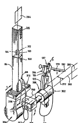
A preferred ' ~';~ of the articulated arm
according to the invention is illustrated in Figure 1. The
arm comprises an arcuate member in the ~orm of an arch 124
(see=Figure 7) slidably mounted on a first movable support
member in the form of an-arch support 300. A tool holder in
the form of a carriage 137 (see Figure 3), for holding a
medical tool in an operational position, iE carried by the
arch 124. A first drive assembly in the form of arch drive
aEsembly 302 (see Figure 8) is provided on said arch support
300 for slidably moving the arch 124 with said carriage 137
so that the carriage can be moved along an arcuate path so as
to alter the operational position of the tool in a
predetermined manner. A second drive assembly in the form of
a pivot length drive assembly 304 is provided in connection
with the carriage 137 for moving the carriage along a linear
path. Advantageously, the linear path along which the
carriage 137 can move intersects with the centre of curvature
of the arch 124.
The arch drive support 300 of the articulated arm
is in turn carried by a second movable support men7ber in the
form of a vertical support column 84 by means of a coupling
275 The support column 84 is provided with a third drive
219 ~3
W097/00649 ~ PCT/SG95/00009
-- 8
assernbly in the form of a ring drive assembly 306 in
cnnn~c~;on therewith, for pivoting the cQ--rling 275 about a
ring drive axis 180. The rollrling 275 creates an offset 272
between a longitudinal axis 308 of the arch support 300 and
the axis of rotation 180 of the ring drive assembly 306. The
ring drive assembly 306 is in turn mounted in r~nn~ct;on with
a fourth drive assembly in the form of a head travel drive
assembly 310 (see Figure 11) provided at the base of support
column 84. The head travel drive assembly is adapted to move
the ring drive assembly 306 and all of the previously
;rm~ drive ~rr ' li~5 together with the arch support 300
and arch 124 along a linear path in a generally horizontal
direction.
The articulated arm of the preferred '~~;
provides freedom- of motion in four in~ nr1~ directions.
They are namely the head travel direction 1, the pivot length
direction 2, the arch r v. direction 3, and the ring
rotation 4. The art;cnlat~ arm typically has a pivot point
182 (Figures 1 and 15), which is the centre of ~uLv~LULe of
the arch 124 of the arch drive asse~bly 302. The pivot
point, through which a central axis of the tool carried by
the arm preferably passes, advantageously lies on the ring
rotational axis 180. This is achieved by having the arch
displaced at an offset 272 from the arch drive assembly 302.
Surgical or diagno8tic tool8 can be ~tt~ch~d onto the
articulated arm via the carriage 137, (Figure 3). It is
usually desirable that the central, or desired, axis of the~e
tools can be arranged to pass through the pivot point at all
times and all configurations. This way, the tool axis forms
the radial axis of the arch. Though the pivot point is fixed
with respect to the arch 124, its position with respect to
the tool body is variable. This is desirable in optimising
the cutting strategy to be described later.
The pivot length drive assembly 304 (Figure 3) is
the nearest to the patient. It consists of the carriage 137,
a motor housing 144, a servo motor 139, a printed circuit
board 140, guide rails 145, ball bnrh;ng~ 143, locking levers
_ _ _ _ _ _ . . . . _
~ wO97/00649 219 ~ ~ 8 3 PCT/SC9S/0oO09
g _
150, a pair of set screws 147, and a rack 135 and pinion 136.
The carriage is shaped in such a way that clearances/offsets
134 are provided to clear the penis head and the resectoscope
body. The centre line, which is also the central axis of the
resectoscope, is arranged to align with the ring rotational
axis 180.
The carriage 137 is supported on the guide rails
145 using ball bllRh;ngR 143 housed in the motor hou~ing. As
i8 clearly illuatrated in Figure 3, the servo motor 139 i8
partly enclosed in its housing, with its shaft protruding
m~rnf-~th. The rack and pinion a~ .y~ 135, 136 allows
the motor 139 to drive the carriage and move it back and
forth. The motor housing 144 remains stationary and is
secured firmly by means of slot 142 onto an ear 299 milled on
the arch 124, via three screws 298 (see Figure 7). Any
- ~, of the arch will carry the entire pivot length drive
assembly 304 with it.
The printed circuit board 140 houses a differential
line driver for the encoder signals to make them less
susceptible to noise. The board is properly shielded to
suppress ele~L~ 3n~tic interference (notably from a
electro-cauterisation unit commonly u8ed in the operating
theatre), and is mounted beside the motor housing 144. this
board allow8 ea8y r~rl~f t of the motor, by providing
electrical ~nnn~rtnrs on board.
The carriage 137 has two, or more, pairs of grooves
138 cut in a manner shown in Figure 3. A selected pair of
these grooves can be used to engage a C-bracket 151. The
choice of groove depends on the desired location of the pivot
point 182 with respect to a resectoscope body 179. The C-
bracket lSl (see Figure 4) is secured semi-permanently, using
screws, onto a suitable portion of the resectoscope 179 and
is sterilised together with the resectoscope. The C-bracket
151 forms one part of a bracket assembly, with another
bracket 157. With a 81ight gap between the two parts of the
bracket assembly, a suff;ri~ntly firm grip can be nht~;nr~
when they are assembled onto a cylindrical portion of a tool
W097/00649 ~ 9 8 3 PCT~G95loO009
-- 10 --
such a3 an ~n~srope. Other forms of brackets may be readily
made to fix onto other tools whose bodies are not
cylindrical.
A protruding section on the bracket 157, as shown
in Figure 4, has a slot 159 through which a spindle 154, of
corr~pnn~;ng diameter can pass. Recesses 160 are made on
both sides of the thickness of the protruding section. A set
of locking keys 156, constituting two halves, can be pushed
manually, or under spring action, toward the slot 159 or away
from it (see Figure 4). The spindle 154 already mounted on
the tip of a retractor (for example, a Greenberg retractor),
is slid into the slot 159 of the protruding section. The
entrance of the slot is slightly chamfered to ease entry.
The spindle 154 is provided with a keyway 155. The spindle,
or the C-bracket, can be rotated 90 that the keyway is
directly in the path of the locking keys 156. Once aligned,
the locking keys are pushed (manually) down to engage with
the keyway 155 milled on the spindle, or, the spring action
pushes the locking keys 156 into the keyway 155. This
removes two degrees of freedom, 286 and 287 (see Figure 4),
so that there is no relative v~ between the endoscope
and the tool holder. ~ence the endoscope can only be
--n;r~ t~ in ways permitted by the holder. To remove the
endO8COpe i8 achieved simply by lifting the locking key8 156
upward 80 that they are not in the keyway 155. The spindle,
and the holder it ~tt~h~ onto, can now be removed from the
protruding section; and ~t~h~d from the ~n~ ope
Semi-circular grooves 152 cut on the C-bracket 151
are for the purpose of locking the bracket assembly when the
C-bracket is engaged into grooves 138 cut on the carriage
137. The locking levers 150 have a flat 149 milled over a
suitable length of its body. The levers are inserted into
drilled holes 146 whose centres are offset 80 that the levers
can be rotated to a poaition where the flat is flush with the
grooves 138. When flush, the C-bracket can be inserted. The
locking levers are prevented from dropping out by the set
screws 147. Once the C-bracket is in place, the locking
~ 2~9~gg3
WO 97/00649 ~ r~.~s~.~s,~
-- 11 --
levers are turned 90 degrees or more 80 that the rounded
portion of the levers now fill the grooves lS2 cut on the C-
bracket, thus locking it to the carriage 137.
One ' ~';~ of an arch drive assembly is
5 illu5trated in Figure 8. As can be seen in Figure 8, the
arch drive consist8 of a drive shaft 130, bearing supports
118 and 129, a differential optical encoder 106 (or other
form of position sensor), an ele~:LL~ _ ~ ;c brake 131
(preferably of normally locked type~, bearing housing 117 and
p~~nAimn 113, and a servo motor 119. The bearing housing ~-
117 and extension 113 together form the arch support 300.
Axial grooves (not shown in Figure 8) are cut on the annular
surface of the housing 117 for the passage of wires. These
wires, from the arch drive motor 119, encoder 106, brake 131,
15 and from the arch 124 itself (limit switches or other
additions), are collected at the recess where another branch
of wires, (from the pivot length drive), t~orm;nRte via an
electrical connector 108. The wires 116 leading to the pivot
length drive are bundled in a highly flexible insulated
20 conduit 115, and with extra overhang to allow r ,vc t of the
arch 124.
Wires collected within a recess 288 are all
arranged to pass through a central hole 112 in the housing
extension 113 at the end of which is another ~1 ~ctr; mAl
25 C.~,nn_~t~~ir 109. The use of connectors, as described above, is
to facilitate changing of the .~Qllpl ;ng 275 to accommodate
different ~1; t~r offsets 272 and tool lengths. From the
end of the housing extension 113 ~Figures 7 and 8), another
flexible insulated and shielded conduit 107, with overhang to
30 allow for ring rotation, bundles the wires and leads them to
the vertical column 84 (Figure 4) where it enters with a
stress relief.
The arch drive assembly 302 carries and drives the - ~-
arch 124 in the arch assembly (see Figures 7 and 8). The
35 arch assembly consists of the arch 124 which has an external
gear profile 120 cut on its circumference and is in the shape
of an arc of a circle (of suitable radius and subtended
W097/00649 219 ~ 9 g ~ PCT/SG95/00009
- 12 -
angle), stiffeners 121 and 125, a cross shaft 126, a bevel
gear set 128, a stopper 297, bearings 123, bearing tPn~inninrJ
screws 301, limit switches 294 (inside the stiffener 125, not
visible), and a bearing housing 127. As v ~nnPd before,
the arch 124 has an ear 299 for the ; ng of the pivot
length drive assev.,bly. When the arch drive motor 119 rotates
the drive shaft 130, the motion is transmitted via the bevel
gear set, 128 and 120, to the arch 124. The tool, mounted on
the carriage 137 of the pivot length drive assembly, is
caused to move along an arcuate path and the pivot point,
formed by the centre of curvature of the arch, is at a
suitable location along the tool body. This location can be
adjusted either by moving the carriage 137 using the pivot
length drive or by placing the tool with the C-bracket
manually onto another available pair of grooves 138 on the
carriage.
In the manual option, one can select the right pair
of grooves 138 on the carriage, which can be one of several
pairs limited by the length of the carriage, to engage the C-
bracket. The other option is motorised and . _~Prised, inthat the pivot length drive can move the tool it carries with
respect to the arch and hence its centre. Furthermore, the
ol~te position of the pivot point in space can be adjusted
by moving the head travel drive assev.,bly 310 after the
articulated arm is locked onto the c~llntrrh~l ~nr~ support
system.
The stiffeners 121, 125 act to strengthen the arch
support and to give bearing surfaces for the smooth sliding
~ t of the arch 124. The arch has a series of holes 293
and 294 cut on its body at specific radial distances or
intervals. The holes 293 allow the screwing on of a hard
stop to positively prevent the arch 124 from overrun. Two
hard stops are preferably provided to limit the arch travel
in both directions. One stop is fixed, the rl~ of the
other into the holes is ~PpPn~Pnt on the size of the target
and is le ~P~ by the YofL~Le. The total arch travel is
therefore adjustable. If a larger travel is needed, an
. _ _ _ _ _ _ _ _ _ _ _ _ _ _ _
=
219~983
w~s7~00649
- 13 -
arcuate member of larger subtended angle and/or having a
different radius of euLvdLuLe can be used. Indeed, the
arcuate member may have a radius of ~uLvdLuLe from anywhere
between 5cm to ;nf;n;tP (in the latter case the arcuate
mernber is aubs~nt;~lly straight so that the tool holder will
be movable along a linear path). The optimum travel for
treating the prostate, using electro-TURP is zero to 40
degrees with a radius of euLvdLuLe of 250 to 300 mm.
An alternative means of ac -' ~;ng different
radii, rather than using an arcuate mernber having a different
radius of eULVd8ULe, is to use a tool holder or carriage 137
that has ~ n~d length to provide more slots 138. Each of
these slots carries the tool at a different radius of
curvature.
A preferred ~~' '; ' of a ring drive assembly
will now be described with reference to Figure 9. The ring
drive consists of a motor 96, bearings 98 and 105, a housing
94, tr~n ~si~n shafts 97 and 100, a position sensor 103, a
gear train 95 and 102, and a brake 104. The ring drive
assembly carries the arch support 300, the arch 124 and hence
the pivot length drive, via a rollrl;ng 275 (Figure lo). The
rota~i~n~l r ~. provided by the ring drive is limited to
+/- 180 degrees. As the ring drive rotates the arch drive
assernbly and pivot length drive as~ernbly, it brings the tool
to address different angular positions 181 with respect to
the patient ~Figure 15). Wires 289 from the ring drive join
a separate insulated and ~h;~ d conduit 290 and t~nmin~te
at the lower portion of the column 84.
The arch drive axis 308 together with the pivot
length drive axis, are arranged to be c~nf;n~d in a plane 285
(Figure 1). This is to save space. When the ring drive
operates, this plane rotates and forms an angle with a plane
284 that c~nt~;n~ the head drive and ring drive axea. The
planes are ~;~plac~d by an offset 272 which can be alterable.
The offset 272 is alterable by means of the
coupling 275 illustrated in Figure 10. The P~n~;~n 113 of
i~ ~ 9 ~ ~ ~3
W097/00649 rc.,~ sc~ 3
- 14 -
arch support 300 is non-rotatably received in recess 274 of
~onrl;ng 275, whilst the tr~nr~ inn shaft 100 of the ring
drive assembly i8 non-rotatably received in the recess 273 of
the collrl; ng, Hence, rotation of shaft 100 will produce a
pivoting - VG ' of the coupling 275, which in turn produces
an orbital motion of the arch support and its associated
ass ~l;~r about the ring drive axis 180. The radius of
orbit, corresponding to the offset 272 can be altered simply
by replacing rollrlinrJ 275 with another rollrl;ng of different
length. Alternatively~ an adjustable offset of variable
length may be employed
The length of the ~t~n~inn arm 113 of arch support
300 can be altered to ac ' ~e different tool lengths. A
telescopic arm can be used. The rOllrl; ng void 274 can
a~ te a limited difference in length7 by sliding the
arm in it Also, 113 is made modular 80 that a different
length of 113 can be selected. However, too long an arm will
not be desirable as the bending r v~ about the bail joint
163 will be great. Although 163 can be modified
dimensionally and/or texturally to suit the need, h~n~l ;ng
the art;~nl~t~ arm becomes prohibitively difficult.
Finally, the head travel drive assembly 310, which
is provided at the bottom of column 84, will be described
with reference to Figure 11. The head travel drive assembly
consists of a motor 89, a po8ition encoder (not shown), a
rack 81 and pinion 91, a guide rail 79, ~n~rl~t~ 87, housing
83, limit switches 86 and base plate 88 (Figure 11). The
head travel drive moves all the as8emblies described in the
preceding p~L~yl~hs in a horizontal fashion, sagitally 1
(Figure 1) with respect to the patient. This allows
different prostate lengths to be administered. All conduit~
and wires t~r~;n~ing at the column 84 are connected to four
or five sockets 291 at the upper end of the column.
Co.,~ ;nrJ plugs affixed on the Y-arm of the
count~rh~l ~nre support system can be connected to these
sockets to prepare the robot for use.
At the upper end of the column 84, a bolt with a
~ ~19Sg~3''''
W097/00649 PcT~GsS/OOOOs
- 15 -
spherical head 164 i8 secured firmly. The spherical head 164
is to engage into a ball joint assembly 312 ~P~ignPd to
facilitate rapid rnnnPrtinn/~;~rnnnpct;nn of the art;c~ tP~
arm to a ~o~ntPrh~l~n~P support 6ystem (see Figure 13). The
head travel has a movable limit 6tops 78 that poaitively (by
way of jam bolts 85 and limit switches 80) prevents the tool,
being carried by the arm, from going beyond a safe limit,
even when there is a di~L~cu~y in the software driving the
head travel motor.
lo AdvAnt~geou~ly, the pivot length drive is 6elf-
cnnt~;nPd and can be ~Pt~hPd for a stand alone ~ppliratinn
It is therefore capable of being held by a -~h~n; ~P~ holder
such as, but not limited to, an Flmed retractor to perform
such useful tasks as urodynamics study where the diagnostic
probe, usually ultrasound, has to be moved at fixed and
accurate intervals. These lineari v. l'~ can be delivered
by the pivot length drive alone. The same argument extends
to other axes of the articulated arm, either individually or
in , - n~t; nn, aa they are modular by design.
In a preferred '~'i-- of the current invention,
the articulated arm is sn~pPn~Pd on a rountPrh~llnnP support
system (Figure 13) via a ball joint 164 (Figure l) which is
manually lockable by means of a handle 168. The
conntPrh~l~nre support system provides three degrees of
linear freedom, 8 (X), 7 (Y) and 6 (Z), plus a rot~t;nn~l
swing 5 (R). The latter allows a quick swing away of the
robot when human intervention is ~PcP~ry in an . yGn~y.
The Z I ~G ' 6 is collntPrh~l~nrPd by a suitable
number of deadweights 248 hidden in the main column 28. (see
Figure 12). The conntPrh~l~nre support system ;n~ P~ a
horizontal Y-arm 17, a horizontal X-arm 18 from which the
art;~nl~tPd arm is suspended, and a vertical main column 28.
When ~L~Liately tuned, the total weight of the Y-arm 17,
X-arm 18 and the art;rnlite~ arm (Figure 2) is h~lAncPd.
Hence, very little effort is required by the human operator
in order to move the ~r- ' 1 i PS during a setup pL~ceduLG.
The X, Y and Z , v. '~ of the conntPrh~l~nee
.... .. . ~
lgS~3 '"
WO 97/00649 ~ PCT/.~:C:9 ~ 9
- 16 -
support system provide cnnt;nnm~C -- v. along the
respective axes. The physical aL-_ _ is such that the
X arm 18 is carried by the Y arm 17 which is in turn carried
by a column rotation aasembly, which in turn is carried by
the Z arm ~on the main column 28) via a swing bracket
aasembly 19 (see Figure 13). The load being carried ride9 on
a bearing guide 30a and 30b (Figure 12), 240a and 240b
(Figure 14) which is able to with~t~n~ the bending moment and
torsional torques. In the swing bracket assembly 19, the Y
arm 17 rotates on a sliding bearing about a awivel shaft 29,
(Figure 13). Unlike the linear axes, the rot~tinn~l axis has
intermittent steps of rotation. A plunger 36 is spring
loaded and falls into a series of tapered holes 39 to give
very positive locking action. To unlock, either a solenoid
35 i8 activated, or the plunger button 32 is compressed, both
lift the plunger off its seat, thus allowing swivel action of
the horizontal Y-arm 17. (Figure 13).
Within each of the X arm 17, Y arm 18 and main
column 28 means ior slidably moving the art;cnl~ted arm in
the X, Y and Z directions respectively are provided Figure
14 provides a cut away view of the X arm 18 illustrating a
preferred means of effecting the sliding - v. . A cable
and pulley system is used comprising an endless cable loop
239 received on guide pulleys 237 provided at both ends of
the arm. A bearing mounted slide unit 240b is slidably
mounted on a linear guide 240a and is provided with a ball
joint atta~' 241 to which the ball joint assembly 312 is
connected. The slide unit 240b is fixed to the cable 239,
and its sliding , ,v. t on the linear guide 240a is limited
by rubber stoppers 243. Along the cable path, one or two
locking r -hAn;l can be ;nct~llP~, ~PpPn~;ng on the stroke
length. As a fail safe feature, the cable, and hence the
load being carried, is normally prevented from moving. Thi8
is accomplished by a cl _; n5 lever 268, pivot pin 266 and
spring 269, the cable 239 is gripped by the lever under the
spring force acting at a pivot distance 265. To unlock the
cable, the solPnnid 236 (Figure 14) can be activated
_ _ _ _ . _ _ _ _ _ _ . _ . .. . .. . . . . ..
~ 2195g~:3',,'
W097/00649 PCT/sG95loouos
- 17 -
electrically, which pulls the lever away from the cable, thus
freeing it. A manual override lever 34 is provided to allow
manual override, in case of power failure or unforr~e~hle
circumstances. The cable and pulley aLL_ , is repeated
in all the linear axes, with slight alterations, particularly
stroke length. (see Figure 12, solenoid 249 and manual lever
247).
In the illustrated : ' ' t of the current
invention, the nn-lnt~rh~l~nr~ support system is in the form
o~ a trolley system that can be wheeled around for
transportation or storage purpose. The trolley is equipped
with shelves 25, 26 and 27 (Figure 13) for the housing of
personal ~ ~r systems, surgical in~LLl t ~ and a motion
control system. The trolley has a base 24 that can be
lowered such that the entire system, including the robot,
rests on the floor on four footings. The weight of the
system ensures that, even with the d~yare~ ve~ g of the
Y-arm the system is sufficiently rigid and stable with
respect to the patient tl~uuylluuL an op~rAt;nn or np~n~t;r,n~.
When the operation is complete or not required, the robot and
its trolley (rollnt~h~l~nre support) system can be wheeled
away. This is achieved by applying a force on a foot pedal
254 to lift up the base and have the wheels 257 engage with
the floor and bear the weight of the overall system.
In a setup procedure for TURP, the target to be
est~hli~hPd as the reference position of the robot is the
ve-l , which is the junction of the ej~nnl~tory duct
and the prostatic urethra. The veL, ~nnm is i~nt;~;r~
under direct endoscopic vision. When it is found, the
endoscope/cystoscope/resectoscope is fixed in space by an
iate holder such as a Greenberg retractor or an Elmed
retractor. It should be appreciated that the orientation of
~ the endoscope is arbitrary when the veL, t: is found.
When the endoscope i5 fixed, the art;c~leted arm is then
brought into ~nga~ with the ~ ~n~r~pe.
Moving the nri~ntat;r,n and position of the
art~rnl~t~ arm, relative to the patient who is fixed under
2 ~ 8 3
W097/00649 PCT/SG95/00009
- 18 -
his o~n weight or with some suitable method of strapping (see
Figure 2), is facilitated by the ~-mlnt~rh~l~n~ support
8y8tem, mainly by its three linear axes which must first be
nnlorkPd by activating the corr~pn"~;ng solenoids, such as
8o1Pnn;~q 236, 249 and the ball joint. In the first
~y~l~a~h~ the surgeon ~-n;rlll~tP~ the articulated arm,
lifting it or lowering it, ahifting it ~;.d I y~ swivelling
it about the ball joint, until the C-bracket 151 enters into
the grooves 138 cut on the carriage 137 of the art~rnl~tPd
arm, ~see Figure 3). Alternatively, to avoid the use of a
passive holder, the articulated arm and the tool holder are
brought close to the endoscope, which is then engaged onto
the carriage 137, disturbing it slightly from the reference
position; followed by moving the articnl~tP~ arm together
with the Pn~n~cope (as one body without moving the robot's
axes) to re-establish the V~L ' ~ lo~t;nn~ These two
approaches are both valid, their use depending on the
surgeon~ 8 preference and skill.
The rmlntPrh~l~n~e gantry can be provided with a
plurality of cameras at the same level as the x-arm 18. Thi~
is to enhance the safety of the art;rlll~t~ arm during an
operation. The cameras are to observe markers or stripes
strlt~g;r~lly placed on to the robot and are properly
calibrated 80 as to obtain optimal accuracy. Knowing
directly the joints rotation or tr~n~lat;nn using machine
vision, and with known k;r ~~;C~ and geometry of the arm,
the tip of the cutter can be ensured to always move within a
prP~Pf;nPd safe working envelope. Any r~ t outgide this
envelope will trigger a suspension of motion and an alarm to
warn the user.
The C-bracket 151 is previously fastened onto the
resectoscope/endoscope and sterilised together with it.
Securing of the C-bracket 151, hence the resectoscope, is
achieved by turning the two levers 150, preventing it from
dropping out of the grooves 138 of the c~rr;~ge 137. The X,
Y, z and R axes of the countprh~l~n~p support system are then
locked by de-activation of their respective solenoids 236,
w097/00649 2 i 9~ ~ PCT~C95/oooog
-- 19 --
249 and 35. This is a fail safe feature in that the axes are
normally locked when the s~l~nni~ are not powered. The
retractor i9 then removed by l~nl nr~; n~ the lever 156 and
sliding the pin 154 away from the C-bracket. The setup
procedure is then , ~lete. Although the vc~ ~mlm has
been used in the above ~ yL~phs to show a setup procedure
for TURP, it should be appreciated that other targets, to be
i~nt;f;~ ~n~n~copjcally and referenced, or other kinds of
intervention, can benefit from the same setup yl~ed~L~
The articl~1~teA arm, like other known robot
systems, may be controlled by a motion controller 47 which in
turn is supervised by a portabie personal c ~r 48, via
RS232 communication 76 (see Figure 17). Motorisation 73 of
the respective axes (positioning, velocity, acceleration, and
coo~dination) are controlled using a conv~ntinn~l PID control
loop impl~ t~ digitally on a state of the art
microprocessor based multi-axis motion control system 47.
Position sensors, such as, but not limited to, a differential
optical encoder 71, can be employed to provide both
positional and velocity ;nfor~-t;nn for the control loop.
The motion control system is also capable of a number of
signal input and output functions 59. These 80 called l/o
functions are normally used to capture the status of limit
switches, 74 and 75, for the purpose of ~homing~', turn on or
off ~q~ working in conjunction with the robot send off
alarm or light ;n~;cat;nn to serve as a warning of danger or
other important events etc.. The above r ~; nn~d motion
control system is well known and is widely available
commercially and will not, therefore, be described in detail
here.
sarrel cavity g~n~t;nn/ as shown in Figure 15, is
a typical form of resection capable of being performed by the
articulated arm design. In this cutting strategy, one or
more axes of ~ ,vc are involved at any one time.
Coordinated , ~. ' of the arch and pivot length drive to
trace a, 80 called, sagittal line 185 being the ideal.
~ ~ reconstruction of ultrasound images facilitates the
2195~g3
w097/0~9 ~ ,j r~
- 20 -
fl~t~rm;nA~inn of a series of sagittal lines in a round the
clock fashion 181. The ring axis brings the r~nertn~ope to
address each of these ~agitt2l lines. In the case of
ele~L~ r;n2t~nn, tracing, and hence cutting, along the
sagittal line is repeated clockwise and counter-clockwise and
progres8ively outwards till the surgical capsule 279 (Figure
15) i8 reached. In the case of Laser T~RP (such as VLAP,
vi8ual laser ~hl2t;nn of the Prostate), the speed of
withfl 1 of the laser fibre and its spatial power pattern
can be controlled on the fly (i.e. dyn~m;rAlly) so as to
achieve optimal cutting, not guite possible by a human
operator. Similar ~L' ' extends to TUNA (transurethral
needle Ahlat; nn of the Prostate) where the needles are to
protrude/retract at different clock positions to different
depths and the power setting is to be controlled in a manner
quite cumbersome for a human operator.
Figure 17 is a circuit block diagram showing
typical ~ _ ~nt n which the linked arm can work in
conjunction with to carry out useful and safe activities.
These , ~ n are:
An nn;nt~rruptable power supply 45 to supply power
to the system cnnt;nllAlly. It sustains the system to operate
for at least 10 minutes (fl~pPnfl;ng on the capacity of standby
battery 44) in the event of a power failure.
A 4-axis motion controller 47 with input-output
functions 59, that can be interrupt driven. The motion
controller can be stand alone or mounted on an ~p2nRi~n slot
in a personal computer 48. r ;cat;on with the PC in the
stand alone case is via a RS232 line, while the latter is via
a local bus.
A personal c ~ ~r 48 with a usual di8play monitor
56, hard disk 58, floppy disk drive, RAM and ROM operating
memories, op~rAt;ng system, printer 57, and optional e~rn2l
storage devices such as a compact disk drive 60, and an
optical di~k drive 61.
A diagnostic ultrasound 8~Ann;ng 8ystem 49. Either
transrectal or LL~.su,~thral probe or both can be used. A
_ _ _ _ _ _ _ _ _ _ _ ~ .. . . : .. . .. . _ ... _ . . _ . . .
~ ~195~3
W097/00649 PcTlsGs5/oooos
- 21 -
frame grabber 62, ;n#tAllr~ on the PC 48, captures the
ultrasound images from the ultrAAolln~ scanner via a set of
co-axial video signal cables. Communication between the
ultrasound scanner and the ~r is optional, but can be~ estAhl;Ah~ using the GPIB standard conformed to IEEE 488,77.
A video rAAsette recorder 50, to archive ultrasound
and/or endoscopic images, intra, pre- or postop~rAt;vely.
A remote light source 51, supplying cool light via
optical fibres to the operating site and the scene u~8uL~d
by CCD (charged coupled device) camera 69, for maximum
efficiency. The image, in the form of electronic signals,
can be processed and ~nh~nred and finally displayed onto an
endoscopic monitor screen 70.
Energy source 52 for cutting. In the case of
electlu~uly~Ly, this is the 80 called ~;~th~rmy unit. In the
case of laser surgery, this will be a gQn~rAtor for high
intensity laser light source.
A hard logic safety monitor 53, r~t~;nR
yLuyL hl e logic arrays that will monitor logic status of
critical paL ~rs and send out interrupting signals to
suspend the robot action, or warning signals to alert the
user of any untoward events during an operation.
The personal computer ~cnt~A an interface
~ uL~w~Le 191, called APUI (~lt~ t~ Pro8tAtect~ y U8er
Tnt~rfAre), which carries out tasks as ol~tl~n~d in Pigure 16.
Each task to be performed by the user interface is designed
to occupy a page of screen display, with pop-up windows and
pull down menus to offer versatile s~lertl~nA.
Support 192, displays a acreen with control buttons
and status boxes to control the activation ~or deactivation)
of locks of the colmtQrh~lAnre support sygtem, in, ';n~tirn
or individually. It also displays the lock status of each of
the axes of the rrllnt~rhalance support system, (except B,
200) X, 201, Y, 202, A, 203, R, 204, which correspond to the
axes of the collnt~rhAlAnre support system (Figure 13), 8, 7,
5 and 6 respectively.
A homing seguence of the robot, after or before the
. . . _ _ _ _ _
w097/00649 219 5 ~ ~ 3 r~ ,3
- 22 -
setup procedure i8 accomplished, i8 carried out by a
subroutine in the interface called HOME, 193. This routine
brings each of the axes of the robot to a suitable reference
poaition ;nf1u~nr~d by the size and orientation of the target
that i8 to be addressed by the robot. HOME occupies a screen
display page, again with pop-up windows and pull down menus
to f~r;1;tate a~lert;nn of a large variety, including
redefinition of home poaition of the axes.
A frame grabber board F.GR3 62 and biaxial cable
take and tran8fer image8 respectively from the ultrasound
scanner. A state-of-the-art ~fLw~le for image processing,
together with customised software routine3, process the
images. The end results are used to guide the operation to
be performed by the articulated arm.
The desired~ v~ t., or ~ ~;n~t;nn Of
to achieve the ;nt~n~ed work envelope within the patient, and
events like switching, adjusting the power setting, etc , are
f- l~t~d into 'R understood by the motion controller
and sent to it by the personal ~ ~ ~r via RS232 (or serial)
lines ~6. The s~f LW~Le routines that handle this translation
are grouped under BARRE~ 198.
The articulated arm is also capable of manual
control where ~oysticks, foot or finger switches, at
dp~L~iate location8, are provided for fl~;hl~ control of
the ~ s of the robot manually. To interpret the
controls from joysticks and the like, routines in MAN 199
undertake the tr~n~]at;~n~
Patients' biodata are ~LuLed u8ing PAT 196, of
the APUI. These data can be the amount of tissue removed,
30 time taken, age of patient, etc., which are of interest to
researchers. St~t;~t;r~l tools, either standard or
customised, can be integrated with PAT 196.
Power supply to the robot system is monitored by
PWR.M 197, of the APUI. It P~L~LI.~ the n~c~ ry actiona to
ensure cont;nnrl~ operation of the robot ayctem without
~ t;nn, in the event of a power failure. Graceful
degradation of the system will be ;n;t;~t~ after a
_ _ _ _ _ _ _ . . . _ . _
w097/OO~g 2 1 ~ 5 9 8 3 . ~ 93,~ ~
- 23 -
pr~f;nPd period of time.
ERROR hAn~l; ng routines 195 are to handle both
foreseeable (normal error h2n~l;ng - for example, memory
overflow, 1088 of communication etc.) and nn~Y~ct~d
(exception hAn~l ;ng) errors. Recovery procedures will be
activated for the first type of errors to restore system
operation. Attempts to recover will be activated for
nn~rected errors, but will generally lead to a graceful
shutdown. In addition, 195 also comprises safety monitoring
routines. For example, when the tool holder axis is not
moving, cutting power will not be allowed to switch on.
In order to place the.work envelope of the cutting
tool, or other forms of implement carried by the robot, in a
desired location within the patient, and to know the limit of
the work envelope, it is desirable that some form of image
guidance be used for the articulated arm. Ultrasound
imaging, both transrectal and tL~n~u~èLhral, have been found
adequate and safe for such a task. To avoid the problem of
patient registration, that is the r-tnh;ns of pre- and intra-
( ~ even post-) operative data, it is best that the
imaging process shares the same reference frame as the robot
system. This means the imaging process has to be done
intraoperatively with some form of adaptor for the imaging
probe and the cutting tool (or other implements). seing
intraoperative, ~i ~;nn~l and lo~t;nnAl mea~u~ ~ of
the target have to be accomplished quickly 80 that the
process does not add significantly to the overall operating
time. ~his can be achieved by using computer image
processing techniques.
To facilitate the above method, it is a preferred
aspect of the current invention that provision is allowed for
both transrectal and transurethral intraoperative scanning of
the target to be treated. Transurethral scanning is possible
by leaving the outer sheath of the ~Ldosc~pe, which is
attached to the carriage (or tool holder) of the articulated
arm, in the patient. In the place of the usual working
element is the t~ u-ethral ult~A~olln~ probe 186 (see Figure
219~983
w097/00649 ~ 9
- 24 -
5). During srAnn;ng, only the head travel i8 moved, bringing
the probe in and out of the patient to obtain the needed
tl~nnv~L~e scan data. The probe is replaced by the working
element when the scanning is completed, thus sharing of the
3ame frame of reference is realised. The arch stays put at
its zero degree, or horizontal, position t~Luu~huuL the
8~nnn; ng process.
Tran8rectal scanning is pon~;hle by way of an
~ttA~' ~ (8ee Figure 6), which has an extension 188 with
one end adapted to the outer sheath of the Pn~nnrope and the
other a holder 187 for the probe body 190. The extension is
adjustable 80 that the ~ntrAnre~ governed by the offset 189,
to the rectum is fairly level. Grabbing of the probe body is
variable along its length to ic_ ~t~ different setup
conditions and pAt;Pntn. As in the case of tL~"nuL~thral
scanning, only the head travel is moved, and the probe and
extension are replaced by the working element when g~nn;ng
is complete.
In a TURP, or procedure to address sPH, the
information to be oh~;n~d in an ultrnno~n~ scanning is the
coordinates of the boundary known as a surgical capsule.
Cutting beyond the surgical capsule is ~ t desirable as it
can lead to ;nr~nt;nPnre~ impotence or torrential bleeding.
The first two cûmplications are due to nerve endings, a fair
amount of which lie just behind the capsule, being damaged as
a result of cutting. The reason for torrential bleP~;ng is
obvious - big vessels are usually situated outside the
capsule.
It is not i ~ tPly obvious in a transver8e
ultrasound scan where the capsule boundary is. It requiree
a trained eye to pick it up and very often not ~ _let~1y and
precisely. The articulated arm can perform an ;ntn~pPrative
ultrasound sc~nn;n~ either semi-~nt: t;~nlly or
automatically. ~ t; n~s in 194 cater for this purpose. In
semi-auto scanning, the surgeon has to outline the boundary
using a digiti8er 8uch a8 a mou8e. In auto srAnn;ng, the
image analysis algorithms will do the job (of course to be
W097/00649 219 ~ 9 8 3 PCT~G9S/00009 ~
- 25 -
ay~L-Jved by the silrgeon when done). The choice of which mode
of Er~nn;ng to use is a matter of convenience versus safety.
Both must not add ~i~n;f;r~ntly to the operating time.
Preferably all the drive ~r ~ 3 can be draped
in a sterilised bag just prior to an npPrat;nn~ The bag can
be conveniently fastened onto the column structure 84 of the
head drive assembly. It has ample sag room to allow
.v~ S of the articulated arm during an operation yet
preserving sterility. The entire system is best arranged in
a manner shown in Figure 2 where the robot is ghown gnsp~n~
on the connt~rhAl~n~ e support structure which, with its
trolley platforms 25, 26 and 27, also houses the computing
and surgical ~ l; t ~n~ngrrp;c view is shown on a
monitor 259 suitably supported 21 to give comfortable
viewing.
To ensure maximum safety and convenience of
operation of the system a control panel 218 is placed next to
the computer console 258 on the trolley platform (see Pigure
2) . There are visual ;n~;c3tnrs on the control panel to
di8play operating status of each axis of the motion
controller. When activated, an ;~ ~el.ey switch cuts off
power supply to the motors and cutting power sources only,
leaving all other system l , -n~5 still functional. Even
80, this is a more drastic measure compared to the keyboard
interrupt feature installed in the user interface routine.
The keyl)~Jald interrupt feature is enabled once any, or a
number of motors, is set into motion. By hitting any key on
the ~ r keyboard, all motor I v~ n~r can be killed or
suspended.
Although it has been shown that an I~Y~ nt target
for the articulated arm is the prostate gland, it should be
appreciated that it can also be used for other targets, for
example, the bladder, the womb, etc. Furthermore, the
treatment modality is not limited to electrocauterisation,
but may include other modalities such as laser, high
intensity ultrasound, TUNA (Trans Urethral Needle Ablation),
etc. The articulated arm is ~.or;gn~d so as to be capable of
~19~983
W097/00649 PcT/sGgs/oooos
- 26 -
holding a wide range of medical tools and to facilitate more
precise and ~rCllrRte medical interventions in an operating
theatre. With the help of ~ _ ~rised motion control, the
system is able to carry the tools, one at a time and to
conduct useful medical interventions. In 80 doing, it
assists the surgeon to perform his/her task better in terms
of time rnnl _Lion, skill, accuracy and hence safety. These
may lead to additional benefits such as low mortality, low
morbidity, $ewer assistants and nursing staff, less
cnn, hl~fi~ and lower risks by avoiding prolonged operating
time.
The arm is typically made such that the drive
Rr_ ~lies are modular. This allows an entirely different
k; n t; r configuration to be nhtR;n~ by replacing the
existing drive and support mer.,bers with new ones. Wires are
self c~ntR;n~ in a drive and connector~ are provided to
enable control and drive signals to reach and leave the
drive.
Now that a preferred rmho~; of the articnl~tr~
arm has been described in detail, numerous variations and
';f;r~t;nnr will 8uggest themselves to persons skilled in
the relevant arts, in addition to those already described,
without departing from the basic inventive concepts. All
such variations and r ';~irat;rnC are to be rnnr;~red within
the scope of the pre8ent invention, the nature of which is to
be ~rtrrm;n~d from the foregoing description and the Rpp~n~
claims.