Note: Descriptions are shown in the official language in which they were submitted.
WO 96/03169 2 1 9 6 0 4 3 Y~"~ e ~05,,~ S
.
FTA~TT'~Tn COTTAPSIRTT~ VA~CU~AR CAT~TTTP
Technical Field __ __ _ __ _
The present invention relates generally to catheters
inserted into the vascular system for extended periods of
time, and more particularly, to a collapsible catheter for
insertion into a blood vessel, and methods for placing such
collapsible catheter into the blood vessel.
Backqround Art _ __ _ __ __ _ _
Insertion of catheters into the vascular system of humans
and animals is a commonly performed procedure. These
catheters function as a conduit for infusion of fluids or
drugs. When a catheter needs to be in place for greater than
three or four days, it is common to place a so-called central
line catheter, and to locate the leading tip of the catheter
in one of the major veins at the top of the chest leading to
the heart, such as the subclavian vein or the major veins of
the mediastinum. In some instances, but less commonly,
catheters are passed from the lower half of the body into the
inferior vena cava. Central catheters are usually passed into
the subclavian vein, jugular vein, or into an antecubital vein
at the elbow. Such central catheters may have single or
multiple lumens, and are typically made from a relatively
rigid plastic material with a standard, round cross-section,
both to facilitate placement of the catheter into the vein and
to prevent the catheter lumens from collapsing within the
vein. Generally speaking, these catheters are constructed in
such a way that the lumen or lumens extending therethrough
retain their cross-sectional configuration unless an external
mechanical force compresses the catheter.
A complication of placing a central line catheter is the
formation of clots on the wall of the catheter located in the
vascular system. Blood clots form for several reasons. The
presence of any object occupying space within a blood vessel
causes turbulence and slowing of the blood flow through the
vessel, and these factors induce the formation of clots.
SUBSTITUTE SHEE~ (RULE 26~
WO96/03169 r~~
21 ~6043
Generally, the greater the cross-sectional area of the
catheter relative to the blood vessel, the greater the induced
turbulence and slowing of the blood. In addition, the
catheter is a foreign body, and the surface of the catheter in
contact with blood acts as a nidus for clot formation. Once
again, the greater the amount of surface area of the catheter
or other foreign body in contact with the blood, the more
likely that clots will form.
Such clots can break away and flow in the blood stream to
the heart and lungs, causing severe complications.
Furthermore, the formation of clots can often cause such veins
to become irreversibly damaged and thrombose, preventing
further blood flow through such veins. This may ultimately
cause debilitating swelling of the limb being drained by these
veins.
Apart from the risks of forming clots within the blood
vessel, present central line catheters also suffer from
susceptibility to clotting within the catheter itself. In
this regard, blood enters the lumen of the catheter and forms
a clot within the lumen, obstructing the passage of fluids
through the catheter into the vein, and thereby rendering it
unusable. While such clots may not be life threatening to the
patient, blockage of the catheter can require removal and
replacement of the catheter, a procedure which poses an
lnconvenience to both the patient and the attending physician,
and adds to ~he cost of maintaining venous access.
U.S. Patent No. 5,176,659 issued to Mancini discloses an
~Yp~n~hle intravenous catheter which has a lesser diameter
during insertion into a vein, and which is thereafter expanded
following placement to a larger diameter. While such device
simplifies insertion of the catheter, it still maintains a
sizable obstruction within the vein with a significant exposed
surface area, and it still permits blood to enter the lumen of
the catheter in the absence of fluid flow.
U.S. Patent No. 5,106,368 to Uldall et al. discloses a
dual lumen catheter for vascular access. The distal portion
of the catheter includes two tubular members attached to each
SUBS~lTUTE SHEE~ (RULE 26)
~ } ,
Wo96/03169 2 1 9 6 0 4 3 r~l~L~ ,39~
other, only one of which is collapsible. The catheter is
inserted into a blood vessel through a peel-away sheath, and
over both a stiffening cannula and a guide wire. The
collapsible lumen returns to its original circular shape once
placed in the blood vessel_ Thus, no reduction of the cross-
sectional area, or surface area, of the catheter is achieved
after the catheter is placed. In addition, blood can still
enter both lumens of the catheter in the absence of fluid
flow.
U.S. Patent No. 4,406,656 issued to Hattler et al.
discloses a multi-lumen catheter adapted to be inserted
through the center of an insertion needle into the vein of a
patient. The catheter disclosed by Hattler et al. includes
two or more collapsible lumens formed around a flexible, but
non-collapsible, central lumen. The collapsible lumens expand
outwardly under the pressure of fluid flow and collapse to a
smaller cross-sectional area in the absence of fluid flow.
However, the central lumen of the Hattler et al. device is
formed of materials which retain the shape of the central
passageway whether or not fluids flow therethrough. Thus,
even when the collapsible lumens are collapsed, the device
disclosed by Hattler et al. still approximates the cross-
sectional area of a conventional single lumen catheter.
Indeed, Hattler et al. state that the central lumen of the
disclosed multi-lumen catheter requires a certain degree of
stiffness or rigidity to provide sufficient structural support
so that the catheter can be handled as are conventional
catheters. While the device disclosed by Hattler et al.
somewhat reduces the cross-sectional area of a multi-lumen
catheter, it does not reduce the cross-sectional area or
surface area of the catheter below that of a conventional
single lumen catheter, nor does it prevent blood from entering
the central, non-collapsible lumen in the absence of fluid
flow.
Accordingly, it is an object of the present invention to
provide a central line catheter which reduces the likelihood
of the formation of clots within the blood vessel into which
SUBSTITUTE SHEET(RULE 26)
.. . . _ . _ .. . . . . .. . . . .. . . ... . . . .. . . . . . . . . .
WO96/03169 I ~I/U~,'.'~9394
21 96043
the catheter is placed. ~ ~
It is another object of the present invention to provide
such a catheter which presents a minimal cross-section
obstruction to the normal flow of blood within the blood
vessel when the catheter is not being used for infusion, while
providing a sati6factory flow path to infused fluids during
infusion procedures.
It is still another object of the present invention to
provide such a catheter which minimizes the surface area of
the catheter exposed to the blood when infusion procedures are
not being performed.
It is a further object of the present invention to
provide such a catheter which minimizes the likelihood of
blood entering the lumen of the catheter and forming a
blockage therein.
A still further object of the present invention is to
provide a method for conveniently placing such a catheter
within the desired blood vessel using commonly available
vascular apparatus.
These and other objects of the present invention will
become more apparent to those skilled in the art as the
description of the present invention proceeds.
~isclosure of Invention _ _ _
Briefly described, and in accordance with the preferred
~mhoa;r nts thereof, the present invention is a collapsible
vascular infusion catheter apparatus for providing an infusion
passage into a blood vessel while occupying minimal space, and
presenting minimal surface area within the blood vessel, when
infusion is not being conducted. The catheter of the present
invention includes at least first and second elongated,
generally flattened strips of flexible material; suitable
flexible material include without limitation plastic sheets
formed of polyethylene, polyethylene teraphthalate, and
polyvinyl chloride. The respective sides of the first and
second strips are joined with each other to form an elongated,
normally-flattened tube having an inner lumen and having first
SU8STITUTE SHEE7' (RULE 26)
2 1 96043
WO 96~03169 r~ )S ~ g4
and second opposing ends. The first, or trailing, end of the
tube is adapted to receive fluid to be infused into a blood
~ vessel of a patient, and the second, or leading, end of the
tube provides an exit port through which fluid received at the
first end of the tube can be introduced into a blood vessel of
a patient.
The normally-flattened tube expands toward a generally
oval shape, depending upon the rate of infusion, when fluid i5
infused into a blood vessel of a patient, thereby providing an
open path for the infusion fluid. When the infusion procedure
is terminated, the tube collapses back to a generally
flattened configuration for lying adjacent a wall of the blood
vessel. In this manner, the tube presents minimal cross-
sectional obstruction to the flow of blood within the blood
vessel, and also presents minimal surface area exposed to the
blood flowing in the blood vessel. In addition, the tube acts
like a valve by sealing the tip of the tube when the tube
collapses to prevent blood from entering the lumen of the
catheter.
The preferred embodiment of the above-described catheter
includes a radiopaque marker to allow the catheter to be
viewed by X-ray or fluoroscope to ensure that the catheter has
been positioned within the selected blood vessel as desired.
This radiopaque marker may take the form of a radiopaque
stripe applied to one face of the tube, a radiopaque marker
located at the leading end of the tube, or a radiopaque wire
extending along one seam of the catheter.
Alternate embodiments of the present invention include
similarly collapsible catheters having two or more lumens
formed therein. For example, a second lumen may be provided
by including a third elongated, generally flattened strip of
the same flexible material extending generally along at least
one of the first and second strips, and joining the respective
sides of third strip with those of:the first and/or second
strips to form a second elongated, normally-flattened tube
having a second lumen in parallel with the first lumen.
In order to facilitate handling of the first, or
SUBSTITUTE 5H EET (RULE 26)
:'
WO96/03169 P~~
21 96043
trailing, end of the catheter after placement, and to prevent
unintended damage to the tube at the skin entry point, the
first end of the tube may include a more rigid skin entry
portion extending from the hub of the catheter to the entry
point of the blood vessel.
Another aspect of the invention relates to the apparatus
and method for placing the catheter within the patient's blood
vessel. One such procedure uses a cylindrical guide wire
initially extending through the lumen of the normally-
flattened tube to rigidify the tube and to shape the tube intoa generally oval shape for insertion into a blood vessel of
the patient. The catheter is pre-loaded over the guide wire
prior to insertion, with the tip portion of the guide wire
extending through and beyond the exit port of the tube. An
entry path is established through the patient's skin into a
blood vessel, as by placing an introducer sheath using
standard angiographic techniques. The guide wire and the
leading end of the tube are inserted as a unit through the
entry path and into the blood vessel. The catheter and guide
wire are advanced together through the blood vessel to a
desired location using fluoroscopic, ultrasonic, or X-ray
guidance.
Following insertion in the manner described above, the
introducer sheath is removed (assuming that one was used),
while temporarily leaving both the guide wire and catheter in
place. The guide wire is then removed from the tube while
leaving the second, or leading, end of the tube within the
blood vessel at the desired location and allowing the inserted
portion of the tube to collapse against the wall of the blood
vessel. To facilitate the release of the guide wire from the
lumen of the tube, the present invention may include a
r-~h~n;s~ for temporarily infusing fluid into the first, or
trailing, end of the tube while the guide wire is present
within the catheter for expanding the tube. The infused fluid
expands the tube, freeing the tube from the guide wire,
thereby allowing the guide wire to be more easily withdrawn by
pulling the same from the first end of the tube.
SUBSTIl~ITE SH EEl' (RULE 26)
WO96/03169 2 t q 6 0 4 3 P~ s
~ 7
An alternate procedure for placing the collapsible
catheter of the present invention in a selected blood vessel
~ involves the initial formation of a seal at the second, or
leading, end of the tube for allowing the tube to be inflated
by fluid under pressure. The seal initially formed at the
second end of=the tube is adapted to be broken for providing
the exit port. After establishing an entry path through the
patient's skin into the blood vessel, an introducer sheath is
inserted through the entry path and into the blood vessel.
The introducer sheath provides an entry passageway into a
blood vessel into which the tube is to be placed. Next, a
device for a~pplying a fluid under pressure, such as a syringe,
is releasably coupled to the first end of the normally-
flattened tube to rigidify the tube and to temporarily form
the tube into a more oval shape to facilitate passage of the
tube through the introducer sheath for placement within the
blood vessel. The leading end of the tube is then inserted
into the introducer sheath and into the blood vessel while
maintaining the fluid within the tube under pressure. Once
the catheter has been advanced to the desired location using
fluoroscopic, ultrasonic, or X-ray guidance, the introducer
sheath is removed from the entry path while leaving the tube
within the blood vessel.
Before the catheter placed in the above-described manner
can be used, the seal initially formed at the second end of
the tube must first be broken for allowing fluid within the
tube to exit into the blood vessel. In one embodiment of the
present invention, a seal-breaking apparatus is inserted into
the tube after the syringe or other pressure application
30 r-~h~ni cm is removed. The seal-breaking apparatus is extended
along the length of the tube to a point proximate the second
end of the tube for opening the seal at the second end of the
tube. Such a seal-breaking apparatus may include a simple
guide wire which is inserted into the tube along the length of
the tube to a point proximate the second end of the tube for
piercing the second end of the tube.
In another embodiment of the present invention, the seal
SUBSmUTE SHEET (RULE 26~
... . ... . .. . ... . . ... . . . .
W096/03169 ' 1~~ 'u'~'4
21 96043
formed at the second end of the tube is broken remotely by
further increasing the inflation pressure applied to the tube.
In this embodiment, the seal initially formed at the second
end of the tube includes a weakened break line that ruptures
when fluid pressure within the tube exceeds a predPtPrm;n
value. During placement of the catheter into the blood vessel
through the introducer sheath, the fluid pressure is
maintained below this predetermined value to avoid pL~Iu~Lu~e
rupture of the seal. Once the catheter is properly placed,
the fluid pressure is increased up to the predetermined burst
value to rupture the seal along the weakened break line for
providing the exit port.
Brief Descri~tion of the Drawin~s
Fig. l is a perspective view of a collapsible venous
infusion catheter in accordance with a first embodiment of the
present invention.
Fig. 2 is a partial perspective view of the collapsible
catheter shown in Fig. l with an attached guide wire diaphragm
having a side-mounted infusion port.
Fig. 3A is a cross-sectional view of the collapsible
catheter of Fig. l in its collapsed condition.
Fig. 3B is a cross-sectional view of the collapsible
catheter shown in Fig. l in its expanded condition.
Fig. 3C is a cross-sectional view of the collapsible
catheter shown in Fig. 1 taken through the plane designated by
lines 3C in Fig. l.
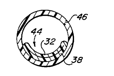
Fig. 3D is a cross-sectional view of the collapsible
catheter collapsed against-the surrounding wall of a blood
vessel into which the catheter has been placed.
Fig. 4 is a cross-sectional view of the infusion end of
the collapsible catheter, and related guide wire
diaphragm/infusion port taken through the plane indicated by
line 4 in Fig. 2.
Fig. 5 is an exploded perspective view of the guide wire
diaphragm/infusion port shown in Figs. l and 2.
Fig. 6 is a perspective view of the second, or leading,
SUBSTITUTE SHEET (RULE 261
WO96/03169 r 2 1 9 6043 r~"v~ 9~
end of the collapsible catheter, and including a radiopaque
6tripe and a weakened break line.
Fig. 7 is a cross-sectional view of the forward tip of
the collapsible catheter shown in Fig. 6 viewed through the
plane designated by lines 7 within Fig. 6.
Fig. 8A is a partially sectioned view of a patient's vein
lying below the skin, and illustrating the placement of a
peal-away introducer sheath along the entry path into the
vein.
Flg. 8B illustrates the introducer sheath within the
patient's vein following removal of its accompanying dilator.
Fig. 8C is a perspective view of a syringe coupled to the
trailing end of the collapsible catheter.
Fig. 8D is a sectional view of the tip of the collapsible
catheter and showing the tip of a guide wire inserted into the
collapsible catheter and about to pierce the sealed leading
end thereof.
Fig. 8E illustrates the entry of the second, or leading,
end of the collapsible catheter into the introducer sheath.
Fig. 8r illustrates the inflated collapsible catheter
following insertion through the introducer sheath.
Fig. 8G illustrates the collapsible catheter following
removal of the sheath, and immediately following bursting of
the initially sealed tip of the catheter.
Fig. 9A iIlustrates a cross-sectional view of the
collapsible catheter and supporting guide wire, as shown in
Fig. 1, immediately following placement of the catheter into a
vein.
Fig. 9B is a cross-sectional view of the collapsible
catheter and supporting guide wire after infusing fluid into
the catheter for allowing the guide wire to be withdrawn.
Fig. lO is a side view showing a portion of syringe
coupled to the side-mounted infusion port of the guide wire
diaphragm device for infusing fluid into the collapsible
catheter to free the guide wire from the walls of the
catheter.
Fig. 11 is a cross-sectional view of a dual lumen
SUBSTITUTE SHEET (RULE 26~
. . . _ _ ~
WO96103169 2 1 9 6 0 4 3 ~ c~5~
collapsible catheter constructed in accordance with the
teachings of the present invention, supported by a stiffening
guide wire, and including a radiopaque marking wire.
Fig. 12 is a triple lumen collapsible catheter
constructed in accordance with the teachings of=the present
invention and including a supporting insertion guide wire.
Best ~ode for O~rr~i~q Out the Invention
Fig. 1 illustrates a first embodiment of a fully-
collapsible venous infusion catheter apparatus for providing
an infusion passage into a vein in accordance with the
teachings of the present invention. While the preferred
embodiment of the present invention described herein is placed
in a vein, the present invention is not intended to be limited
to use with veins, but should be understood to extend to any
blood vessel. The collapsible catheter is designated
generally by reference numeral 20 within Fiq. 1, and includes
a leading open end 22 and an opposing trailing end 24.
Leading end 22 provides an exit port through which fluid
received at trailing end 24 can be introduced into a vein of a
patient. Trailing end 24 terminates in a conventional rigid
plastic body 26 having a knurled collar or hub 28 to
facilitate handling. A conventional luer lock connector
fitting 30 is provided at the end of body 26 for connection to
syringes, infusion lines and the like for receiving fluid to
be passed through catheter 20 to exit port 22.
~eferring to Fig. 3A and 3B, the collapsible portion of
catheter 20 includes a first elongated, generally flattened
strip 32 of flexible material having first and second opposing
sides 34 and 36. First strip 32 extends the length of
catheter ~o from trailing end 24 to leading end 22. The
collapsible portion of catheter 20 also includes a second
elongated, generally flattened strip 38 of flexible material
having first and second opposing sides 40 and 42, and like
first strip 32, extends the length of catheter 20 from
trailing end 24 to leading end 22. First side 40 of second
strip 38 is joined with the first side 34 of first strip 32;
SUBSTITUTE SHEET (RULE 26)
WO 96tO3169 2 1 9 6 0 4 3 P~ '394
11
likewise, second side 42 of second strip 38 is joined with the
second side 36 of first strip 32 to form an elongated,
normally-flattened, collapsible tube 44 having opposing ends
22 and 24.
Tube 44 is shown in its collapsed configuration in Fig.
3A prior to placement in a vein. Within Fig. 3D, tube 44 is
shown collapsed against the inner wall of vein 46 following
placement in the vein, and in the absence of fluid flow
therethrough. This is the configuration which the inventor
anticipates tube 44 will assume in the vein in the absence of
the flow of infusion fluid therethrough. In this collapsed
configuration, tube 44 occupies minimal space within the vein
when infusion is not being conducted, and therefore causes
minimal turbulence and slowing of blood flow within vein 46.
In addition, because almost one-half of the surface of tube 44
lies adjacent the wall of vein 46 when in the collapsed
condition shown in Fig. 3D, the amount of surface area of tube
44 exposed to blood flow within vein 46 is minimized. All of
these features lessen the likelihood of blood clot formation
within vein 46. In addition, since exit port 22 of tube 44
also assumes the collapsed configuration shown in Fig. 3D in
the absence of fluid flow, blood is prevented from entering
the lumen of tube 44 between infusion procedures, thereby
lessening the possibility for clots to form within the lumen,
and the resulting blockage of infusion fluid. Thus, the
colIapsible leading end of tube 44 functions like a one-way
flap valve to permit infusion fluid to escape therefrom and to
prevent blood from entering therein.
As shown in Figs. 3B and 98, tube 44 can be expanded by
infusing fluid through tube 44. The normally-flattened tube
44 expands to a generally oval shape when fluid is infused
into a vein of a patient, thereby providing a sizable cross-
sectional path for fluid to be passed into the vein. When
infusion is terminated, tube 44 collapses back to the
generally flattened configuration shown in Fig. 3D for lying
adjacent the wall of the vein.
While strips 32 and 38 have been described as discrete
SUBSTITUTE SHEET(RULE26)
WO96/031G9 Pc~
2196043
12
strips joined along their respective sides, it should be
understood that strips 32 and 38 may be integrally formed with
each other, and that the described joinder of the side edges
of such strips may, in fact, constitute the formation of
pleats or folds in what is otherwise a single, smooth
continuous surface. Suitable materials for forming strips 32
and 38 include strong but flexible plastic films, including
those made of polyethylene, polyethylene teraphthalate, and
polyvinyl chloride. In the preferred embodiment of the
present invention, these plastic films are inelastic, although
plastic films which exhibit elasticity might also be used.
Referring back to Fig. l, tube 44 includes a skin entry
portion 48 extending adjacent trailing end 24 of tube 44, and
adjacent hub 28; this skin entry portion ultimately extends
through the skin of the patient at the point of entry
following placement of the catheter. If desired, this skin
entry portion 48 of tube 44 may be made relatively rigid for a
length of approximately eight to ten centimeters, as measured
from hub 28, to facilitate handling of the catheter by medical
personnel following ~placement, and to prevent damage to
catheter 20 from long term manipulation.
As noted above, it is desired to make the majority of
tube 44 that lies within the vein fully collapsible. However,
a catheter that has no rigidity is almost impossible to insert
into a vein as compared with a catheter which has rigidity.
Accordingly, another aspect of the present invention relates
to the apparatus and method used to place such a fully
collapsible catheter within a vein. One such apparatus and
method is shown in Figs. 1 and 3C, wherein catheter 20 is pre-
loaded onto a cylindrical guide wire 50 that initially extendsthrough normally-flattened tube 44 to rigidify tube 44 and to
temporarily shape it into a generally oval shape for insertion
into the vein of a patient. As used in this specification and
within the claims which follow, the term generally-oval should
be understood to include cylindrical shapes. Figs. 3C, 9A,
and 9B illustrate the space within tube 44 surrounding guide
wire 50 as being relatively large for clarity and to simplify
SUBSTITUTE 5HEET (RULE 26)
WO 96103169 2 1 9 6 0 4 3 P.,l,v~ -'/~,." I
13
the drawings. In practice, the inventor anticipates that
guide wire 50 would closely approximate the internal
"diameter" of tube 44, thereby providing a relatively close
fit between tube 44 and guide wire 50 to avoid bunching of
'~ 5 tube 44 along guide wire 50 during insertion. Guide wire 50
has a tip portion 52 which extends through and beyond exit
port 22 of tube 44 during insertion.
Prior to insertion of catheter 20 and guide wire 50 into
the vein of the patient, a guide wire diaphragm/side infusion
port device 54 is slid over the trailing end of guide wire 50,
as shown in Fig. 1. As shown best in Figs. 4 and 5, device 54
includes a split seal diaphraym 56 secured thereto by a
threaded cap 58 having a central bore 60 formed therein.
Guide wire 50 extends through bore 60 and is wipingly engaged
by split seal diaphragm 56 to prevent the loss of blood or
infused fluid around guide wire 50. Device 54 also includes a
slde port 62 which is preferably provided with a luer lock
connector fitting 64 for receiving a syringe or other source
of infusion fluid. As shown in Figs. 2 and 4, device 54 also
includes a luer lock fitting 65 which is engaged with mating
luer lock fitting 30 of catheter 20 to form a fluid tight seal
therebetween.
Next, an entry path is established through the skin.
Such an entry path may be established, by way of example,
using the Seldinger technique or modified Seldinger technique,
both of which are well known to those skilled in the art. For
example, using the modified Seldinger technique, an introducer
sheath is inserted through the skin into the vein, providing a
convenient passage for inserting the guide wire 50 and
catheter 20, as a unit, into the vein. Proper placement of
the leading end 22 of the catheter can be confirmed using X-
rays, fluoroscopy, or ultrasound provided that a radiopaque
marker stripe 66, like that shown in Figs. 6 and 7, is formed
upon and along one of flattened strips 32 or 38 of tube 44.
Alternatively, a radiopaque wire 6~ can be incorporated within
a seam or pleat of tube 44 for extending along the tube, as
shown in Fig. 11.
SUBSTITUTE SHEET ~RULE 26)
.: ~ .. . . . . . .. _ . ~ _ _ _ _ _
WO96/03169 1~ a~
2 1 ~ 6043
14
Once proper placement of the catheter tip is confirmed,
guide wire 50 is removed. However, as shown in Figs. 3C and
9A, guide wire 50lclosely approximates the internal diameter
of tube 44, and excessive friction between guide wire 50 and
tube 44 could dislodge tube 44 from its desired position
within the vein and/or cause kinks in catheter 20.
Accordingly, prior to removal of guide wire 50, a syringe 70
or other mech~nism for injecting a fluid is coupled to side
port 62 of diaphragm device 54 r as shown in Fig. 10, for
injecting fluid into the lumen of tube 44. As indicated in
Fig. 9B, the injected fluid 72 further expands tube 44 and
moves the internal walls thereof away from guide wire 50 while
additionally lubricating guide wire 50, thus allowing guide
wire 50 to be withdrawn from catheter 20 without dislodging
catheter 20 within the vein or creating kinks therein. Upon
removal of guide wire 50, device 54 is removed from luer lock
connector fitting 30 of catheter 20. Tube 44 then collapses
against the wall of the vein, as shown in Fig. 3D, until an
infusion procedure is initiated.
A second method of rigidifying the catheter for insertion
avoids the need for a guide wire and instead uses a
pressurized fluid to inflate tube 44 for purposes of
insertion. This second method requires that the leading end
22 of catheter 20 is initially sealed, as shown in Figs. 6 and
7, rather than being open as described with respect to Fig. 1.
As shown in Figs. 6 and 7, leading end 22 of tube 44 is
initially sealed, but the seal formed at the second end of
tube 44 preferably includes a weakened break line 74 which is
adapted to be broken for providing an exit port. As described
in greater detail below, this seal is later broken either
locally or remotely after the catheter is properly placed.
Prior to placement of catheter 20 using the pressurized
fluid method, an introducer sheath is inserted into the vein,
in the manner shown in Fig. 8A. The introducer sheath
includes a stiffening dilator 76 and a pull-apart sheath 78.
The introducer sheath assembly is itself guided into vein 46
over a guide wire (not shown). As indicated in Fig. 8B, the
SU8STITUTE 5HEET (RULE 26)
WO96/03169 r~ s~ y4
~ 21 ~6043
. 15
rigid dilator 76 is then removed, leaving the pull-apart
sheath extending through the skin 47 and into vein 46, thereby
providing an entry passageway into a vein into which tube 44
is to be placed.
The next step is to pressurize tube 44 with fluid to
rigidify tube 44 and make it more oval. As indicated in Fig.
8C, an angiographic syringe 80 filled with contrast dye is
releasably secured to luer lock fitting 30 of catheter 20, and
the plunger of syringe 80 is depressed sufficiently to inflate
tube 44 with contrast dye fluid. The materials suggested
above for use in forming tube 44 are easily capable of
withstanding a pressure of 5 Atmospheres without bursting, and
such pressure is adequate to temporarily rigidify tube 44 for
placement within the vein. While not illustrated, syringes
which include pressure gauges are available and well known to
those physicians practicing in the art. As indicated in Figs.
8E and 8F, tube 44 of catheter 20 is then inserted into sheath
78 and advanced therethrough into vein 46 until sealed end 22
is po5itioned at a desired location within the vein, while
maintaining pressure on the fluid within the tube. The
presence of the contrast dye within tube 44, and the
radiopaque markings on the tube, allows the catheter to be
visible in X-rays or on a fluoroscope.
After properly positioning catheter 20, pull-apart sheath
78 is withdrawn from the entry path while leaving tube 44
within the vein. Fig. 8F shows catheter 20 within vein 46
following removal of sheath 78 but before pressure has been
released from tube 44. The final step is to break the seal at
the seal at the leading end 22 of tube 44 for allowing
infusion fluid within the tube to exit into the vein. Two
preferred methods of breaking the seal will now be described.
In the first seal breaking method, syringe 80 is removed
from luer lock connector 30 of catheter 20, and a seal-
breaking apparatus is inserted into tube 44 along the length
of the tube to a point proximate leading end 22 of the tube
for opening the seal therein. For example, as shown in Fig.
8D, the so-called seal-breaking apparatus consists of a guide
SUBSTITUTE SHEE~ (RULE 26~
= =
WO96/03169 I~~ 0~l
21 96043
16
wire 82 inserted into tube 44 along the length of the tube to
a point proximate leading end 22; the leading tip portion of
guide wire 82 is advanced into the sealed end of tube 44 for
piercing the sealed end of the tube to create the exit port.
S The second method for breaking the seal at the leading
end of tube 44, after the tube is properly positioned within
the vein, involves raising the fluid pressure within tube 44
beyond the burst strength of the weakened break line at the
second end of the tube. As mentioned above, syringe 80 (see
Fig. 8C) normally applies no more than 5 Atmospheres of
pressure to the contrast fluid dye in tube 44 during insertion
of catheter 20 to avoid premature rupture of the seal.
However, syringe 80 is capable of applying at least 10
Atmospheres of pressure to the contrast dye fluid injected
into tube 44. This higher pressure is adequate to rupture the
seal along the weakened break line 74 at leading end 22 of
tube 44 for providing the exit port. Confirmation of the
successful rupture of the seal using this method can be
confirmed using a fluoroscope by observing a puff of contrast
dye emitted from the tip of the catheter.
While the embodiments of the invention described thus far
provide a catheter having only a single lumen, a fully
collapsible multi-lumen catheter may also be constructed in
accordance with the teachings of the present invention.
Referring to Fig. 11, a third elongated, generally flattened
strip 84 of the same flexible material as strips 32 and 38 can
be secured along its Gide edges with the respective side edges
of first strip 32 to form a second elongated, normally-
flattened tube in parallel with tube 44; the collapsed lumen
of such second tube is designated in Fig. 11 by reference
numeral 86. The original lumen of the first normally-
flattened tube is pre-loaded upon guide wire 50, as shown in
Fig. 11, prior to insertion into the vein. The third strip 84
may, if desired, be made of the same length as strips 32 and
38 to provide a second lumen 86 having an exit port at its
leading end disposed at approximately the same point in the
vein as the exit port of the first lumen. Alternatively,
SUBSTITUTE SHEET (RULE 26)
Wo96103169 2 1 9 6 0 4 3 r~ b~ Js4
1~
third strip 84 may be made shorter in length than strips 32
and 38 to create a shorter second lumen 86 having an exit port
that is longitudinally displaced from the exit port of the
first lumen. Each of the two tubes may be provided with its
own catheter hub (not shown) at the trailing end of such tubes
in order to allow for separate control over the fluids infused
therethrough.
Likewise, in Fig. 12, a fourth elongated, generally
flattened strip 88 of the same flexible material as strips 32,
38, and 84 can be secured along its side edges with the
respective side edges of second strip 38 to form a third
elongated, normalIy-flattened tube in parallel with tube 44;
the collapsed lumen of such third tube is designated in Fig.
12 by reference numeral 90. ~he above-described insertion
methods for catheter 20 apply equally well to the double and
triple lumen catheters shown in Figs. 11 and 12, respectively.
Those skilled in the art will now appreciate that an
improved, fully collapsible venous infusion catheter has been
described which presents minimal obstruction to blood flow
within a vein, which presents minimal surface area in contact
with blood flowing in the vein, and which prevents blood from
enterLng the infusion lumen between infusion cycles, yet which
expands to provide a relatively large infusion path during
infusion procedures. While the present invention has been
described with respect to several preferred embodiments
thereof, such description is for illustrative purposes only,
and is not to be construed as limiting the scope of the
invention. Various modifications and changes may be made to
the described ~ho~;r-ntS by those skilled in the art without
departing from the true spirit and scope of the invention as
defined by the appended claims.
SUBSmUTE SHEET (RULE 263