Note: Descriptions are shown in the official language in which they were submitted.
2196237
VIBRATING STENT FOR OPENING CALCIFIED LESIONS
The invention relates generally to percutaneous
transluminal coronary angioplasty (PTCA) in which a dilatation
catheter is used to cross a lesion and dilate the lesion area
to restore blood flow to the artery. More specifically, the
invention relates to a catheter and stmt assembly adapted to
provide vibratory energy to assist in crossing and dilating
calcified lesions.
In typical PTCA procedures, a guiding catheter having
a pre-shaped distal tip is percutaneously introduced into the
cardiovascular system of a patient and advanced until the pre
shaped distal tip thereof is disposed within the aorta adjacent
to the ostium of the desired coronary artery. The guiding
catheter is twisted or torqued from the proximal end to turn
the distal tip of the guiding catheter so that it can be guided
into the coronary ostium. A dilatation catheter having a
balloon on its distal end and a guide wire slidably disposed
within an inner lumen of the dilatation catheter are introduced
into and advanced through the guiding catheter to its distal
tip. The distal tip of the guide wire is usually manually
shaped (i.e., curved) before the guide wire is introduced into
the guiding catheter along with the dilatation catheter. The
guide wire is first advanced out the distal tip of the guiding
catheter into the coronary artery of the patient, and torque is
applied to the proximal end of the guide wire, which extends
out of the patient, to guide the curved or otherwise-shaped
distal end of the guide wire as the guide wire is advanced
within the coronary anatomy until the shaped distal end of the
guide wire enters the desired artery. The advancement of the
guide wire within the selected artery continues until its
distal end crosses the lesion to be dilated. The dilatation
catheter then is advanced out of the distal tip of the guiding
catheter, over the previously advanced guide wire, until the
balloon on the distal extremity of the dilatation catheter is
properly positioned across the lesion. Once properly
positioned, the dilatation balloon is inflated to a
2~96~37
-2-
predetermined size with radiopaque liquid at relatively high
pressures (e-Q., 3.95-11.84 bars (4-12 atmospheres)) to dilate
the stenosed region of the diseased artery. The balloon then
is deflated so that the dilatation catheter can be removed from
the dilated stenosis and blood flow can resume through the
dilated artery.
Further details of guiding catheters, dilatation
catheters, guide wires, and other devices for angioplasty
procedures can be found in U.S. Patent No. 4,323,071 (Simpson-
Robert); U.S. Patent N. 4,439,185 (Lundquist); U.S. Patent No.
4,468,224 (Enzmann et al.); U.S. Patent No. 4,516,972 (Samson);
U.S. Patent No. 4,438,622 (Samson et al.); U.S. Patent No.
4,554,929 (Samson et al.); U.S. Patent No. 4,582,185 (Samson);
U.S. Patent No. 4,616,652 (Simpson); U.S. Patent No. 4,638,805
(Powell); U.S. Patent No. 4,748,986 (Morrison et al.); U.S.
Patent No. 4,898,577 (Badger et al.); and Canadian Patent No.
1,307,179 (Taylor et al.).
Several notable improvements have recently been made
in balloon angioplasty catheters. One such modification,
commonly referred to as a rapid-exchange catheter, is described
in U.S. Patent No. 4,748,982 (Horzewski et al.), wherein a
short sleeve or inner lumen at least about 10 cm in length is
provided within the distal section of the catheter body which
extends from a first port proximal to the balloon to a second
port in the distal end of the catheter and which is adapted to
slidably receive a guide wire. The proximal port is not less
than about 10 cm and not more than about 40 cm from the distal
end of the catheter. Preferably, a slit is provided in the
catheter body extending from the proximal port to a location
proximal to the proximal end of the balloon to facilitate the
removal of the catheter from the proximal end of the guide wire
which extends out of the pattern.
Another modification, which was introduced into the
marketplace by the assignee of the present application
(Advanced Cardiovascular Systems, Inc.), is perfusion-type
dilatation catheters which allow for long-term dilatations to
repair arterial dissections and other arterial damage. These
2196237
-3-
perfusion catheters have a plurality of perfusion ports in the
wall forming at least part of the catheter body proximal to the
balloon which are in fluid communication with an inner lumen
extending to the distal end of the catheter body. A plurality
of perfusion ports are preferably provided in the catheter body
distal to the balloon which are also in fluid communication
with the inner lumen extending to the distal end of the
catheter body. When the balloon on the distal extremity of the
dilatation catheter is inflated to dilate a stenosis,
oxygenated blood in the artery or the aorta or both, depending
upon the location of the dilatation catheter within the
coronary anatomy, is forced to pass through the proximal
perfusion ports, through the inner lumen of the catheter body
and out the distal perfusion ports. This provides oxygenated
blood downstream from the inflated balloon to thereby prevent
or minimize ischemic conditions in tissue distal to the
catheter to thereby facilitate long-term dilatations. As a
result, care should be exercised in sizing the perfusion ports
and the inner lumen to ensure that there is adequate flow of
oxygenated blood to tissue distal to the catheter to eliminate
or minimize ischemic conditions. Commercially available
perfusion catheters generally have relatively large profiles
due to the size of the inner tubular member which extends
through the interior of the balloon which prevents their use in
many distal coronary locations.
A major and on-going thrust of development work in
the field of intravascular catheters, particularly coronary
angioplasty catheters, has been to reduce the profile, i.e.,
transverse dimensions, of the aforementioned catheters and to
improve the flexibility thereof without detrimentally affecting
the pushability, particularly in the distal portion of such
catheters . A reduction in profile with little or no loss in
pushability allows a dilatation catheter to be advanced much
further into the coronary vasculature of a patient and to cross
much tighter lesions.
While the foregoing methods and devices are suitable
in most instances to perform PTCA, especially the prior art
2196231
-4-
low-profile catheters, there exist certain conditions which
preclude or at least make PTCA procedures extremely difficult
to perform with the prior art devices. For example, when the
stenosis (or lesion) in the coronary artery is a near total
occlusion, or when the plaque is calcified and essentially
blocking almost all blood flow, conventional guide wires and
dilatation catheters are unable to cross the stenosis.
Complications also can arise if the physician tries to force
the guide wire or dilatation catheter through the plaque . Very
often, plaque has only one opening through which blood flows,
but there are a number of fissures in the plaque. If the
physician tries to force the guide wire through a tight lesion,
and instead the guide wire follows one of the fissures, then
the artery might be perforated as the guide wire follows the
fissure instead of the blood flow path. Assuming the guide
wire and balloon can cross the stenosis, hard lesions may have
calcium in them and typically will require very high balloon
pressures to "crack" the lesion and restore blood flow.
Assuming the guide wire is able to cross a tight
lesion, there is no guarantee the dilatation catheter will be
able to cross, and even if it does cross, it may be difficult
or dangerous to the patient to inflate the dilatation balloon
at high pressures. The prior art devices offer no solution to
this problem of tight lesions, other than to withdraw the guide
wire and catheter and then consider alternative procedures such
as cardiopulmonary bypass surgery. The present invention is
designed to cross nearly occluded arteries and allow the
balloon to dilate a calcified lesion more easily and at lower
pressures.
SUMMARY OF THE INVENTION
The invention provides a catheter and stmt assembly
adapted to open calcified lesions using vibratory energy.
The intravascular catheter assembly of the invention
includes an elongated tubular member with proximal and distal
ends and an expandable member (balloon) near the distal end.
CA 02196237 2000-06-27
_5_
An intravascular stmt is mounted on the balloon and is
crimped down in a first collapsed condition. The balloon
and stmt are positioned at a stenosed region that is
difficult to cross and formed of a calcified or otherwise
hardened plaque. A flexible elongated member, such as a
wire, extends from outside the patient, through the
catheter, and its distal end is positioned near or is in
contact with the stmt. A vibratory energy source,
exterior of the patient, provides vibratory energy along
the flexible wire to the stmt. The vibratory energy
transferred to the stmt vibrates the hardened plaque
making it easier for the balloon-and-stmt portion of the
catheter assembly to dilate the lesion. The vibrating even
may partially break up or pulverize the plaque into small
particles which harmlessly will be carried away with
increased blood flow.
Accordingly, the present invention provides an
apparatus for imparting vibratory energy to a stenosed
region in a body lumen, comprising:
a generally tubular and radially expandable stmt
having a first collapsed condition and a second expanded
condition, and adapted to be positioned in contact with the
stenosed region of the body lumen;
a source of vibratory energy; and
a flexible elongated member having a proximal end
and a distal end and coupled at the proximal end exterior
of the body to the energy source and at the distal end to
the stmt, whereby vibratory energy from the source is
transmitted through the flexible elongated member and the
stmt to the stenosed region.
The present invention also provides an apparatus
for imparting vibratory energy to a stenosed region in a
body lumen, comprising:
a generally tubular and radially expandable stmt
having a first collapsed condition and a second expanded
condition, and adapted to be positioned in contact with the
stenosed region of the body lumen;
a vibratory energy source for providing energy;
CA 02196237 2000-06-27
-5a-
a catheter having a distal end, a proximal end,
an expandable region at the catheter distal end, and a
fluid lumen extending through the catheter and in fluid
communication with the expandable region, the stmt being
mounted on the expandable region in the first collapsed
condition; and
a flexible elongated member having a proximal end
and a distal end, the flexible elongated member coupled at
the proximal end exterior of the body to the vibratory
energy source, and the flexible elongated member distal end
terminates within the expandable region, whereby inflation
liquid is introduced through the fluid lumen to expand the
expandable region and thereby expand the stmt from the
first collapsed condition to the second expanded condition
and whereby vibratory energy from the vibratory energy
source transfers at least some of the vibratory energy
through the inflation fluid in the expandable region to the
stmt and therefore tot he stenosed region.
The vibratory energy can be supplied by
ultrasound energy that provides continuous energy, pulsed
energy, or irregular, non-repetitive energy waves to the
flexible waves to the flexible wire and hence to the stmt.
The vibratory energy source also can be a mechanical device
that produces sufficiently high frequency vibrations to
transmit energy along the flexible wire to the stmt and
thus to the plaque region.
It is desirable to removably attach the flexible
wire to the stmt, so that after the balloon and stmt have
crossed the lesion and the stmt has been implanted in the
coronary artery, the wire can be detached from the
implanted stmt and the catheter assembly with the wire
withdrawn from the patient.
In one embodiment, the vibratory energy is
generated by an audio sound generating device which
transmits sound waves through the inflation fluid in the
inflation lumen in the Catheter. After inflation fluid is
injected into the inflation lumen and partially into the
balloon, the audio energy source provides vibratory energy
CA 02196237 2000-06-27
-5b-
to the inflation fluid and hence to the balloon and stmt
mounted thereon. The vibratory energy again permits the
balloon and stmt to crack the plaque and more easily
dilate the lesion, and may even pulverize a portion of the
plaque in the process.
~~96237
-6-
In the preferred method of using the vibratory energy
to help dilate the stenosed region, the catheter, with the
stent mounted thereon, is first positioned within the stenosed
region. A vibratory energy source is supplied to the stmt
while it is in its collapsed condition on the balloon portion
of the catheter, thereby transmitting at least a portion of the
vibratory energy through the stent and into the stenosed
region. As the stenosed region begins to break up and
otherwise provide more of an opening for the distal end of the
catheter and the stmt, the catheter can be advanced distally
so that the balloon and stmt are completely positioned within
the stenosed region. A continued supply of vibratory energy
will facilitate the expansion of the balloon and stent and the
opening of the body lumen to permit blood flow therethrough.
At the end of the procedure the balloon portion of the catheter
is deflated and the catheter and balloon are withdrawn from the
body lumen, leaving the stmt implanted to assist in holding
open the lumen.
These and other advantages of the invention will
become more apparent from the following detailed description of
the invention when taken in conjunction with the accompanying
exemplary drawings.
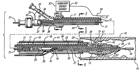
FIGURE 1 is an elevational view, partially in
section, of a prior art dilatation catheter known as a rapid
exchange-type catheter.
FIG. 2 is an elevational view, partially in section,
of a prior art dilatation catheter having perfusion
capabilities.
FIG. 3 is a cross-sectional view of a catheter and
stent assembly incorporating features of the invention.
2 96237
FIG. 4 is a transverse, cross-sectional view of the
catheter shown in FIG. 3 taken along lines 4-4.
FIG. 5 is a transverse, cross-sectional view of the
catheter shown in FIG. 3 taken along lines 5-5.
FIG. 6 is a transverse, cross-section view of the
catheter shown in FIG. 3 taken along line 6-6.
FIG. 7 is an elevational view, partially in section,
of a rapid-exchange-type catheter embodying features of the
invention.
to FIG. 8 is an elevational view, partially in section,
of a rapid-exchange-type catheter depicting a vibratory energy
source for vibrating an expandable stent.
FIG. 1 illustrates a prior art rapid-exchange-type
dilatation catheter 10 for use in PTCA procedures which allows
for the exchange of a catheter while the guide wire remains in
place within the arterial system of a patient to avoid loss of
the arterial position. This dilatation catheter is typical of
the types of catheters used to open tight lesions or partially
occluded lesions. Another prior art catheter, as shown in
FIG. 2, also can open tight lesions, and has the added feature
of being able to perfuse blood while the balloon portion of the
catheter is expanded during the PTCA procedure . When the prior
art catheters are unable to expand or open a tight or hardened
lesion, the present invention can be employed.
Referring to FIGS. 3-7, a preferred embodiment of the
catheter and stmt system using vibratory energy is depicted.
The catheter 10 generally comprises an elongated catheter shaft
11, an inflation lumen 12 adapted to direct inflation fluid
from the proximal end of the catheter shaft to the interior of
an inflatable balloon 13 on a distal portion of the catheter
~~96237
-8_
shaft and a guide wire-receiving inner lumen 14 extending
therein from the proximal end of the catheter shaft to a first
guide wire port 15 in the distal end of the catheter shaft. A
second guide wire port 16 which is also in communication with
the guide wire lumen 14 , is provided in the wall forming at
least in part catheter shaft 11 at a location of about 10-50 cm
from the distal end of the catheter shaft and a substantial
distance from the proximal end of the catheter shaft.
As shown in FIGS. 3-7, the proximal section 17 and
the distal section 18 of the catheter shaft 11 are of a dual
lumen construction with the inflation lumen 12 and guide wire
receiving lumen 14 having circular transverse cross-sections.
The inflation lumen 12 terminates at the proximal end of the
balloon 13 and is in fluid communication with the interior of
the balloon. A tubular extension 20 of the catheter shaft 11,
which defines in part the guide wire-receiving lumen 14,
extends to the distal end of the catheter shaft 11. The distal
end of the balloon 13 is sealingly secured to the distal end of
the extension 20 by suitable means such as heat bonding or an
adhesive. The inflation lumen 12 within the proximal section
17 preferably is provided with a supporting inner tubular
member 21 formed of a high strength material such as a
polyamide, stainless steel, or a suitable superelastic nickel-
titanium (NiTi) alloy. The distal part 23 of the supporting
inner tubular member 21 may be formed of a tubular stock with
a thinner wall as shown in FIG. 6. The proximal waist 22 of
the balloon 13 is secured in a suitable manner, such as heat
bonding or by an adhesive, to the exterior of the distal
section 18 of the shaft 11.
A proximal section 17 of the catheter shaft 11 is
provided with a proximal slit 24, which extends from the
proximal end of the shaft il to a location proximal to the
guide wire guiding member 25. This construction is typical of
a convertible over-the-wire/rapid exchange catheter. The
distal catheter shaft section 18 also is provided with a distal
slit 26, which extends from the second or proximal guide wire
2196231
-9-
port 16 to a location 27 proximal to the proximal waist 22 of
the balloon 13.
A guide wire 28, which is slidably disposed within
the inner guide wire lumen 14, has a coil 29 on its distal end,
which is shown in FIG. 3 extending out of the first guide wire
port 15, and an elongated core member 30, which is shown
extending through the guide wire-receiving lumen 14 and out of
the second guide wire port 16, as would be utilized in a rapid
exchange mode. A replacement guide wire 31 is shown within the
guide wire lumen 14 in the proximal portion of the catheter
shaft 11.
A multi-arm adapter 32, which is provided on the
proximal end 17 of the catheter shaft 11, has one arm 33 with
an inner lumen 34 which is adapted to introduce inflation fluid
into the inflation lumen 12 and a second arm 35 with an inner
lumen 36 which is adapted to receive the replacement guide wire
31 to be guided into the guide wire receiving lumen 14 within
the catheter shaft 11. The proximal end of the catheter shaft
11 is provided with an insert 37 which fits into the interior
of the adapter 32 as shown. The second arm 35 of adapter 32 is
provided with a slit 38 and the insert 37 is provided with a
slit 39, both of the slits being continuous with the slit 24
and the proximal section 17 of the catheter shaft 11. A
portion of the insert 37 sealingly connects the inner lumen 34
with the inner inflation lumen 12 within the catheter shaft 11.
The insert 37 may be formed as a separate element and then
secured to the proximal end of the catheter shaft 11 or it may
be formed as part of the catheter shaft.
As depicted in FIG. 3, the balloon 13 is in its
deflated state to provide a low profile for crossing tight
lesions. An expandable stent 40 is mounted on the balloon 13,
usually by compressing the stent by known means so that it is
tightly compressed onto the balloon. A protective sheath 41 is
provided to cover the stent 40 and protect the body lumen 42
from any sharp edges on the stmt 40, and to help secure the
stmt 40 to the balloon 13. The protective sheath 41 is
particularly important when advancing a catheter past a tight
2196237
-10-
calcified lesion 43 as depicted in FIG. 3. Protective sheaths
41 are known in the art and are more fully described in
commonly-owned Canadian Application No. 2,060,067-5 (see also
U.S. Patent No. 5,458,615) (Klemm et al.). If a protective
sheath is used with a rapid exchange catheter, the sheath will
require a slit for the guide wire to pass through, and an
opening where the guide wire exits the catheter, at guide wire
port 16.
In keeping with the invention, as depicted in FIGS.
3-7, a means is shown for providing vibratory energy to the
stmt (and hence to the calcified lesion 43). A flexible wire
50 is provided for removable connection at its distal end 51 to
the stent 40. The proximal end 52 of the flexible wire is
connected to vibratory energy source 53 located external to the
patient. The vibratory energy source can be an ultrasound
device that imparts continuous energy to the flexible wire 50,
or it can impart pulsed energy to the flexible wire 50. The
flexible wire 50 can be any metallic wire, such as stainless
steel or nickel titanium, for example, which materials are
capable of transmitting vibratory energy. The frequency of the
vibratory energy is a matter of choice and depends on numerous
factors, including the hardness of calcified lesion 43 and
other conditions specific to individual patients. It also is
envisioned that the vibratory energy source 53 alternatively
provide irregular, non-repetitive energy waves to flexible wire
50, which waves then are transmitted to the stent 40 and to the
calcified lesion 43.
The distal end 51 of the wire 50 can be adhesively
bonded to the balloon 13 and then the stent 40 can be crimped
onto the balloon over the wire 50. After the vibratory energy
is provided and the lesion is dilated, the stmt remains
implanted while the balloon is deflated and, along with the
wire 50, the catheter is removed from the patient.
In another embodiment of the invention, depicted in
FIG. 8, the vibratory energy source 53 provides vibratory
energy in the form of audio sound waves. The audio sound waves
are transmitted from the vibratory energy source 53 through the
~~9b237
-11-
inflation fluid in the inflation lumen 12. The inflation fluid
will transmit audio sound waves through to the balloon 13 and
to the stent 40, which then will transmit at least partial
vibratory energy to the calcified lesion 43.
In keeping with the method of use of the invention,
a catheter system embodying the invention can be inserted into
the patient in conventional rapid exchange fashion, with the
guide wire 28 pre-loaded within the inner lumen 14 in the
distal section 18 of the catheter shaft and extending
proximally out of the proximal guide wire port 16.
Alternatively, a catheter system embodying the invention can be
inserted in a conventional over-the-wire fashion, with the
guide wire extending through the entire length of the guide
wire lumen 14 and out the second arm 35 of the adapter 32. The
guide wire 28 and the catheter 10 are advanced into the body
lumen 42, e-cr., one of the coronary arteries, and the
combination is advanced to a point up to the calcified lesion
43. As is depicted in FIG. 3, the catheter and guide wire
further are advanced to be positioned within the calcified
lesion 43 prior to inflation of the balloon 13. Thereafter the
balloon 13 is inflated, which will expand the stmt 40 and
dilate the calcified lesion 43. As the dilatation procedure
commences, vibratory energy from the vibratory energy source 53
is transmitted through the flexible wire 50, or audio sound
waves are transmitted through the inflation lumen (FIG. 8) to
assist in partially pulverizing the calcified lesion 43, and
making inflation of the balloon and stmt an easier process.
As the balloon 13 and the stmt 40 become fully expanded, as
shown, for example, in FIG. 7, the calcified lesion 43 has been
expanded radially outwardly and, because of the vibratory
energy transmitted through the stent 40, the calcified lesion
43 has been at least partially, pulverized and disintegrated.
After the body lumen 42 is dilated and the stmt 40 is fully
expanded and implanted, the balloon 13 is deflated by
withdrawing the inflation fluid, and the catheter and guide
wire are withdrawn from the patient.
219G?.37
-12-
The catheter body 11 can be formed by conventional
techniques, e-Q. , by extrusion from materials which already are
known to be useful in making intravascular catheters, such as
polyethylene, polyvinyl chloride, polyesters and composite
materials. The various components of the catheter can be
joined by a suitable adhesive such as the acrylonitrile-based
adhesive sold under the tradename "LOCTITE 405" by the Loctite
Corporation. Heat shrinking or heat bonding also may be
employed where appropriate.
The size of the catheter body 11 and of the guide
wire-receiving-inner lumen 14, to a large extent, are
determined by the size of the guide wire 28 and replacement
guide wire 31 to be employed and the size of the artery or
other body lumen through which the catheter must pass. The
catheter body 11 is sufficiently long to extend from outside
the proximal end of a guiding catheter to a stenosis to be
treated within the vascular system of the patient (or other
desired location in the body of the patient), from about 100 to
150 cm when a Sledinger approach through the femoral artery is
employed to introduce the catheter 10 into the vasculature.
The wall forming the catheter must be of sufficient thickness
and strength so that it can be pushed over the guide wire 28
(or replacement guide wire 31) to the desired location within
the blood vessel.
It is to be understood that while PTCA procedures
have been discussed herein in connection with particular
embodiments of the invention, any body lumen can be treated
according to the claimed method and apparatus. Thus,
embodiments of the invention can be used to treat calcified or
tight lesions in arteries, veins, blood vessels, coronary
arteries, carotid arteries, peripheral veins, bile ducts, the
aorta, and virtually any body lumen.
While the invention has been described herein in
terms of certain presently preferred embodiments directed to
catheters for opening calcified lesions and for implanting a
stent therein, those skilled in the art will recognize that the
catheter of the invention may be used in a variety of body
296237
-13-
lumens. Further, although a rapid-exchange and perfusion-type
catheter was described herein, other types of catheters, such
as over-the-wire catheters, can be employed for use with the
invention for vibrating calcified lesions. Other modifications
and improvements may be made to the invention without departing
from the scope thereof.