Note: Descriptions are shown in the official language in which they were submitted.
~~9~~25
RADIATION MEASURING CATHETER APPARATUS AND METHOD
Technical Field
This invention pertains to an apparatus and method
for measuring radiation levels in a region of interest in
a body of a subject and, more particularly, to a
radiation measurement apparatus including a fiber optic-
equipped catheter having a radiation sensitive
scintillation material disposed within a distal portion
of the catheter, the distal portion of the catheter
adapted to be inserted and maneuvered through a blood
vessel to the region of interest.
Backcrround Art
"Tagged drugs" are widely used in nuclear medicine
to locate infected or damaged portions of blood vessels
and internal body organs, for example, the liver or
kidneys. A tagged drug includes radioactive isotopes
attached to carrier molecules. The radioactive isotopes
in tagged drugs generally have a short half life (6 hours
is typical) and emit low energy gamma rays and beta rays.
When a tagged drug is injected into a blood vessel, the
carrier molecules attach themselves to cells in the in
bloodstream. The carrier molecules of certain tagged
drugs attach themselves to red blood cells, while other
tagged drugs have carrier molecules which attach to white
blood cells.
Depending on the medical condition to be diagnosed,
a red or white blood cell attaching tagged drug is
chosen. For example, if the diagnoses involves locating
an infected portion of a blood vessel, a white blood cell
attaching tagged drug is used since white blood cells
will congregate at the site of the infection to fight the
infection. Therefore, a greater concentration of tagged
white blood cells will be found at the infected portion
of the blood vessel. This results in a higher level of
gamma and beta ray radiation at the infection site. As
C,~-lSG
219682
r
2
such, the infection site may be pinpointed with a
suitable radiation measurement device.
An exemplary use of a red blood cell attaching
tagged drug involves locating the position of an aneurism
in a blood vessel wall. An aneurism is a weakening of a
portion of a blood vessel wall. In the aneurism area,
the wall may have a small a hole that allows blood to
escape the vessel or the wall may be stretched so thin
that blood cells are able to seep through the wall.
As the tagged red blood cells flow through the
bloodstream, some of the tagged cells will seep or leak
out of the blood vessel in the aneurism area and
accumulate outside the vessel wall. A higher level of
gamma and beta ray radiation will be detected at the
aneurism location due to the accumulation of radioactive
isotopes outside the weakened blood vessel wall portion.
Tagged drugs can also be used to located the
position of a damaged or diseased portion of an internal
organ. An appropriate tagged drugged is injected
"upstream" of the organ and, as described above, a higher
concentration of tagged blood cells will be found at the
situs of the disease or injury.
Conventionally, the level of radiation emitted by
the tagged drugs in the body is visualized by a physician
or nuclear medicine technician using a fluoroscopy
device, typically a gamma camera. As its name implies,
the gamma camera detects gamma ray radiation and provides
a visual "map" of the levels of radiation emitted from an
area of the body viewed on the viewing screen. The gamma
camera is positioned externally to the body and includes
a large single scintillation crystal or a multiplicity of
smaller scintillation crystals. When a photon associated
with a gamma, beta ray impacts and is absorbed by a
scintillation crystal, the crystal scintillates and emits
a light pulse at the location of impact.
The gamma camera also includes one or more
photodetector tubes, associated circuitry and a viewing
~196~2~
3
screen which converts the pulses of light emitted by the
scintillation crystal into a radiation level "map" of the
area of the body the gamma camera is positioned adjacent
to. The image represented on the viewing screen
represents levels of radiation over the area monitored.
The darker an area on the viewing screen, the greater the
level of measured radiation. The darkest regions on the
viewing screen indicate high levels of radiation and are
commonly called "hot spots." A hot spot represents a
concentration of tagged blood cells and is found at the
location of the diseased or damaged blood vessel.
While the gamma camera, used in combination with
tagged drugs, has proven to be a useful medical
diagnostic tool, its ability to precisely locate a
position of damaged or diseased portions of a blood
vessel or internal organ is compromised by virtue of the
gamma camera being external to the subject's body and,
therefore, a significant distance from the source of the
radiation in the body. For health reasons, the quantity
and strength of the radioactive isotopes incorporated
into a dosage of a tagged drug must be minimized to avoid
the deleterious effects of radiation to the patient. The
emitted radiation from the tagged drug isotopes are
relatively weak (normally on the order of 100's of kilo
electron volts (keVs)).
All other things being equal, the closer to the
source of radiation a measuring device is, the more
accurate the measurement of that radiation. The strength
3o of the radiation emitted ("signal") by a source of
radiation decreases proportionately with the square of
the distance from the source. Therefore, the closer to
the source of radiation a radiation measuring device is,
the stronger the "signal" it will receive and the more
precisely the location of the radiation can be
pinpointed. With the gamma camera external to the body,
the relatively weak radiation generated by the isotopes
will often be insufficient to allow the physician or
219625
4
nuclear medicine technologist to accurately pinpoint the
location of the problem area.
The present invention effectively moves the
radiation measurement device adjacent to the source of
radiation, thereby facilitating accurate and efficient
measurement of radiation levels and permitting precise
determination of the damaged or diseased area of the
blood vessel or internal organ.
Summary of the Invention
The apparatus of the present invention is adapted to
accurately measure radiation at a region of interest
inside a body. The apparatus comprises a catheter
adapted to be inserted into a blood vessel, a length of
the catheter is sufficient to extend to the region of
interest while a proximal end of the catheter remains
outside the body.
The apparatus includes a luminescent scintillation
material disposed in a distal portion of a lumen of the
catheter. When a photon associated with an alpha, beta,
gamma or X-ray strikes and is absorbed by the
scintillation material, the scintillation material
luminesces and emits a pulse of electromagnetic radiation
in the visible spectrum,.i.e., a pulse of light. A fiber
optic light pipe is also disposed in the lumen of the
catheter proximal to the scintillation material. An
index matching material optically couples the
scintillation material to a distal end of a fiber optic
light pipe. The generated pulses of light are received
by and transmitted along the fiber optic light pipe.
The portion of the fiber optic light pipe extending
beyond a proximal end of the catheter is coupled to a
photomultiplier tube. The generated pulses of light
traversing the fiber optic light pipe are received by the
photomultiplier tube and converted to electric signals.
The signals are input to a signal processor analyzer
which.converts the photomultiplier signals into a
-- 2196~2~
relative measure of radiation. The signal processor
analyzer in turn is coupled to an output display which
permits a physician or nuclear medicine technologist to
continuously monitor the radiation level at the distal
5 end of the catheter as the distal end is advanced into
and through the region of interest.
The scintillation material is preferably
cylindrically shaped to snugly fit within the cylindrical
catheter lumen. To make the apparatus directionally
l0 sensitive, radiation blocking material is disposed in the
catheter lumen adjacent distal and proximal end walls of
the scintillation material. The radiation blocking
material blocks photons emitted by sources of radiation
in front of or behind the scintillation material, that
is, sources of radiation in axial alignment with a major
or longitudinal axis of the scintillation material for
the most part will not be detected or measured. Only
photons traveling along paths that intersect the side
wall of the scintillation material will impact the
scintillation material and be detected. Thus, the
apparatus detects sources of radiation radially outwardly
of the scintillation material side wall and blocks
detection of sources of radiation axially aligned with
the scintillation material end walls.
The scintillation material preferably comprises a
scintillation crystal. If the apparatus is to be used to
measure gamma radiation, a scintillation crystal
comprising Cesium Iodide doped with Thallium (CsI(T1)) is
preferable. A CsI(T1) scintillation crystal is non-
hygroscopic, has a relatively high absorption efficiency
and when energized produces easily detectable pulses of
light. The absorption efficiency of a scintillation
material is the efficiency with which the crystal absorbs
gamma energy and converts the energy to scintillations of
light. Alternately, a scintillation crystal comprising
Sodium Iodide doped with Thallium (NaI(T1)) may be
employed to measure gamma radiation. A plastic
2196~2~
6
scintillation phosphor may be utilized in the apparatus
in lieu of a scintillation crystal. Such a plastic
scintillation material is best suited to the
measurement
of beta radiation.
A method of measuring radiation in a region of
interest inside a body using the radiation measuring
apparatus of the present invention is also disclosed
.
The steps of the method comprise: providing a radiation
measuring apparatus including a catheter with a fiber
l0 optic light pipe extending through a lumen of the
catheter, the fiber optic light pipe being optically
coupled to a scintillation material disposed in the
catheter lumen in a distal portion of the catheter;
inserting the distal portion of the catheter through an
opening in the body; maneuvering the catheter to position
the distal portion adjacent a region of interest, the
scintillation material generating pulses of
electromagnetic radiation upon being impacted by and
absorbing photons associated with radioactivity, the
2~ generated pulses of electromagnetic radiation traversing
the fiber optic light pipe; sensing the pulses of
electromagnetic radiation traversing the fiber optic
light pipe; and converting the sensed bursts of
electromagnetic radiation,into a measure of radiation at
the region of interest. The step of converting the
sensed pulses of electromagnetic radiation into
a measure
of radiation includes the substep of convertin
th
g
e
pulses of electromagnetic radiation to electric pulses.
Additional features of the invention will become
apparent and a fuller understanding obtained by reading
the following detailed description made in connection
with the accompanying drawings.
Brief Describtion of the Drawings
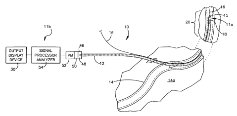
Figure 1 is a schematic representation of a
radiation measurement apparatus of the present invention
in use, a radiation detecting portion of a catheter
2196825
extends into a patient's blood vessel while the radiation
measurement portion remains outside the patient's body;
Figure 2 is an enlarged fragmentary front elevation
view of the radiation measurement apparatus of Figure 1;
Figure 3 is a section view of the radiation
measurement apparatus of Figure 1 as seen from the plane
indicated by line 3-3 in Figure 2;
Figure 4 is a section view of the radiation
measurement apparatus of Figure 1 as seen from the plane
indicated by line 4-4 in Figure 2; and
Figure 5 is an enlarged longitudinal sectional view
of a distal portion of the radiation measurement
apparatus of Figure 1.
Detailed Description of the Invention
Turning to the drawings, Figure 1 illustrates a
radiation measurement apparatus of the present invention,
shown generally at l0, in use. The apparatus 10 includes
a radiation detecting assembly lla and a radiation
measurement assembly llb (best seen in Figure 5). The
radiation detecting assembly lla is disposed in a lumen
of a catheter 12 and bulges a wall defining the lumen
slightly outwardly (as can best be seen in Figures 2 and
5). A portion of the catheter 12 extends into a
~
subject's blood vessel l4
through an opening in the
subject's skin 14a.
A guidewire 16 is used to guide a distal portion 18
of the catheter 12, including the radiation detecting
assembly lla, to a region of interest 20 of the blood
vessel 14. (Only a portion of the total guidewire 16 i
s
shown in the Figures.) The region of interest 20 is a
portion of the blood vessel 14 along which a level of
radiation is desired to be measured. The distal portion
18 of the catheter 12 slides along the guidewire 16 to
the region of interest 20 of the blood vessel 14. The
guidewire 16 is comprised of a tightly wound stainless
steel coil coated with Teflon~ or other low coefficient
z19fi825
8
of friction material. The guidewire 16 has an outer
diameter of approximately .5 mm to 1.0 mm. (.02 in. to
.04 in.).
The guidewire 16 is inserted using a guidewire
insertion catheter (not shown). When the guidewire is
properly positioned to extend somewhat beyond the region
of interest 14b of the blood vessel 14, the guidewire
insertion catheter is withdrawn, leaving the guidewire 16
in place. The distal portion 18 of the catheter 12
l0 slides along the guidewire 15.
As can best be seen in Figures 2 and 3, the
guidewire 16 is threaded through a section of tube 22
which is affixed to the catheter by a length of plastic
heat shrink tubing 24. Heat is applied to the tubing 24
to snugly secure the section of tube 22 to the catheter
12. The guidewire 16 is further threaded through an
angled aperture 26 (best seen in Figure 5) in a nipple
portion 28 of the catheter 12. The nipple portion 20 is
located at an end of the catheter distal portion 18. The
angled aperture 26 is rounded and extends from a side
wall of the nipple portion 28 to a distal end of the
nipple portion 20.
As the distal portion 18 of the catheter 12 is
advanced along the guidewire 16 through the blood vessel
14, the apparatus 10 provides a continuous measure of
radiation intensity detected by the radiation detection
assembly lla. The detected radiation is converted to
a
relative measure of radiation by the radiation
measurement assembly llb which is outside the subject's
body. The radiation measurement assembly 11b includes an
output display device or monitor 30. The monitor 30
permits a physician or nuclear medical technologist to
continuously monitor changes in radiation level as the
distal portion 18 of the catheter 12 is advanced through
the blood vessel 14.
Additionally, the advancement of the distal portion
18 of the catheter 12 may be viewed on a.fluoroscopy
219682
9
screen. A "hot spot" of high radiation produced by
photons emitted by the radioactive isotopes of a tagged
drug is indicative of a diseased or damaged portion of
the blood vessel 14. When such a "hot spot" is
encountered, the location and length of the "hot spot"
area is accurately noted and, upon removal of the
catheter 12, appropriate treatment may be instituted.
Because the detection of radiation is taken in close
proximity to the source or sources of the radiation in
the body, the measurement of radiation intensity or level
by the apparatus 10 is very accurate.
The radiation detecting assembly lla and the
radiation measuring assembly 11b are optically coupled by
a fiber optic light pipe 32. The fiber optic light pipe
32 is preferably comprised of plastic for greater
flexibility, although it should be appreciated that a
quartz fiber optic light pipe could also be utilized.
The fiber optic light pipe 32 has an outside diameter of
between 0.25 mm. and 0.75 mm. (0.010 in. to 0.030 in.).
A suitable quartz fiber optic light pipe may be purchased
from Ceramoptec of Enfield, Connecticut. A suitable PIMA
plastic fiber optic light pipe may be purchased from
Toray of Japan.
As can best be seen. in Figure 5, the radiation
detecting assembly lia is disposed within a distal
section of the lumen of the catheter 12. The radiation
detecting assembly lla includes a scintillation material
34, an index matching material 36, a pair of radiation
blocking members 38, 40 and a section of plastic heat
shrink tubing 42 which overlies the other components of
the radiation detecting assembly lla.
The scintillation material 34 is cylindrical in
shape sized to snugly fit in the lumen of the catheter
12. The scintillation material 34 is approximately 1 mm
.
(0.04 in.) in length and approximately 1 mm
to 2 m
.
m.
(0.04 in. to 0.08 in.) in diameter. When the
scintillation material 34 is struck by an alpha, beta,
~19~~2~
gamma, or X-ray, that radiation is absorbed by the
scintillation material, the scintillation material
scintillates or luminesces, that is, the material
generates a pulse of electromagnetic radiation. If the
5 generated pulse of electromagnetic radiation has a
wavelength in the visible spectrum, the pulse of
electromagnetic radiation is a pulse of visible light.
Not all radiation striking the scintillation material 34
is absorbed by the material and results in the generation
10 of a pulse of electromagnetic radiation.
The "absorption efficiency" of the scintillation
material 34 is a measure of the percent of energy
absorbed by a scintillation material of a given thickness
when a parallel beam of radiation is directed at the
scintillation material. The scintillation material 34
for the apparatus 10 preferably is a scintillation
crystal comprised of Cesium Iodide doped with Thallium
(CsI(T1)). A CsI(T1) scintillation crystal is
characterized by a good absorption efficiency with
2o respect to gamma rays. A CsI(T1) scintillation crystal 1
mm. (0.04 in.) thick would absorb approximately 35% of
the energy of a 100 keV gamma or x-ray normally incident
to the crystal. Additionally, a CsI(T1) scintillation
crystal generates pulses of electromagnetic radiation
having a wavelength of approximately 580 nanometers
(nm.), such pulses constitute easily detectable pulses of
visible light. Further, a CsI(T1) scintillation crystal
is non-hygroscopic, that is, the crystal does not react
with water. A CsI(T1) scintillation crystal is
commercially available from NE Technology Ltd. of
Edinburgh, Scotland. NE Technology Ltd. is a division of
Bicron of Newbury, Ohio.
Alternately, the scintillation material 34 may be
comprised of a Sodium Iodide crystal doped with Thallium
(NaI(T1)) which also is suitable for detecting gamma rays
and produces pulses of light having a wavelength of
approximately 413 nm. If beta radiation is being
2196825
detected, a plastic phosphor scintillation material is
preferable. An appropriate phosphor scintillation
material is available from NE Technology of Edinburgh,
Scotland, part number NE102A.
The index matching material 36 optically couples the
scintillation material 34 to a necked down distal portion
44 the fiber optic light pipe 32. The necked down distal
portion 37 has an outer diameter of approximately .055
mm. (0.0022 in.). The index matching material 36
l0 facilitates the transfer of light pulses generated by the
scintillation material 34 to the fiber optic light pipe
32 and minimizes reflection of light pulses away from the
fiber optic light pipe. The index matching material is
preferably a flexible optical gel or grease. An
15 appropriate flexible optical grease is available from
Bicron_of Newbury, Ohio, part number BC630.
The first radiation blocking member 38 is comprised
of a ring of radiation blocking metal having a length of
about 2 mm. (0.08 in.). Platinum and iridium are
20 suitable materials. The member 38 overlies the necked
down distal portion 44 of the optical fiber light pipe 32
and the index matching material 36 and abuts a proximal
end wall of the scintillation material 34. The second
radiation blocking member 40 is cylindrically shaped
25 polyurethane doped with bismuth trioxide (Bi03)
(approximately 60% by volume). The blocking member has a
length of about 2 mm. (0.08 in.). The first and second
radiation blocking members 38, 40 function to make the
radiation detection of the apparatus 10 directionally
30 sensitive.
The radiation blocking members 38, 40 block most of
the rays emitted by sources of radiation in front of or
behind the scintillation material 34, that is, sources of
radiation in axial alignment with a major or longitudinal
35 axis of the scintillation material. Onl
y photons
traveling along paths that intersect a side wall of the
scintillation material 34 will impact the scintillation
219625
12
material and cause a pulse of light to be generated.
Thus, the apparatus 10 detects and measures sources of
radiation radially outwardly of the scintillation
material side wall and blocks detection of sources of
radiation axially aligned with the scintillation material
end walls.
The plastic heat shrink tubing 42 overlies the
radiation detecting components lla and is heated to
shrink thereby securing the components into a unitary
structure that the catheter 12 can be "pulled over" .
during assembly of the apparatus 12. The tubing 42 also
prevents the index matching material 36 from leaking
along the lumen of the catheter 12. The tubing 42
extends from the optical fiber light pipe 32 proximal to
the necked down portion 37 to about half way along the
length of the second radiation blocking member 40.
A coating comprising titanium oxide (TiOz) is
applied to the tubing and outer radial surfaces of the
scintillation material 34 contacted by the tubing. The
Ti02 is highly light reflective and functions to
integrate or colluminate the pulses of light generated by
the scintillation material 34 into the index matching
material 36 and ultimately the distal portion 44 of the
optical fiber light pipe.32. The TiOZ permits the
photons to pass through to the scintillation material 34
with no significant absorption or attenuation.
Alternately, instead of applying the Ti02 coating to the
outer surface of the radiation detecting components, the
tubing 42 may be doped with TiOZ particles.
The catheter 12 is comprised of soft nylon or
polyurethane material which is doped with 40% TioZ by
volume. The TiOZ doping provides for light reflectivity
to minimize losses of pulses of light generated by a
scintillation material 34 and transmitted through the
optical fiber light pipe 32. The Ti02 dopant particles
function as a barrier to keep light from outside the
catheter 12 from being transmitted through the catheter
219682
13
wall into the lumen and minimizes loss of pulses of light
traveling along the optical fiber light pipe 32. The
doped catheter wall provides a relatively non-absorptive,
non-attenuating shield for the gamma and beta rays
emitted by radioactive isotopes to penetrate and excludes
water and other ambient substances which would adversely
affect the radiation detecting assembly components
disposed within the catheter lumen.
Approximate dimensions of the catheter 12 and the
tube section 20, labeled with reference letters A through
I in Figures 2 and 3 are as follows:
Label Description Lenath or Diameter
A Catheter overall length 160 cm. (63.o in.)
B Tube section 15 cm. (5.9 in.)
C Catheter section length between 2.5 cm. (1.0 in.)
tube section and radiation
detecting assembly
D Catheter radiation detecting 5 mm. (0.20 in.)
section length
Catheter nipple section length 5 mm. (0.20 in.)
F ~ Catheter outside diameter 1 mm. (0.04 in.)
Catheter inside diameter 0.5 mm. (0.02 in.)
H Tube section outside diameter 0.55 mm. (0.024
in.)
Tube section inside diameter 0.5 mm. (0.02 in.)
A portion of the catheter 12 overlying the radiation
detecting assembly lla is bulged or stretched radially
outwardly as can be seen in Figures 2 and 5. As noted
above the scintillation material 34 has an outside
diameter of approximately 1 mm. (.04 in.). Thus, lumen
of the catheter 12 must stretch radially outwardly from
its normal diameter of approximately 0.5 mm. (0.02 in.)
to accommodate the scintillation material 34 when the
catheter 12 is "pulled over" the radiation detecting
assembly.
219682
14
The pulses of light generated by the scintillation
material 34 are transmitted though the index matching
material 36 and into the distal portion 44 of the fiber
optic light pipe 32. The generated pulses of light
traverse the fiber optic light pipe 32. A proximal
portion 48 of the fiber optic light pipe 32 is optically
coupled to the radiation measuring assembly llb, which
convert the pulses of light into electric signals which
provide a relative measure of the intensity of the
radiation detected by the radiation detecting assembly
lla.
The radiation measuring assembly llb includes an
optical fiber adaptor (FC type) 46, a socket assembly 50
and a photomultiplier tube 52. These components function
to convert the pulses of light transmitted along the
fiber optic light pipe 32 into electric signals. A
signal processor analyzer 54 is coupled to the
photomultiplier tube 52 and converts the electrical
signals output by the photomultiplier tube 52 into a
measure of radiation. Finally, as noted previously, the
display monitor 30 is coupled to the signal processor
analyzer 54 and provides a visual display of the measure
of radiation.
An appropriate optical fiber adaptor 46 is sold by
,
Hammamatsu Phototronics K
.K., part number E5775
As
.
can
be seen in Fig. 1, the proximal end portion 48 of the
fiber optic light pipe 32 extends into an end of the
adaptor 46. A D-type socket assembly 50 is received in
an opposite end of the adaptor 46. A suitable D-type
socket assembly, also sold by Hammamatsu, is part number
E5780. The D-type socket assembly includes sockets
adapted to receive the terminal pins (not shown) of a
metal can type photomultiplier tube 52. A suitable
photomultiplier tube is Hammamatsu~s part number 85600.
The photomultiplier tube 52 converts and amplifies
the pulses of light emanating from the proximal end 48 of
the fiber optic light pipe into electrical signals. The
2196825
output signals of the photomultiplier tube 52 are coupled
to a signal processor analyzer 54. The signal processor
analyzer 54 converts the output signals of the
photomultiplier tube 54 into a signal which corresponds
5 to a measure of radiation detected by the radiation
detecting assembly lla.
Preferably, the signal processor analyzer 54
comprises a Tennelec/Nucleus PCA-P spectroscopy software
package which runs on a suitable personal computer (PC)
10 system. The PCA-P software provides a complete NaI(T1)
or CsI(T1) spectroscopy system. The spectral data are
displayed on the display monitor 30.
A suitable personal computer (PC) system for the
signal processor analyzer 30 includes at least 512K bytes
15 of RAM memory and an MS, DOS 3.0 (or higher] operating
system. To provide color output, the PC system includes
an EGA graphics display card and with the display monitor
30 being an EGA monitor. Alternately, a VGA graphics
display card may be utilized with the monitor 30 being a
VGA monitor.
The PCA-P software is resident on a half-length card
which plugs in a single eight bit slot on the PC system
motherboard. The PCA-P card includes a high voltage
power supply, a charge-sensitive preamplifier, a shaping
amplifier and a 1024 channel, 80 MHz Wilkinson Analog to
Digital converter with single channel analyzer. The PCA-
P card circuitry further includes built-in digital gain
stabilization.
Although the invention has been described in its
preferred form with a certain degree of particularity, it
is understood that the present disclosure of the
preferred form has been made only by way of example and
that numerous changes in the details of construction,
operation and combination and arrangement of parts may be
resorted to without departing from the spirit and the
scope of the invention as hereinafter claimed.