Note: Descriptions are shown in the official language in which they were submitted.
~ W096/0735~2 1 9 7 6 8 2 PCT~S95/10746
METHOD AND APPARATUS
FOR ~OCATIO~ OF A CATHETER TIP
Field of the Invention
5The present invention relates to methods and
apparatus for locating the tip of a catheter positioned
in biological tissue.
Backqround
The treatment of certain diseases often
re¢uires infusion of drugs or medicants into the body.
When infusing such medicants, a caregiver will often
make a venipuncture with a cannula to access a vein, and
thread a catheter through the vein to a desired
location. The catheter ; n~l lld~.c a distal end, a
proximal end and one or more lu.mens therebetween. The
distal end of the catheter may be placed at a variety of
locations including the superior vena cava. The
proximal end of the catheter is then coupled to a portal
within the body or passes exterior of the body.
Generally, portals are sealed reservoirs
accessible through a septum. The portal is connected to
the proximal end of the catheter and subcutaneously
implanted within the patient. Catheters which pass
external of the body include: PICC (Peripherally
Inserted Central Catheter) catheters, which access the
superior vena cava through the cephalic vein in the arm;
and chest catheters (often referred to as Hickman
catheters) which pass through a tunnel bored into the
chest then into the cephalic vein.
The prior art provides for methods and devices
to locate the distal end of the catheter within the body
as the catheter is inserted into the vein. For example,
commonly assigned U.S. Patent No. 4,905,698 to Strohl et :1
al describes a method and apparatus for catheter
location determination. The method and apparatus of
Strohl utilizes a source which generates an alternating
magnetic field, and a detector positioned proximate the
distal end of the catheter. The detector generates a
voltage in response to the proximity of the field
W096/073s2 2 1 q 1 6 8 2 PCT~S951107~6
generated by the source. The phase of the voltage in
the detector shifts as the source passes over it. By
referencing anatomical landmarks at the time the phase
shift occurs, the caregiver can ~t~rm;.n~ the exact
location of the detector, and thus the location of the
tip of the cathete~.
The method and apparatus of S~rohl are
advantageous because the tip of the catheter can be
located without use of fluoroscopy. Fluoroscopy
involves usi~g x-rays to locate the distal end of the
catheter
After the catheter i6 implanted, medicants are
delivered therethrough to the desired location
Medicants are introduced either directly into the
catheter, as-in the case of a PICC catheter or chest
catheter, or=through bolus injection or continuous
infusion into the portal. Each techni~ue for delivering
medicants has associated advantages which a caregiver
evaluates in ~t~rm;n;ng how medicants will be delivered
to a patient.
In the prior art, catheters are sold in
predetermined length6. Often, the length of the
catheter purchased does no~ meet the exact needs of the
patient. In the case of catheters used in conjunction
with portals, the caregiver positions the distal end of
the catheter within the patient, for example at the
superior vena cava, and trims the proximal end so that
the catheter is the proper length. This is especially
necessary when the length of the catheter is greater
than the distance between the superior vena cava and the
location of the portal. The distal end will be located,
and the proximal end will be trimmed prior to connection
with the portal.
If the device of Strohl has been used to
locate the distal end of the catheter, in connection
with placement of a portal, the caregiver will remove
the device of Strohl prior to trimming the proximal end
~ W096/07352 21 97 ~8 2 PCT~595ll0746
of the catheter. The device of Strohl is removed by
disconnecting a coupling positioned on an associated
junction box The caregiver then trims the proximal end
of the catheter, fits the portal to the proximal end of
the catheter and places the portal within the patient.
Chest type and PICC catheters are also sold in
predetermined lengths which do not often meet the needs
of the patient. To place a chest or PICC catheter the
caregiver would lay the catheter over the patient to
determine the proper length and trim the distal end.
The catheter would then be placed using fluoroscopy.
In the prior art, use of devices of the type
described in Strohl U.S. Patent No. 4,905,698 are not
used with either chest or PICC type catheters. Use of
these devices in the prior art are limited to use with
portals. In particular, the device of Strohl is secured
within the lumen of the catheter so that the detector o~
Strohl is positioned at the distal end of the catheter
When the distal end of the catheter has been located the
proximal end is trimmed. soth PICC and chest catheters
include permanent fittings on their proximal ends which
cannot be trimmed. The device and method taught by
Strohl cannot be used with catheters which include
fittings at the proximal end, such as PICC catheters or -
chest catheters, because the proximal end of the
catheter cannot be trimmed Therefore, a need has
arisen to provide an apparatus and method which permits
the safe and convenient use of the devicç of Strohl or
other related devices with PICC and chest catheters.
SummarY of thç Invention
The present invention provides a method for
det~r~;n;ng the location of a tip of a catheter located
in biological tissue. The catheter includes a dlstal
end, a proximal end, and a lumen positioned
therebetween. The method includes providing a source ~ -:
which develops an external alternating magnetic field,
W096107352 21 976~2 PCT~3951107~6 ~
to locate a detectort which is operable in generating a
voltage when the source is proximate the detector. ~he
detector is locked remote from the distal end of the
lumen of the catheter.
The method further includes the steps of
trimming the distal end of the catheter. The detector
is then unlocked from within the lumen of the catheter
and positioned proximate the trimmed dissal end of the
catheter ~he catheter is then positioned within the
biological t ssue and the source is placed proximate the
detector toslocate the trimmed distal end of the
catheter. : ~
~ he present invention also provides a catheter
for use with a catheter tip location device. ~he
catheter includes a proximal end, a distal end, and a
lumen positioned therebetween, while the catheter tip
location device includes a source pro~i~ing an
alternating magnetic current and a detector positioned
within the lumen of the catheter. ~he catheter tip
location devlce is operable in detecting the position of
the detecto~positioned within the lumen of the
catheter. A lock i~ positioned at the proximal end o~
the catheter, the lock being selectively movable among a
variety of locked positions, the lock operable in
selectively locking the detector re~ote from the distal
end in any position chosen by the caregiver or
manufacturer.
Brief Description of the Drawinqs
Figure 1 is a front view of a dual lumen chest
catheter.
Figure 2 is a cross-sectional view of the
chest cathetçr taken along the line 2-2.
Figure 3 is a front view of the catheter shown
in Figure 1~=showing its distal end being trimmed.
W096107352 ~ 1 9 7 6 82 PCTIUS95110746
Figure 4 is a front view of a patient with the
chest catheter of Figure 3 in place prior to removal of
a detector.
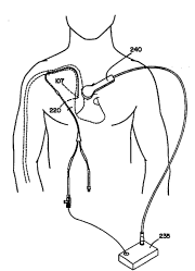
Figure 5 is a front view of a patient with the ~
chest catheter of Figure 3 in place, with a device : ~_
operative in locating the tip of the catheter in use.
Figure 5A is a front view of a patient with
the chest catheter o~ Figure l, with the catheter in
operation.
Figure 6 is a front view of a PIC~ catheter
having its distal end trimmed.
Figure 6A is a front view of a patient with
the PICC catheter of Figure 6 in place, prior to removal
of the detector.
Figure 7 is a cross sectional view of a
detector assembly operative in locating the tip of a
catheter.
Detailed Descri~tion of the Preferred ~mborl;r t.':
The present invention is directed toward a
method and apparatus for placing a chest catheter or
PICC catheter within a patient. The catheter includes a
distal end, a proximal end, and at least one lumen
positioned therebetween. The proximal end of the
catheter includes a lock fittil~Lg. The method involves
the steps of providing a catheter having a lumen and a
detector locked within the lumen of the catheter where
the detector is positioned remote from the distal end of -
the catheter. The detector is operable in generating a
voltage when a source is proximate the detector. ~ ~-
The catheter is placed external to the body as
to determine the proper length of the catheter. The
distal end of the catheter is trimmed to the proper - ~ -
length, and then the detector is nnl~ k~rl from within
the lumen of the catheter. The detector is then
repositioned proximate to the trlmmed distal end of the
catheter and locked in place using the lock ~itting.
W096/07352 PCT~S95/10746
21~7682 ~
After the detector is repositioned, the catheter is
positioned within the biological tissue~and the source
is placed proximate the detector to locate the trimmed
distal end of the catheter.
Referring now to the Figures in which like
elements are numbered alike, the present invention is
shown. The catheter shown in Figure 1 is a dual lumen
chest catheter, while the catheter shown in Figure 6 is
a PICC catheter. A significant difference i~ structure
between the chest catheter of Figure 1 and the PICC
catheter of Figure 6 is dimension and the number of
lumens each m~ay contain. For example, the greater
diameter of the chest catheter shown in Figure 1 permits
the multiple lumens disclosed in Figure 2. However, the
method and apparatus of the present invention is not
precluded fro~m use in multi-lumen PICCs.
With initial reference to Figure 1, there is
shown a catheter loo having a first proximal end 102, a
second proximal end 104 and a distal end 106. ~atheter
loo ~f the f~rst preferred em~odiment includes three
portions, which are connected by a Y shaped member 108.
Generally these portions are a lower portion 11~ and two
pigtail porti~ns 112, lls.
Lower portion llO of catheter lO0, as shown in
Figure 2, includes a ~irst lumen 116 and a sec~nd lumen
118 separated by a septum 119. First lumen 116 connects
distal end lo~ to a first pigtail and first proximal end
102 and seco~d lumen 116 connects distal end 106 to
second pigtail and second proximal end lOs. In one
preferred embodiment, first lumen 116 has a larger lumen
than that of second lumen llo.
Positioned on lower portion llO between Y
member 10~ and distal end 106 is a cuff 120.
Preferably, cuff 120 is constructed from Dacrbn, a
3~ registered tra~m~rk of E. I. Dupont De ~emours.
(Dacron is a~polyester fiber typically made from
polyethylene~terephthalate.) Cuff 120 functions to
W096/073~2 2 1 9 7 6 82 PCT~S9~ll0746
anchor catheter lD0 within the biological tissue after :~
placement. Specifically, and with reference to Figure
4, there is shown a chest catheter, which when implanted
within a patient passes through a tunnel 250 bored
within the chest of the patient. Catheter 100 is
anchored by the biological tissue surrounding cuff 120
in tunnel Z50 which grows into cuff 12Q. Cuff 120 then
assists in preventing infection within the patient.
Referring to Figures 1 and 3, a pair of
fittings 132 and 134 are attached to first and second
proximal ends 102, 104 of catheter 100, respectively.
Fittings 132 and 134 facilitate connection of catheter
100 to other medical devices, such as an infusion pump
(not shown), which operates to deliver medicants through
~ 15 catheter lOD to the superior vena cava, for example.
In the first preferred embodiment of the
present invention, a locking fitting 140 is attached to
first proximal end 102 of catheter 100 at fitting 132. ~~
~ocking fitting 140 is preferably a locking hemostasis
type valve which includes an aperture which decreases in .
size in response to twisting the valve. Uemostasis type
valves ordinarily are used to prevent blood flow out of -
the patient through the catheter Specifically, locking
fitting 140 operates to lock a detector assembly 200, of
25 an apparatus for locating the tip of the catheter, into --
place. The apparatus for locating the tip of the
catheter is of the type described in commonly assigned
U.S. Patent No. 4,905,698 the disclosure of which is
ful~y incorporated herein by reference.
Referring to Figures 5 and 7, detector
assembly 200 generally includes a detector 220 which has
a magnetic core 210 wrapped by a wire 212. Wire 212
includes a first end and a second end both of which are : :
connected to an electronic controller 230. Magnetic
core 210 including wire 212 which is wrapped therearound :h
is generally referred to as detector 220. The detector
assembly 200, as described in U.S. Patent 4,905,698, is
. . . ~
W096/07352 PCT~S951107~6
2i q76~2 ~~
located in hollow jacket 225, while the detector is
located at the tip of the hollow jacket.
In the preferred embodiment o~ the present
inventlon detector assembly 200 is locked within ~irst
lumen 116 of ~ower portion lIQ of catheter 100 proximate
cuff 120 Detector assembly 200 is locked into position
by locking ~itting 140. In the ~ir~t preferred
embodiment, aetector assembly 200 passes through locking
fitting 140, which operates to capture hollow jacket 225 :-
of detector assembly 200.
As shown in Figure 3 fittings 130 and 132 are
positioned on the proximal ends 102 and~10s of catheter
100. Becaus-e fittings 130 and 132 are positioned on
proximal ends 102 and lQ4 of catheter 100, a caregiver
cannot first:place catheter 100 and then trim the excess
catheter le~gth off of the proximal end as taught by the
priQr art. In the present invention, the caregiver
first lays catheter 100 on the patient to determine the
approximate length of catheter 100 nece~sary for the
specific patient. The proper length is determined by
the caregiver through reference to anatomical landmarks
on the patient.
The caregiver then trims distal end 106 of
catheter 100, as to define a trimmed distal end 107, as
shown in Figure 3 so that it i8 the proper length. Once
catheter 100 is trimmed to the proper length, the
caregiver car~ loosen locking fitting 140 and reposition
detector 220 of detector assembly 200 at trimmed distal
end 107 of catheter 100.
The catheter can then be placed into the
biological tissue using the techni~ues of commonly
assigned U.5~ Patent No. 4,905,698, which has been
previously incorporated by reference. The method and
apparatus described in U.S. Patent ~o. 4,905,698
involves the use of detector 220, a source, shown as 235
in Figure 5, and a locator 240 Source~;.235 generates an
alternating magnetic field which is placed proximate
~ W096/07352 21 9 7 6 8 2 PCT~S95110746
detector 220 by a locator 240. Detector 220 generates a S --
voltage in response to the magnetic field when locator
240 is proximate detector 220. As stated above,
detector is positioned proximate trim~ed distal end 107
of catheter 100. The phase of the voltage generated by
detector 220 shifts as locator 240 passes over it. By
referencing anatomical landmarks at the time the phase ~ -
shift occurs, the caregiver can determine the location
of detector 220, and thus the location of trimmed distal
end 107 of catheter 100_
Once the trimmed distal end 107 of catheter
100 is placed, and located, locking fitting 140 is
loosened and detector assembly 110 is removed. Detector
assembly is removed by pulling it through lumen 116 of
catheter 100.
As shown in Figure 5A, fittings 130 and 132
may each then be connected to a mating fitting 133 from
a pump (not shown) or another catheter (not shown). In
this fashion medicants may be delivered through the
catheter to the patient.
It is to be understood that the above
described method and constructlon is intended for use : -
with any catheter, regardless of how many pigtail ends,
which has a fitting or other structure on its proximal
end and therefore prohibits the trimming of its proximal
end after pl~pm~n~ Further, it is to be understood
that the above-described method includes alternate
methods of catheter tip location which include insertion
or removal of a detector --
With reference to Figures 6 and 6A, an example
of a second type of catheter which in~ln~q a fitting at
its proximal end is shown. Specifically, the catheter
shown in Figure 6 is a PICC catheter 300 PICC catheter
300 accesses the cephalic vein proximate the arm of the
patient and travels therethrough to the superior vena
cava, where medicants may be delivered. As above, PICC : 1
catheter 300 ;n~ q a detector assembly 200 placed
. . . _ _ . .. ... ... .. . .. . . _ _ _ _ _
W096/07352 PCTNS95110746 ~b
21 976~2 ''
within its lumen, not separately shown, with detector
220 of detect:or assembly 200 positioned distal from the
distal end of PICC catheter 300.
Generally, and with reference to ~igure 6,
PICC catheter 300 does not include pigtails as the
diameter of the lumen of PICC catheter 300 is too small.
PICC catheter 300 includes a distal end 304 having a
fitting 330 and a proxim-al end ~0~. To place PICC
catheter 30~ using the method of the present invention,
a caregiver-places PICC catheter 300 over the pasient to
estimate the approximate length of PICC catheter 300, in
a fashion similar to that described above. As shown in
Figure 6, the caregivër then trims distal end 304 as to
define a trimmed distal end 305 of PICC catheter 300.
On-ce the distal end of PICC catheter 300 has
been trimmed, a locking fitting 341, preferably a
locking hemostasis type valve, may be loosed and
detector 220 of detector assembly 2Q0 may be placed
proximate trimmed distal end 305 of PICC catheter 300.
PICC catheter 30Q may then be placed using the method
taught in U.S. patent 4,905,698 to Strohl, previously
incorporated by reference.
The present invention includes significant
advantages over the prior art. These advantages include
minimization of steps necessary in positioning a
catheter. Minimization of the steps reduces the
opportunity or error to occur. Errors which may occur
in the placement of the catheter include cutting the
detector assembly and nnne~ss~ry placement of catheters
which have an improper length.
Additionally, PICC type catheters which travel
a relatively great distance within the body can now be
tracked using the non-intrusive method taught by United
States Patent 4,905,698 to Strohl. This is a
significant advantage in view of the various body
pathway "intersections" which a PICC type catheter must
pass, on its way to the superior vena cava, in which the
~ W096/07352 PCT~SgS/10746
21 q7682
11
catheter can deviate from its intended path during
placement. If such deviation goes unchecked it can
result in delivering much needed medicants to the wrong - -~
location. ~ -
While the foregoing detailed description of
the present invention describes the invention of the
preferred em'oodiments, it will be appreciated that it is
the intent of the invention to include all modifications
and equlvalent designs. Accordingly, the scope of the
present invention is intended to ~e limited only ~y the
claims which are appended hereto. :=