Note: Descriptions are shown in the official language in which they were submitted.
WO 9G/19259 PCTIU59i1I6264
-1-
IMPLANTABLE ENDOCARDIAL LEAD WITH
SELF-HEALING RETRACTABLE FIXATION APPARATUS
Technical Field
The present invention relates generally to cardiac stimulation, and more
particularly to an implantable endocardial lead for stimulation or sensing
electrical activity of the heart in connection with an implantable pacemaker.
The lead employs a retractable fixation mechanism which can be repeatedly
exposed to or shielded from tissue during the process of securing the lead to
cardiac tissue.
Background Art
There are generally two types of body implantable leads used with
cardiac pacemakers -- one which requires surgery to expose the myocardial
tissue to which an electrode is affixed and another which can be inserted
through a body vessel, such as a vein, into the heart where an electrode
contacts the endocardial tissue. In the latter type, the endocardial lead is
often
secured to the heart through the endothelial lining by a helix affixed to a
distal
end of the lead. When the end of the lead contacts the lining of the heart at
a desired location, the lead may be secured in place by rotating the lead,
thus
screwing the helix into the heart tissue.
A helix system has been relatively effective in securing an endocardial
lead once the initial location of the lead has been achieved. However. it is
undesirable to expose the helix while the Lead is being inserted through a
blood vessel into the heart. Moreover, it is difficult to precisely place an
endocardial lead on the first attempt. It is common for a physician to
repeatedly attempt to place an endocardial lead having a helix securing
means. It is desirable, therefore, to be able to shield the helix during the
WO 96119259 ,~ PCTlUS9511G2G4 i
_2_
insertion of the lead through the vein and between attempts to implant the
lead
on the heart lining. In the prior art, various apparatus have been proposed
for
achieving the desired result. For example, U.S. Patent No, 3,974,834 to
Lawrence M. Kane, discloses an implantable intervascu(ar lead having an
accordion-fold sleeve surrounding a helix. The sleeve is retractable to expose
the helix and re-expandable to cover the . helix in the event the helix is
unscrewed and withdrawn. An object of the invention is to permit the lead to
be inserted into and guided through a body vessel withaut snagging the body
vessel.
Another attempt at solving these problems is disclosed in U.S. Patenf
No. 4,146,036 #o Robert G. butcher and Albert S. Benjamin. This patent
discloses a body implantable, intervascular lead, having a helix fixation
means.
Apparatus for shielding the helix comprises a moveable piston or shaft located
within the coils of the helix. The shaft is spring-loaded in a retracted
position
by the action of an elastomeric boot which also serves to seal oft body fluids
from the interior of the lead. A stylet passes through a lumen in the lead and
acts against a proximal end of the shaft to force the shaft forward through
the
helix thus forming a partial barrier and inhibiting the helix fram coming in
contact with tissue, at least in the axial direction.
In U.S. Patent No. 4,649,938 to William A. McArthur, an endocardial
lead with an extendiblelretractable helix fixation means is described. The
helix
is mounted on a bobbin carried within the electrode tip. The bobbin and helix
are retracted into the electrode tip by the action of a spring and are
extended
out of the tip by pressure from the end of the stylet inserted through a lumen
in the lead.
In U.S. Patent No. 5,056,516 to Spehr described an endocardial lead
with a flexible, tubular lanyard. The lanyard passed through a lumen from a
proximal end of the lead to a distal end of the lead, where the lanyard was
aftached to a sliding member supporting a helix. When the helix was in an
CA 02198100 2000-06-27
-3-
exposed position, torque could be transmitted from the proximal end of the
lanyard to the distal end thereof through a piston and thence to the helix to
screw the helix into the endocardial tissue. To stiffen the lead during
implantation, a stylet could be inserted into the lumen in the lanyard. The
invention of this later patent has been assigned to the same assignee as our
present invention. The patent was designated as an improvement on an
invention of Bradshaw, U.S. Patent No. 4,913,164.
A removable lanyard and stylet was disclosed by Spehr and Foster in
U.S. Patent 5,129,404. Brewer described a lead with a removable threaded
stylet in U.S. Patent 4,924,881.
Disclosure of Invention
In one aspect of the invention there is provided a lead for
implantation in a patient, the lead including a catheter adapted for insertion
into a chamber of the patient's heart having a proximal end and a distal
end and a lumen extending therethrough from the proximal end to the
distal end. An electrode is slidingly received in the lumen of the distal end
of the catheter. A coil conductor has at least one wire attached to the
electrode and is disposed within the lumen and adapted to transmit
electrical impulses between the electrode and the proximal end of the
catheter. The coil conductor has an inside diameter and defines an
access. Fixation means is attached distally to the electrode for securing
the electrode to the lining of the heart chamber, the fixation means being
adapted to fit within the lumen of the catheter. A wire coil segment is
attached proximally to the electrode and is adapted for connection to a
stylet. The coil segment has an expandable and retractable inside
diameter, the inside diameter being smaller than the inside diameter of the
coil conductor when retracted and having an axis parallel to the axis of the
coil conductor. The wire coil segment forms a female thread within the coil
conductor.
CA 02198100 2000-06-27
-3a-
In other words, the present invention provides an implantable
endocardial lead with retractable fixation means. In a preferred
embodiment, the fixation means comprises a helix which can be
repeatedly both retracted within a distal end of the lead and displaced
outside the distal end of the lead. The lead defines a lumen from its
proximal to its distal end. A specialized stylet can be inserted into the
lumen at the proximal end and passed through the lead to the distal end.
Located at the distal end of the lead is a piston supporting the helix. The
piston is preferably, but not necessarily, constrained to slide along the axis
of the lead. The piston is attached to a coiled trifilar conductor and has an
electrode adjacent the helix. Immediately adjacent the piston proximally an
additional first short coil of wire is interlocked between the wires of the
trifilar conductor. The short coil has about five to ten turns proximally from
the piston and a smaller diameter than the trifilar conductor.
The stylet comprises a wire having a distal end or tip. Preferably,
the distal tip has a slightly reduced section adjacent to the tip which is
more flexible than the remaining stylet. Adjacent this section, we have
attached
a______________________________________________________________________________
__________
WO 96119259 ~ ~ PCTlUS9Sl162G4
-4-
second single strand short coil segment. This coil segment is spot welded to
the stylet. In operatian, the stylet is inserted into the lead and rotated to
screw
the second shod coil segmene on the stylet into the first short coil adjacent
the
piston. The piston can then be pushed by the stylet to expose the helix, or
putted by the engagement of the two short coils to withdraw the helix into the
distal end of the lead. If too much tension is placed on the piston through
the
stylet, the short coils will disengage by the first coil being forced between
the
wires of the trifilar conductor. When the stylet is withdrawn so that the
distal
end does not overlap the first short coil, the first short coil will again
collapse
to its smaller diameter, thus making the apparatus self-healing. The stylet
can
then be rethreacied into the first short coil.
It is a principal object to provide an implantable endocardial lead with
retractable fixation means wherein the fixation means can be repeatedly
shielded and exposed during the implantation process.
A further object is to provide a lead wherein the ftxation means is
selectively shielded within a distal end of the lead and wherein the fixation
means is selectively exposed and shielded by the acfion of a removable stylet.
Another abject is to provide an implantable lead with a retractable
fixation means which is self-healing.
These and ofher objects and features will be apparent from the detailed
description taken with reference to the accompanying drawings.
W0961192_59 ~ PCTlUS95116264
_5_
Brief Description of Drawings
FIG. 1 is a prospective view of an implantable endocardial lead
according to our invention.
' 5
FIG. 2 is a sectional view of a distal tip of the lead taken along line 2-2
of FIG. 1.
FIG. 3 is a plan view of a stylet for use with the lead of FIG 1.
FIG. 4 is an enlarged sectional view of a portion of the distal tip of FIG.
2 with the stylet of FIG. 3 inserted therein.
Best Mode of Carrying Out the Invention
Reference is now made to the drawings, wherein like numerals
designate like parts throughout. FIG. 1 shows an endocardial lead, generally
designated 10. The lead 10 has a suture sleeve 12 which slides along the
lead 10 and which can be attached at an entrance into a vein of a patient in
a conventional manner. The lead 10 also has an electrode 14 located at a
distal end 16 of the lead.
As shown in FIG. 2, the lead 10 comprises a polyurethane sheath or
catheter 18 which defines a lumen 20 along a longitudinal axis of the lead 10.
Within the lumen 20, there is a coil conductor 22 for transmitting electrical
impulses between the electrode 14 and a proximal end 24 of the lead 10. In
the illustrated embodiment, a trifilar conductor is shown as the coil
conductor
22. The coil conductor 22 wraps around a crimp plug 26 at the distal end 16
of the lead 10.
The electrode 14 comprises a contact 28 and a conductive sleeve 30.
The conductive sleeve 30 fits over the crimp plug 26 and the coil conductor
22.
WO 96119259 r~ ~ ~ ~ PCTIGS951162h:1
-6-
A first single strand short coil segment 32 is interposed between the coils of
the trifilar conductor 22 and adjacent crimp plug 26. As can best be seen in
FIG. 4, the short coil segment 32 has a smaller normal interior diameter than
does the trifilar conductor 22. This causes the short coil segment 32 to
effectively form a thread within the trifilar conductor 22. The short coil
segment 32 ext~:nds along the crimp plug 26 and for preferably between five
and ten turns proximally from the crimp plug -26. The four elements just
mentioned are secured together by a crimp 34 in the conductive sleeve 30.
A silicone rubber sheath 36 houses the electrode 14. The silicone
rubber sheath 36 comprises a distal edge 38 wtrich is open to allow the
electrode to be exposed. Near the distaff edge 38 is a radiopaque ring 40
which is useful to a physician in placing the lead 10 within the heart of the
patient . Proximal from the radiopaque ring 40 is an hexagonal chamber 42.
The hexagonal chamber 42 houses a male hexagonal piston 44. The silicone
rubber sheath 36 is connected to the polyurethane sheath 18 by a
polyurethane splice tube 46. The silicone sheath 36 is glued to the
polyurethane sheath 18 and the polyurethane splice tube 46 with silicone
medical adhesive. Polyurethane adhesive bonds the polyurethane sheath 18
to the polyurethane splice tube 46.
In the illustrated embodiment, a fixation means is illustrated by a helix
48. The piston 44 and contact 28 support the helix 48 in relatively constant
alignment along the longitudinal axis of the lead 10. The piston 44 slidably
engages the hexagonal chamber 42 of the silicone sheath 36. In our preferred
embodiment, the hexagonal chamber 42 and the hexagonal piston 44 permit
the helix 48 to be rotated by rotating the entire lead. However, it is equally
possible to omit this feature and permit the piston to rotate inside the
electrode. Torque would be applied to the helix through a stylef, to be ,
described hereafter. Such a configuration would be especially well adapted
for use in atrial leads, which frequently have a "J'° shape which
prevents the
entire lead from being turned.
11'096!19259 q ~~ PCTIUS95I1G26a
-7-
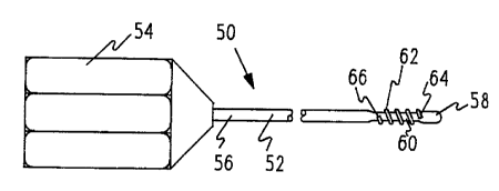
A stylet 50 is illustrated in FIG. 3. The stylet 50 comprises a stylet wire
52. The stylet wire 52 has a handle 54 at a proximal end 56. A distal end 58
~ is inserted info the lead 10. Adjacent the distal end 58 is a reduced
diameter
segment 60 which renders the stylet wire 52 slightly more flexible distally. A
second single strand coil segment 62 of wire is wound along the reduced
segment 60. The single strand coil segment 62 has a distal end 64 and a
proximal end 66. These ends 64, 66 are attached to the stylet, preferably by
welding. The second coiled segment 62 has a similar pitch and inner
diameter as the first short coil segment 32. The second coil segment 62 is
preferably about 4 or 5 coils long. The first short segment 32 and the second
Gail segment 62 thus form female and male threads respectively. Clearly, the
short coil segment 62 could be replaced by actual threads, as suggested by
Brewer, U. S. Patent 4,924,881.
The two short coil segments 32, 62 can threadably engage each other.
The stylet 50 passes through the coil 22 within the lumen 20 from the proximal
end 24 to the distal end 16 of the lead 10. The distal end 58 of the stylet
can
then be threaded into the short coil segment 32. By pushing on the stylet a
physician can expose the helix 48 outside of the distal end 16 of the lead 10.
By pulling on the stylet, the helix 48 can be withdrawn within the lead 10. If
excessive force is used to withdraw the helix 48, the first short tail segment
32 will be expanded into the tri6lar conductor 22. However, as soon as the
distal end 58 of the stylet has disengaged from the first coil segment 32, fhe
first coil segment 32 will resume its previous smaller diameter, thus making
this apparatus self-healing. During implantation, the helix 48 can be
repeatedly moved into and out of the distal end 16 of the lead 10 until proper
placement has been achieved. Then the physician can withdraw the stylet 50.
The styiet 50 can be replaced in the lead or withdrawn therefrom as often as
desired. It can also be replaced in the lead after the lead has be implanted
for
a period of time, should it become necessary to reposition the lead and if it
is
desired to retract the helix within the lead.