Note: Descriptions are shown in the official language in which they were submitted.
') 96/06679 PCT/US95/11162
X19860
CENTRIFUGE SYRINGE APPARATUS AND METHOD
10 1. FIELD OF THE INVENTION
The present invention relates to a centrifugable cell-separation apparatus
that is
useful in density-gradient separation of cells and other biological materials.
2. BACKGROUND
The prior art contains a number of devices that provide for the extraction of
fluid
samples as well as their centrifugation. For example, U.S. Patent No.
4,459,997 to
Sarstedt discloses a blood extraction and centrifugation device that provides
for the
withdrawal of blood from a patient into a tube that can be used for
centrifugation. The
centrifugation tube is a simple straight-walled tube that does not contain a
constricted region
or provide for the use of density gradient material.
f U.S. Patent No. 4,020,831 to Adler discloses a syringe that can draw a
specimen,
and then allow disassembling of certain parts of the syringe so that the
portion of the
' syringe holding the specimen can be placed in a centrifuge. The syringe also
contains a
plug of a specific density. During centrifugation, the specimen will separate
so that lighter
phases are above the plug, and heavier phases are below the plug. This device
does not
PCT/US95111162
WO 96/06679
2
provide for easy removal of the separated phases, and does not provide for the
use of a
density gradient material.
In addition, U.S. Patent No. 3,965,889 to Sachs discloses an apparatus for the
sampling of blood and the separation of plasma. The syringe includes a
thermosealable
walled container with a medial restriction into which blood is drawn. After
the blood is
drawn into the container, the container is removed and placed in a carrier for
centrifugation, after which the container can be sealed at the restriction to
separate the
phases of blood. This device requires the removal of the specimen container to
a different
carrier for centrifugation, thereby increasing the risk of contamination of
the specimen.
There is a need in the art for a centrifugation tube that can be used to
separate
components of a cellular mixture in a manner such that cells present in the
supernatant can
be readily and quantitatively collected by decantation, without disturbance to
or
contamination from higher density cells present in the lower phases and the
pellet. In
particular there is a need for a device that can be used in conjunction with a
density
gradient material to effect separation and collection of relatively rare cells
from a mixture.
There is also a need in the art for a syringe that can be used to separate
materials of
different densities which is an integrated unit that does not require transfer
of sample to a
different container for centrifugation and therefore reduces risk of
contamination. The
present invention provides these features and a sterile environment in which
all required
cell-sorting manipulations can be carried out.
3. SUMMARY OF THE INVENTION
In one aspect, the present invention is directed to a centrifugable cell-
separation
apparatus. The apparatus includes a container and a plunger slidably disposed
within the
container. In a preferred embodiment, the plunger includes a cylindrical
housing. In this
embodiment, the outer diameter of the housing makes a seal with the inner
diameter of the
apparatus container.
The container has an orifice that provides for fluid flow into the container.
The
orifice preferably includes a fitting which allows for sterile transfer of
fluid into the
apparatus. In preferred embodiments, fluid transfer through the orifice can be
introduced
by way of a sterile needle or a tubing that is further adapted to connect to a
reservoir.
The plunger forms a fluid receiving space. The top wall of the plunger is a
constriction member that defines an opening through which fluid flows into the
space. The
constriction member is constructed in such a way that fluid is retained in the
plunger when
CA 02198606 2000-02-14
WO 96J06679 PCTIUS95/11162
3
the plunger is inverted. In a preferred embodiment, the opening defined by the
plunger top
wall is annular; however, the opening can assume a number of different shapes,
including
star-shaped, oval, rectangular and the like. Alternatively, the opening can be
a plurality of
openings or can be covered by a mesh or grid.
The apparatus also includes means for sliding the plunger within the apparatus
container. In a preferred embodiment, the sliding means is an elongated.
member secured to
the plunger. The elongated member passes through a central orifice in the
other end of the
container. In a preferred embodiment, the elongated member is removable
secured to the
plunger bottom wall. In another preferred embodiment, the member may be
reattached to
the bottom wall.
In a specific embodiment, the invention includes a centrifuge syringe that
provides
an integral syringe and centrifugation tube in one apparatus and further
provides for the use
of density gradient material to enhance its cell-separation capabilities. The
apparatus has a
specimen container with one end having a fitting covering an orifice adapted
for the sterile
introduction or ejection of fluids, and the opposite end having a central
orifice for the
sealing engagement with a handle of a plunger. The handle is connected to a
plunger at one
end, which is located within the container. The opposite end of the handle
remains outside
the specimen container, and is used to move the plunger longitudinally within
the container.
i In another embodiment, the fluid receiving space of the plunger is filled
with a den
sity gradient material. The density gradient material preferably extends to a
level above the
top wall constriction member, filling part of the upper portion of the
container.
In another aspect, the invention includes a closed system for analysis of
fluid. Such
a system is particularly useful when cells to be separated can be drawn from a
patient and
directly separated in the apparatus. Alternatively, the cell mixture will be
stored in a sterile
bag, prior to extraction therefrom and separation by use of the cell
separation apparatus.
Thus, this embodiment of the invention will include, in addition to the cell
separation appa-
ratus, a fluid sample reservoir, and tubing sterilely connected
between the fluid sample reservoir and the apparatus.
In a further aspect, the present invention will be seen to encompass a kit.
The kit
includes a cell separation apparatus, as described above, and a quantity of
density gradient
material sufficient to fill the fluid receiving space in the plunger and to
further fill the
container to a level above the plunger top wall.
In yet a further aspect, the invention includes a method of extracting and
centrifu-
ging a fluid specimen, to separate components, such as cells, present in
the~specimen.
WO 96/06679 a ~ PCT/US95/11162
4
According to the method, a centrifugation apparatus as described above is
filled with a
density gradient material to a level above the top wall of the plunger. The
specimen
sample is then into the apparatus and onto the density gradient material in
the apparatus.
Centrifugal force is then applied to the apparatus to pull the sample toward
the lower end of
the apparatus container. The portion of sample remaining above the top wall of
the plunger
is then removed. Preferably, the desired specimen component will be found in
this top
fraction; however, the method can also be used to separate sedimenting
materials, by
further extracting the portion of the separated specimen that remains within
the plunger after
centrifugation.
4. BRIEF DESCRIPTION OF THE DRAWINGS
FIG. 1 shows a cross-sectional view of a centrifuge syringe of the invention
before
the extraction of a specimen;
FIG. 2 shows a cross-sectional view of the centrifuge syringe of FIG. 1 upon
introduction of the specimen;
FIG. 3 shows a cross-sectional view of the centrifuge syringe of FIG. 1 after
centrifugation and removal of the handle;
FIG. 4 shows a cross-sectional view of the centrifuge syringe of FIG. 1 to
which
the handle has been re-attached, and from which the specimen has been removed;
FIG. 5 is a cross-sectional view of an alternative embodiment of the
centrifuge
syringe according to the invention;
FIG. 6 is a perspective view of the plunger of the alternative embodiment of
FIG.
5;
FIG. 7 is an enlarged view of the plunger of FIG. 5;
FIG. 8 is a cross-sectional view of an alternative embodiment of the
centrifuge
syringe plunger having a valve;
FIGS. 9A-F are cross-sectional views of alternative embodiments of the plunger
of
the centrifuge syringe;
FIG. 10 is a cross-sectional view of an alternative embodiment of the
centrifuge
syringe having multiple constriction members; and
FIG. 11 is diagrammatic illustration of a closed system for blood analysis
using a
centrifuge syringe according to the present invention.
'-',196/06679 219 s s o s p~~S95/11162
5. DETAILED DESCRIPTION OF THE INVENTION
5.1 Centrifuge-Shoe and Methods of Use
The present invention is directed to a centrifugable cell separation apparatus
that is particularly adapted to separation of cells from bodily fluids.
Generally, the
5 apparatus is a syringe that has a specialized plunger slidably disposed
within the syringe
chamber. The plunger has a fluid-receiving space into which fluid can be
delivered. An
important feature of the plunger is that it retains the fluid contained within
its fluid
receiving space when the syringe is inverted.
During operation, the plunger slides between the two ends of the syringe
container
or barrel. Such sliding can be effected by conventional methods, such as by
attaching to
the plunger an elongated member or handle which protrudes through one end of
the syringe
barrel and applying pressure thereto. Alternatively, it will be understood
that the plunger
can be made to slide by force, such as fluid force acting on the fluid
receiving end of the
plunger, or by suction or negative pressure applied to the bottom wall of the
plunger. The
plunger can also be moved within the syringe barrel by externally applied
means, such as
by electromagnetic means.
For operation, the plunger is initially disposed in the top portion of the
syringe
container. As the plunger is drawn toward the bottom of the syringe, fluid is
drawn into
the syringe and plunger. Alternatively, the plunger, and a portion of the
syringe container
above the plunger constriction are pre-filled with a cell separation material,
such as a
density gradient material, and fluid that is drawn into the syringe is layered
onto this
material. When the syringe is sufficiently filled with material, the handle is
removed from
the plunger of the syringe for centrifugation. An essentially fluid-tight seal
between some
portion of the plunger and the inner wall of the centrifugation chamber
ensures that
sufficiently dense materials in the fluid will flow through the constricted
opening in the
plunger and pellet in the bottom portion of the plunger.
The foregoing general description of the centrifugable syringe and its method
of use
is illustrated by the particular embodiments that follow.
5.2. Specific Embodiments
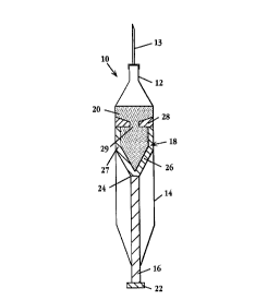
One embodiment of centrifuge syringe 10 according to the invention is
illustrated in
FIG. 1. Centrifuge syringe 10 includes a specimen container 14 with a central
orifice
formed by fitting 12 adapted for receiving a needle 13, a handle 16 and a
plunger 18.
Fitting 12 may be any type of locking tip adapted to hold a needle, for
example, a Luer-
WO 96/06679 ' PCT/US95111162
6
LockT" syringe tip. Alternatively, fitting 12 may be a sterile septum adapted
for connection
with sterile fluid bags and tubes, for example a SAFSITEn small wire extension
set with
reflux valve and Spin-Lockn adaptor available from Burron Medical Inc.,
Bethlehem,
Pennsylvania.
Handle 16 further preferably comprises knob 22 and a removable connection 24
to
plunger 18. As shown in FIGS. 1-4, plunger 18 is single piece, machined or
molded from
a plastic material. Known medical grade plastic materials may be used.
Plunger 18 as shown in FIG. 1 has a funnel-shaped bottom wall 26 that is
removable connected to the handle at connection 24. Side wall 27 preferably
closely fits the
container wall to permit sliding movement but provide an essentially fluid-
tight barrier
therearound. A top wall is formed by constriction member 28, which defines
central
opening 29. Alternatively, the outer diameter of side wall 27 may be slightly
undersized to
facilitate sliding and an o-ring seal provided between side wall 27 and
container 14.
Removable connection 24 may take the form of, for example, a screw fitting or
a snap-fit.
Preferably, connection 24 also provides for reattachment of handle 16. If
reattachment is
not desired, connector 24 may be designed such that handle 16 can be broken
off. A
suitable connection can be selected by those of ordinary skill in the art.
For use in density gradient separation plunger 18 is filled with a cell-
separation
density-gradient medium 20 before the introduction of a specimen. As is
understood by
persons of ordinary skill in the art, such materials have specifically defined
densities which
are selected based on the particular sample material being separated. Examples
of cell-
separation density-gradient media include sucrose, albumin and Ficolln. A
preferred
material is available from Pharmacia Fine Chemicals of Piscataway, New Jersey
and
Uppsala, Sweden under the trademark PERCOLLn. Preferably, the density gradient
material is filled to a level above the constriction member, or at least above
the top of
opening 29. For example, when using a standard SOmI syringe, having an inner
diameter
of about 2.8 cm, the gradient material is preferably filled to a level about
lmm or more
above constriction member 28. This fill level will help to prevent formation
during
centrifugation of an interface portion, as explained below, under constriction
member 28.
Referring to FIG. 2, the introduction of the specimen into centrifuge syringe
10 is
illustrated. Specimen 30 is drawn into the syringe through needle 13 secured
to fitting 12,
aided by the vacuum created by handle 16 and plunger 18 as the handle is
pulled out of
container 14, drawing the plunger away from fitting 12. The handle should be
pulled with
sufficiently low force and velocity to avoid mixing of the specimen with the
density gradient
CA 02198606 2000-02-14
W O 96106!>79 PCTlUS95111162
7
material onto which the sample is layered. Preferably, when the handle is
pulled at an
appropriate force, the sample will form a stream which adheres to the side of
the container
as it is drawn in, as shown in FIG. 2. This will reduce unwanted mixing.
Mixing of the
two materials is also minimized by the fact that the density of the specimen
is preferably
significantly lower than the density of the density gradient material. After
specimen 30 is
drawn into container 14, the container is maintained in an upright position
and the sample
lies on top of density gradient material 20.
Using needle 13, a sample such as peripheral blood may be drawn directly from
a
patient for analysis. The present invention thus ensures sterility of such a
sample by
eliminating direct handling of the sample prior to introduction into the
centrifugation
container. Alternatively, as illustrated in FIG. 11, using a sterile septum as
fitting 12,
blood previously collected by known techniques and stored, for example in a
sterile bag 33,
may be drawn into the centrifugation container through sterile tubing 35 or
other known
sterile connection means. The present invention thus ensures a sterile
transfer of sample
material on a larger scale in a completely closed system, again without direct
handling of
sample material.
Referring again to FIG. 2, once the specimen has been completely drawn into
the
container 14, and the handle 16 has been pulled so that the removable
connection 24 is
located at the lower central orifice of the specimen container 14, the handle
16 can be
removed for the centrifugation step.
FIG. 3 illustrates the centrifugation syringe after the centrifugation step
has been
performed. As shown, the handle has been detached from the plunger 18, which
is located
at the bottom end of the container 14. Centrifugation of container 14 has
resulted in a
pellet 32 being formed from the heavier portions of the specimen at the bottom
of the
plunger 18. Density gradient material 20 is located above pellet 32. An
interface portion
34, which contains the cells of interest, is formed between specimen diluent
33 and density
gradient material 20, and above constriction member 28.
Interface portion 34 may be removed from the centrifuge syringe 10 by
reattaching
handle 16 to connector 24 and ejecting the interface 37 and supernatant
diluent material 31,
as well as a portion of density-gradient material 20, as indicated by arrow 37
in FIG. 4.
Such ejection may be carried out while the syringe is in an inverted position,
as illustrated
in FIG. 4, or may be accomplished by ejection in an upright position, to
minimize inclusion
of density cell separation medium in the ejectate. Alternatively, interface
portion 34 can be
removed without reattachment of the handle, by opening the syringe below
fitting 12, such
WO 96/06679 . .; PCT/US95/11162
8
as at 39, and decanting the supernatant and interface material. Such opening
can be
accomplished by cutting or by means of an integral fitting in the container of
the syringe.
Further removal of density gradient material 20 and pellet 32 can be achieved
by
reattaching handle 16 to plunger 18 at connection 24. The handle then can be
pushed into
the container to aid the removal of the material if necessary.
According to one theory, the presence of the constriction member with a
restricted
opening provides a support or nucleus for formation of an intermediate surface
tension
across the container. This surface tension impedes the mixing of upper and
lower regions
(above and below the constriction member) of the tube when, for example, the
contents of
the upper region are ejected from the tube. Accordingly, the dimensions of the
opening
formed by the constriction member are dictated by the ability to form a
surface tension. A
constriction member that is little more than a rim around the interior of the
barrel may be
sufficient to form the necessary surface tension. Hence, the cross-sectional
area of the
opening formed by the constriction member may be as little as about 5~ or as
great as
about 95 9'0 of the horizontal cross-sectional surface area of the syringe. In
an exemplary
embodiment, where the syringe has an inside diameter of about 2.8 cm, an
aperture having
a diameter of about 0.5 cm is suitable.
In many applications, it will be desirable to collect only the supernatant
fraction
containing interface portion 34. In such cases, the pellet is discarded with
the syringe. In
other cases, the pellet can be removed by mechanical manipulation/disruption.
For
example, the syringe can be inverted and subjected to vortex mixing. Such
mixing will
disrupt the pellet into the adjacent liquid phase and will induce movement of
this liquid
phase and disrupted cells from the second or collection chamber of the syringe
into the first
chamber of the syringe.
An alternative embodiment of the present invention is shown in FIGS. 5-7.
Centrifuge syringe 40 has a plunger 42 formed from separate pieces and without
sidewalls.
Plunger 42 has a flat bottom plate 44, which may be formed by a washer formed
from
medical grade plastic such as polycarbonate. Bottom plate 44 is preferably
circumscribed
by a silicone or rubber seal 46 for the creation of an fluid-tight seal
between bottom plate
44 and the inside wall of the specimen container 48. Threaded or snap-fit
connection 51 is
provided in the bottom plate to removable attach handle 50. Plunger 42 has
fittings 52, to
connect bottom plate 44 to annular constriction member 54, which defines
opening 55.
Fittings 52 are preferably made of medical grade plastic, such as
polycarbonate.
Constriction member 54 is funnel-shaped, and preferably made of silicone or
rubber. There
!~ 96/06679 PCT/US95/11162
9
are preferably three fittings 52, as shown in perspective view of the plunger
and handle
portion of the device in FIG. 6, but there may be only two, or more than three
fittings, if
desired. The constriction member can be secured to the fittings by providing
stepped
recesses 56 in the constriction member, as shown in FIG. 7, for retaining
mushroom like-
s heads 57 on the fittings. Fittings 52 may be glued to bottom plate 44
preferably with
medical grade adhesive. Other means for connection may be devised by persons
skilled in
the art. The particular type of connection used is not critical so long as a
secure connection
between the parts is maintained.
An advantage of the present invention is that the low density material above
the
constriction member of the plunger is separated from material beneath by the
simple act of
ejecting it with the aid of the plunger, as described above. With reference to
FIG. 1, if the
opening at fitting 12 is large enough or if the container is opened as
described with
reference to FIG. 3, above, the cells of interest may be poured off. This
contrasts with
many conventional methods of unloading gradient separations using standard
straight-wall
centrifuge tubes, where materials are separated by carefully pipetting out of
the tube or,
alternatively, by puncturing the bottom of the tube and allowing the contents
of the tube to
slowly drip out into collection vessels. Thus, the present invention provides
a convenient,
simple means for unloading differentially separated materials. In addition,
unlike
conventional straight-wall tubes, if the centrifuge syringe is dropped or
accidentally
inverted, the contents of the upper and lower portions will not readily mix
due to the
presence of the constriction member. Moreover, once separation has taken
place, the
solution present above the constriction member can be mixed in the tube,
without disturbing
(or fear of contamination by) the contents of the syringe below the
constriction member.
Preferably this is done with the syringe in an inverted position as shown in
FIG. 4.
The separation of materials may be further enhanced by the addition of valve
60 to
the plunger, as shown in plunger 64, illustrated in FIG. 8. Valve 60 is
located at opening
62 in plunger 64. Valve 60 may be a one-way valve, or a valve that only opens
upon
application of a threshold centrifugal force. The valve can be formed by
providing flaps of
a softer material over hole 62. In a preferred embodiment, the force required
to open valve
60 would be about 850 times the normal force of gravity. Valve 60 thus allows
heavy cells
to pass through during initial centrifugation, and then keeps those cells in
place, allowing
for further processing, such as washing or mixing, of the lighter cells of
interest located
above the valve. In this way complete and final manipulation of the cells can
be performed
in a single sterile container.
21~~~~~
WO 96/06679 - PCT/US95/11162
The shape of opening 29, 55 is not limited to a circular shape, though in
general a
sloped or funnel-shaped constriction member forming a roughly circular shaped
annulus will
be preferred. The opening may assume other configurations, such as an oval
shape, a star
shape or other non-circular shape that allows passage of cells through the
opening.
5 Alternatively, or in addition, the opening may be formed by a plurality of
openings or may
be covered by a grid or mesh that allows passage of cells therethrough. Such a
mesh or
grid arrangement is also referred to herein as a plurality of openings.
FIGS. 9 A-F are illustrations of alternative shapes and designs for the
plunger of the
centrifuge syringe according to the invention. FIG. 9A shows plunger 70 with a
fluid-recei-
10 ving space having a flat bottom wall. FIG. 9B shows plunger 72 with a
pointed bottom
wall. Plunger 72 with the pointed bottom wall will allow the heavier cells to
form a better
pellet, which may be desired if the cells are to be collected. Alternatively,
plunger 74 with
a defined cell-collecting compartment 76 can be utilized to offer collection
of cells.
FIG. 9D shows plunger 70 that includes a cell trapping material 78, such as a
sponge or gel. Material 78 may contain compounds that specifically bind
certain cell types
or toxins that kill specific cell types. Material 78 may also be made of a
magnetic material
if desired.
FIGS. 9E and F show alternative embodiments of the plunger that facilitate
movement of the plunger within the container. FIG. 9E shows plunger 80 with
extending
contact points 82. The plunger 80 will only contact the container at these
points.
Similarly, in FIG. 9F, plunger 84 is shown with extending contact points 86.
FIG. 10 illustrates a further alternative embodiment of the centrifuge syringe
of
FIG. 1 with an additional constriction member. Dual constriction syringe 90
has a bottom
plate 92 connected to a first constriction member 94 by fittings 96. Second
constriction
member 98 is located above first constriction member 94 to create an
additional
compartment, to allow separation of cells of differing densities. Second
fittings 97 may be
used to secure second constriction member 98. Additional constriction members
could also
be added if a sample of several different densities is to be separated.
FIG. 10 also illustrates one embodiment of the removable and re-attachable
connection means between the handle 102 and the bottom plate 92. In this
embodiment, an
internal screw 100 forms the attachment means between the handle and the
bottom plate, so
that the handle 102 can be removed and then reattached after centrifugation.
Preferably, the centrifugation syringe according to the present invention
would be
provided as a sterilized complete unit with the density gradient material
already in place to
PCT/US95/11162
ACTIVAT~ ' CELL THERAPY, INC, et al. VOSSIUS & PARTNER
Our Ref :~-. A 1533 EP PATENTrIUWHI TE
31E3ERT3TFt. 4
2198fi06 3;6,5 NtUNCHE~I ~v, AUK, »g~
11
an appropriate level. In this way, sterility of the syringe Is guaranteed and
the user need
only open the sterile packaging to use the invention. Alternatively, the
syringe can be
provided in kit form with the density gradient solution separately provided
and the needle
and handle disattached. The user would then fill the plunger of the syringe
with density
gradient material, and then assemble the needle and handle before use.
6. EXAMPLES
The following examples illustrate, but in no way are intended to limit the
present
invention.
EXAMPLE 1
MATERIALS FOR ENRICHMENT OF CD34~ CELLS FROM BLOOD CELL
MIXTURE
i5
1. Periy~heral Blood and Eqne Ma~w_
Patients were hydrated and treated with cycIophosphamide (4 glm~(gmlm~)
administered by intravenous (I~ infusion over two hours through a central
venous catheter.
Twenty-four hours after the completion of the cyclophosphamide infusion,
patients were
treated with G-CSF (NEUPOGEN, Amgen, Thousand Oaks, CA) administered by subcu-
taneous (SC) injection at a dose of approximately IO y~glkg/d. Apheresis was
initiated upon
recovery of the white blood cell count (WBC) to equal or more than 1 x lO9IL.
Apheresis
was performed using a Cobe Spectra Cell Separator (Lakewood, Colorado) at. a
rata of
80 mIlmin (mllmln), for 200 min (total volume of I6 L).
Apheresed peripheral blood was applied directly onto the density gradient.
However, complete broad and bone marrow aspirates were processed to a buffy
coat
(removal of red cells) before they were applied onto the density gradient.
2. Preparation of Density radient~
"PERCOLL" solution was purchased from Phatmacia Biotech (Uppsala, Sweden)
and stored at 4°C according to the recommendation of the vendor. A
stock solution was
prepared by mixing 12 parts of "PERCOLL" with 1 part of 10 x calcium and
magnesium-
free phosphate buffered saline (PBS). The pH of the solution was adjusted to
7.4 and the
osmvlality to 280 mOsmlkg Ha0 (mOsmIKg H~0). For use in separating CD34' cells
in a
cell mixture, the stock solution was further diluted with calcium and
magnesium-free P8S to
a density of
AMEfaDEO SHED
~ 96/06679 PCTIUS95/11162
12
1.0605 ~ 0.0005 gr/ml and used at room temperature. By adjusting the density
of the
gradient to an accuracy of within ~0.0005 gr/ml of 1.0605 gr/ml,
reproducibility and
accuracy of cell separation was ensured. This was done using a high precision
digital
density meter such as DMA 48 (Anton PAAR USA, Ashland, VA). All procedures
were
performed under sterile conditions and at room temperature.
EXAMPLE 2
I LATION OF.CD 34+ PROGENITOR HEMATOPOIETIC CEL
USING CENTRIFUGE SYRINGE
The centrifuge syringe and the method of the invention can be used to isolate
CD34+ progenitor cells from patients treated with chemotherapy and granulocyte
colony
stimulating factor (G-CSF) as described in Example 1 above. These cells can
then be used
to repopulate the patient's lymphohematopoietic system.
Human peripheral blood mononuclear cells (PBMC) are obtained by apheresis of
patients treated with daily injections of G-CSF (l0ug/kg/day). Samples are
then processed
according to standard methods understood by persons skilled in the art.
Cells are resuspended in 25 ml of calcium-free, magnesium-free PBS and then
drawn into the syringe on top of 15'ml of PERCOLL~' solution in a 50 ml
conical
centrifuge syringe fitted with a plunger containing a constriction member, as
illustrated in
FIG. 1. This PERCOLLn solution has a density of 1.0605 g/ml (osmolality 28015
mOsm/kg HZO; pH 7.4). The diameter of the opening in the constriction member
of the
syringe preferably is about 0.5 cm. This volume of PERCOLLr" is sufficient
volume to fill
the container to a level higher than about lmm above the constriction member.
After the
sample is drawn in, the needle and plunger are detached. The centrifuge
syringe is then
centrifuged at about 850xg for 30 minutes at room temperature. The upper
fraction
containing CD34+ cells is collected by ejecting the sample into a sterile
container.
Cell type and purity in the collected fraction are tested according to
standard
methods to determine enrichment of functional CD34+ cells. For example, cells
can be
tested for presence of colony forming units (CFU; indicating committed
hematopoietic
progenitor cells), Long term culture initiating cells (LTC-IC; indicating
uncommitted
hematopoietic progenitor cells), natural killer (NK) cells, and natural
suppressor cell activity
in the interface fraction, according to methods known in the art. Using the
apparatus and
''O 96/06679 219 8 6 0 6 p~~S95/11162
13
method described above, the interface contains approximately 70-90% of the
CD34+ cells
and more than 90% of the CFU's.
While the invention has been described with reference to specific methods and
embodiments, it will be appreciated that various modifications and changes may
be made
without departing from the invention.