Note: Descriptions are shown in the official language in which they were submitted.
WO 96/06582 PCTIUS95/10734
SYRINGE FOR USE IN PERFORMING GEL-INJECTION ADJUSTABLE KERATOPLASTY
BACKGROUND OF THE INVENTION
1. Field of Invention
The present invention relates to a technique for infra-operative correction
of refractive error to eliminate the need for eyeglasses and contact lenses.
More
particularly, the invention relates to a technique for surgically correcting
myopia
and astigmatism by controlled interlamellar annular injection of a polymeric
gel
at the corneal periphery so as to modify the corneal curvature, while sparing
the
central optical zone.
2. Description of the Related Art
The ideal surgical procedure in refractive surgery could be defined as one
which allows all the advantages of eyeglasses or contact lenses, that is, one
which
offers effectiveness or a wide range of corrections, allowing correction of
ametropias both large and small; accuracy or predictability, allowing for
correction of a certain amount of ametropia with precision; alterability or
reversibility, so that if ocular refractive changes occur it might be possible
to
adjust the correction again; innocuousness or without complications, that ist
the
procedure does not lead to adverse situations; visual quality without
alterations
in the size of the image or of the visual field; technical simplicity, that
is~ not
requiring sophisticated techniques to be put into practice; availability; low
cost;
and aesthetically acceptable on the part of the patient.
3 0 A number of surgical techniques have been proposed which have the object
of
infra-operative correction of refractive error. Examples are Radial
Keratotomy,
1
WO 96/06582 O 2 1 9 g g ~ 5 PCT/US95/10734
Keratomileusis, Epikeratoplasty, and Excimer Laser Reprofiling of the Corneal
Surface also known as Photo Refractive Keratoplasty (PRK). These methods
work with the characteristics of the cornea in order to modify either its
curvature or its refractive index. Perhaps the more widespread method and the
one which best approaches the objects noted above is radial keratotomy,
basically
because it can be performed at low cost without the need for additional
materials. However this procedure has a number of limitations, including the
presence of adverse situations (glare) and a lack of stability, predictability
(hypercorrection or hypocorrection) and reversibility. The remaining
procedures described and presently in use demand very sophisticated surgical
equipment requiring very specialized training and also the use of syntl'~etic
or
natural materials that reduce the likelihood of the procedure being available
in
the average clinic. Further, with the present surgical techniques it is not
possible
to accurately predict the patient's refractive outcome, due in part to corneal
hydration and subsequent wound healing processes.
Yet a further prior procedure made use of a rubber annular implant
(intrastromal rings) which were surgically inserted to alter corneal
curvature.
However, that procedure, which was introduced in 1986 by the inventor of the
subject procedure, involved stromal delamination of the central optical zone
and, in addition, precluded infra-operative or post-operative adjustment of
the
patient's refractive power.
Therefore, there remains a need for a surgical technique which can achieve
2 5 infra-operative correction of refractive error to eliminate the need for
eyeglasses
and contact lenses by modifying the corneal curvature which avoids
delamination of the central optical zone and permits infra-operative and post-
operativeadjustment of the patient's refractive power, and which is also
reversible. There further remains a need for such a technique wherein the
2
CA 02198905 2002-09-27
68216-7
surgical equipment is relatively inexpensive and only
moderate skills are required.
SUMMARY OF THE INVENTION
The present invention provides a device for use in
injecting a gel into an annular channel formed at a
predetermined depth in the cornea through an incision made
through the surface of the cornea of an eye for the purpose
of altering the radius of curvature of the cornea,
comprising: a tubular member capable of insertion into an
annular channel, said member having a curved end of a
corkscrew shape to facilitate the insertion through the
incision into the cornea and completely into the channel;
means, coupled to said tubular member, for storing a gel,
comprising a presterilized cartridge containing said gel;
and means, associated with said storage means, for
transporting the gel from said storage means through said
tubular member into the channel.
The present invention concerns a technique and set
of surgical instruments which allow for the surgical
correction of myopia and astigmatism by controlled injection
of a polymeric gel or elastomer at the corneal periphery
without interfering with the central cornea and endothelium.
More particularly, the surgical technique of the invention
allows for subtle modification of the corneal curvature by
interlamellar injection of a synthetic or natural polymeric
gel at the corneal periphery while sparing the optical zone.
In accordance with the surgical procedure, a partial depth,
radial incision is made outside the optical zone. A
corkscrew-like delaminator, for example a helicoidal
spatula, is inserted through the incision to form an annular
track centered about the corneal apex. A modified needle is
then fully or partially inserted in the interlamellar track
3
CA 02198905 2002-09-27
68216-7
formed by the delaminator and a transparent gel is manually
injected while retracting the needle, thus filling the
annular channel, or the gel can be directly injected into
the channel. By monitoring the corneal shape with an
automatic keratometer, for example, emmetropia can be
achieved intra-operatively by controlling the amount of gel
injected and by corneal massage. Using a surgical
keratometer mounted to an operational microscope, the final
corneal power is adjusted by removal of a portion of the
gel.
Thus, the use of an injectable substance gives the
method, in addition to technical simplicity, the possibility
of adjusting the quantity of material to obtain the desired
correction. It is also possible to extract or remove this
material to reverse the procedure, or augment its volume if
necessary.
3a
WO 96/06582 ~ ~~ PCT/US95/10734
Other objects, features, and characteristics of the present invention, as well
as the methods of operation and functions of the related elements of the
structure, and the combination of parts and economies of manufacture, will
become more apparent upon consideration of the following description and the
appended claims with reference to the accompanying drawings, all of which form
,
a part of this specification, wherein like reference numerals designate
corresponding parts in the various figures.
BRIEF DESCRIPTION OF THE DRAWINGS
FIGURE 1 is a schematic perspective view showing the use of a caliper to
locate and mark the central cornea and incision site in accordance with the
invention;
FIGURE 2 is a schematic perspective view of an ultrasonic pachymeter for
measuring corneal thickness in accordance with the procedure of the invention;
FIGURE 3 is a perspective view of a diamond knife equipped with a
micrometer;
FIGURE 4 is a schematic cross-sectional view of an incision formed i n
accordance with the invention;
FIGURE 5 is a schematic elevational view showing the incision of the
2 5 cornea in accordance with the invention;
FIGURE 6 is a schematic elevational view showing the insertion of a blunt
spatula in accordance with the invention for separating the lamella;
4
WO 96/06582 ~ ~ ~ PCT/US95/10734
FIGURE 7 is a schematic elevational view of the insertion of a shoehorn-
like device for facilitating insertion of a corkscrew delaminator in
accordance
with the invention.
'J
FIGURE 8 is a schematic elevational view of the insertion of a corkscrew
delaminator;
FIGURE 9 is a perspective view, partly in cross-section and partly broken
away for clarity, illustrating the formation of an annular track with a
corkscrew
delaminator in accordance with the invention;
FIGURE 10 is a schematic elevational view of the incision site following
complete insertion of the corkscrew delaminator;
FIGURE 11 is a schematic cross-sectional view showing the injection of gel
with the needle in accordance with the invention;
FIGURE 12 is a perspective view, partly in cross-section and partly broken
away for clarity, showing the retraction of the needle while the gel is
injected;
FIGURE 13a is a schematic cross-sectional view of the cornea with the
needle within the annular track;
FIGURE 13b illustrates the track with gel so as to bulge the corneal
posterior lamellae and flatten the central corneal optical zone;
FIGURE 14 is a schematic perspective view illustrating localized corneal
shear variations generated at the limbus which produces astigmatism and their
effect on the gel injected in accordance with the invention;
5
W0.96/06582 9 ~ ~ PCT/US95/10734
FIGURE 15 is a schematic top plan view of a modified procedure in
accordance with the invention;
FIGURE 16 is a schematic cross-sectional view of a gel injector suitable for
use in accordance with the invention;
FIGURE 17 is a plan view of a track width equalizer for use in accordance
with the invention;
FIGURE 18a is a perspective view, partly in phantom, of a different
embodiment of the corkscrew delaminator in accordance with the invention;
FIGURE 18b is a cross-sectional view of the helicoidal section of the
corkscrew delaminator of FIGURE 18a;
FIGURE 18c is fragmentary enlarged view of one embodiment of the tip of
the corkscrew delaminator of FIGURE 18a;
2 0 FIGURE 18d is a fragmentary enlarged view of another embodiment of the
tip of the corkscrew delaminator of FIGURE 18a;
FIGURE 18e is a fragmentary enlarged view of still another embodiment of
the tip of the corkscrew delaminator of FIGURE 18a;
FIGURE 19 is a schematic cross-sectional view of the annular track formed
in the cornea with the delaminator shown in FIGURES 18a and 18c;
fi
WO 96/06582 ~ ~ ~ ~ PCT/US95/10734
FIGURE 20 is a schematic cross-sectional view of the annular track formed
in the cornea with the delaminator shown in FIGURES 18a, 18d and 18e;
FIGURE 21a is an elevational view of an alternative embodiment of a
shoehorn-type device for use in the present invention;
FIGURE 21b is a side view of the device shown in FIGURE 21a;
FIGURE 22a is a perspective view of a channel starting device for use i n
the present invention;
FIGURE 22b is a fragmentary enlarged view of the end of the device shown
in FIGURE 22a;
FIGURE 22c is a across-sectional view taken along lines 22c - 22c of FIGURE
22b;
FIGURE 22d is an elevational view of the device shown in FIGURE 22a;
2 0 FIGURE 23a is a perspective view of a marking instrument for use in the
present invention;
FIGURE 23b is a fragmentary view of an alternative embodiment of the
instrument shown in FIGURE 22a; and
FIGURE 24 is a perspective view of a syringe for use in the present
invention.
7
WO 96/06582 8 9 O ~ PCT/US95/10734
DETAILED DESCRIPTION OF THE PRESENTLY
PREFERRED EXEMPLARY EMBODIMENT
The exact physical properties of the cornea and scleral Timbal tissues have
not been determined. However, if the cornea resists stretching, it can easily
be
delaminated. The large differential in modulus of elasticity between the two
orthogonal corneal planes is responsible for the effects observed with the
procedure of the invention.
The corneal lamellae are laid parallel to Bowman and Descemet layers and
have a radial elastic modulus of approximately 5x10⁶ N/m2 and a transverse
elastic modulus of 3x10⁴ N/m2. These lamellae are weakly bounded and can be
easily separated.
The scleral-Timbal region forms an annulus (the corneal limbus) much
more rigid than the cornea and act as a reference frame with respect to
corneal
deformation. With the procedure of the invention, the limbus geometry and
physical characteristics remain stable (unchanged). Therefore, the procedure
of
the invention causes a redistribution of the nominal corneal stresses produced
by
the positive intraocular pressure. This redistribution causes a change in the
corneal shape with a flattening of the central corneal radius of curvature and
a
steepening of the site of gel injection. However, the inner and outer corneal
surface areas are kept constant. Therefore, little change occurs in stress
applied
along the individual lamella. As the laminar ~ network is kept intact except
for
the small incision site, a very minimal wound healing effect is expected.
Thus,
the keratometric changes produced by the procedure of the invention are
expected to be stable in long-term follow up.
8
WO 96/06582 O 5 PCTIUS95/10734
The procedure in accordance with the invention is as follows:
First, the central cornea and the incision site are defined and marked o n
the eyeball 10 with the help of a standard caliper 12 as shown in FIG. 1.
Specifically, the surface of the eyeball is dried with blotting paper and
marked
with a compass, for example a Castroviejo Caliper #E 2404 available from
Storz,
Inc. having blunt needles or points which have been previously soaked with ink
from an epidermic labeler, such as #150 available from Devon, Inc. The ink
soaked needles are placed on the eyeball to mark the corneal center and the
incision site between about 2.5 and 3.5 mm from the apex, a distance
corresponding substantially to the internal radius of the corkscrew
delaminator
used to form the intra-laminar canal in accordance with the invention.
Additional marks may be made at that predetermined distance from the corneal
center to mark a guide for the dissector to follow during the surgical
maneuver.
Once the central cornea and incision site have been selected and marked, the
corneal thickness is determined with a conventional ultrasonic probe and
pachymeter 14 at the corneal center and at the incision site as shown
schematically in FIG. 2.
An instrument for facilitating the marking of the cornea for the incision
site is shown in FIG. 23a. Marking instrument 15 consists of a handle section
15a
and a guide section 15b. Guide section 15b has an opening 15c and a pair of
parallel arms 15d and 15e which are connected by a rounded section 15f.
Parallel
arms 15d and 15e terminate in a pair of points 15g. Instrument 15 is used by
aligning the apex of rounded section 15f with the corneal center, such that
points
15g of parallel ends 15d and 15e will indicate the inner diameter of the
channel
formed by the delaminator. In the present embodiment, the distance between
points 15g is preferably 5 mm. Finally, a raised section 15b on handle section
15a
offers the surgeon a comfortable and secure grip when using instrument 15.
9
WO 96/06582 PCT/US95/10734
~2 ~ gg 905
A second embodiment of instrument 15 is shown in FIG. 23b as 15'.
Insttument 15' contains a central prong 15j located between points 15g. Prong
15j
acts a centering device, which when positioned at the corneal center, locates
points 15g at the proper incision sites.
A partial depth incision 20 is then performed at the incision site with a
diamond knife 16 which is equipped with a micrometer (not shown) having a
footplate 18 (FIG. 3). The blade length is adjusted to about 86% of the
corneal
thickness, as measured by the pachymeter. The knife is then used to form a
radial incision to a depth D of between 75% and about 85% and most preferably
between about 75% and about 80% of the corneal thickness T. To obtain a flat-
bottomed incision with vertical walls, the diamond blade can be reversed and
moved radially with respect to the center of the cornea (FIG. 4). The
foregoing
two step procedure with the diamond blade can be avoided by the use of a flat-
edged blade. The insertion of the diamond knife 16 to the predetermined 80% ~
5% depth of the cornea 22 is illustrated in particular in FIG. 5. The incision
is
performed to a length L of about 1.0 mm or less and most preferably about 0.4
mm which is preferably between 0 to 0.5 mm less than the width of the
delaminator to be used. The incision is preferably made as small as possible
so as
to minimize wound healing time following the procedure.
Once the incision has been formed, a spatula 24 such as, for example, a
Castroviejo cyclodialisis spatula #E 0292 available from Storz, Inc. having
dimensions identical to that of the helical spatula, for example about 1 mm or
less in width and 200 micrometers in thickness and having an end which is
slightly modified by being polished to a point, is inserted through the
incision to
separate the lamella in the plane of the base of the incision (FIG. 6). The
particular width of the spatula used of course depends upon the length of the
WO 96106582 0 2 1 9 8 ~ O ~ pCT~S95/10734
incision. Alternatively, once the incision has been made to the desired depth
in
the cornea, an instrument is inserted into the incision to locate the proper
plane
along which to separate the lamella and form the intralamellar canal i n
accordance with the invention. A channel starting instrument 25 (FIGS. 22a-d)
consists of a handle 25a, a central section 25b, and end section 25c. Central
section
25b is preferably coaxial with the central axis of handle section 25a, and is
formed
from a flat wire. As can be seen in FIG. 22d, end section 25c, which is shown
as
curved but may be linear, is attached to central section 25b at an acute angle
a to
the central axis through sections 25a and 25b, which angle is preferably
between
40° and 45°. End section 25c consists of a curved or arcuate
portion 25d and a
smaller portion 25e which is also curved. Preferably, section 25c is
constructed
having the same radius of curvature of the helical spatula to be used for the
procedure. Portion 25d is formed out of a flat wire, while portion 25e
consists of
a semicircular section of curved wire, as can be most clearly seen in FIG.
22c.
Instrument 25 is used by inserting end section 25c into incision 20 until
section
25c contacts the bottom of the incision. Handle section 25a is then rotated in
a
manner such that the lamella is separated along an arcuate path by curved
portion 25e of end section 25c. Handle section 25a contains a knurled portion
25f
to assist the user in gripping the instrument.
Following the initial separation of lamella with the blunt spatula 24 and
starting the channel with instrument 25, a shoehorn-type device 26 is inserted
through the incision and laterally between the lamella as shown in FIG. 7. The
shoehorn-type device 26 can be made of plastic or stainless steel or any
material
which is more rigid than the radial elastic modulus of the cornea, which
measures approximately 5x10⁶ N/m2. The thickness of device 26 may approach
300 microns, with the preferred range being between approximately 50 to
approximately 100 microns.
11
WO 96/06582 n ' ~ ~ O ~ pCT~S95/10734
An alternative embodiment of shoehorn-type device 26 is more clearly
shown in FIGS. 21a and 21b. Referring now to FIGS. 21a and 21b, device 26' is
a
planar member having an upper horizontal section 26a' and a downwardly
depending extension 26b'. The upper end 26c' of extension 26b', which is
narrower than upper section 26a', depends from the central region of section
26a'
at an approximate angle of 90°, and extension 26b' may be either
slightly curved
or straight as it continues away from upper section 26a' until it reaches its
lower
end ~26d', which end is rounded for ease of insertion into the incision. The
length of extension 26b' may range between 1 and 6mm, while its width should
be less than or equal to the width of the delaminator.
Ideally, the radius of curvature of extension 26b' should match the radius
of curvature of the helical delaminator.
Extension 26b' of device 26' is inserted into the incision until upper section
26a' contacts the upper surface of cornea 22. The curvature of extension 26b'
of
device 26' facilitates the insertion of spatula 28 to the proper plane for
delamination which has been selected and started by instrument 25 and guides
spatula 28 as it begins to form annular chamber 30. The design of shoehorn-
type
device 26' also provides for easier insertion and extraction of device 26'
from the
incision site.
The shoehorn-type instrument is utilized for facilitating insertion of a
corkscrew delaminator or helicoidal spatula 28, for example a custom made
Archimedes-screw dissector which is inserted behind or under shoehorn device
26, as shown in FIG. 8. The flat corkscrew delaminator is used to carve a
circular
canal between the two corneal lamellae in which a gel such as a silicon gel is
subsequently injected. The illustrated corkscrew delaminator consists of a
flat
wire between 0.25 and 4.5 mm, preferably about 1 mm in width as in the
12
WO 96/06582 PCT/US95/10734
preferred embodiment, again depending upon incision length and 0.1 mm in
thickness, and is curved to about 380°, that is superimposed by
20°. The spatula's
edges are blunt or rounded as is its end. The inner diameter of spatula 28 may
fall within the range of 2.0 to 10.5 mm, with the preferred embodiment
measuring approximately 5 mm.
As shown in FIG. 9, with the corkscrew delaminator, an annular track is
made through the cornea at the preselected plane. A full 360° twisting
motion of
the helicoidal spatula 28 delaminates the cornea completely at the radial
location
of the incision, circumferentially of the central corneal zone. However, the
central and paralimbal zones of the cornea are not delaminated as the
helicoidal
spatula 28 limits the delamination to an annular path at the radial locus of
the
incision. As shown in FIG. 10, the annular delaminafion results in the
formation of an annular chamber or canal 30 opening on either side of the
initial
incision 20. Following the annular delaminatzon, the helicoidal spatula 28 is
removed by rotating the same with a reverse 360° twisting motion. When
spatula 28 is removed, it may be necessary to ensure that the canal 30
openings
on either side of the initial incision 20 are uniform to allow for the proper
flow
of gel through canal 30. A track width equalizer 31 (FIG. 1~ is used for this
purpose. Equalizer 31 contains a handle portion 31a, a curved portion 31b, and
a
rounded end portion 31c. End portion 31c, which typically measures 0.5 mm in
diameter, is inserted through incision 20 into each canal 30 opening, thus
insuring that the injected gel will pass through canal 30. Equalizer 31 is
also used
to remove the gel if necessary.
~ w
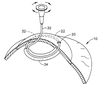
A small corkscrew-like cannula 32, in the range of ~9 to 30 gaug~ in size,
. ,.r
preferably~30 gauge)in the present embodiment, is then inserted into the
annular
channel 30. A syringe, for example filled with a biocompatible gel, is
attached to
the corkscrew cannula 32 following insertion or prior to insertion into the
13
A~ENpE.~ SHEET
WO 96/06582 p 2 1 9 ~ ~ O ~ PCT/US95110734
annular channel 30. Examples of suitable biopolymeric materials for the gel
are
known biocompatible hydrogels (e.g. acrylic hydrogels, polyethylene oxides),
silicone-based cross-linked elastomers, and other biopolymers (e.g. cross-
linked
hyaluronic acid). One gel which has been found to be suitable for use in the
present invention is a crosslinked polyethylene oxide (PEO) gel which is
prepared
by dissolving a sample of PEO in a Balanced Salt Solution (BSS), placing the
solution in a sealed canister, removing any free oxygen from the interior of
the
canister, replacing the oxygen with an inert gas such as argon, irradiating
the
canister to crosslink and sterilize the PEO, and loading the sterilized PEO
into a
sterile syringe. This method is more completely described in co-pending
application Serial No. , which was filed on the same date herewi th
and is assigned commonly herewith. The disclosure of the pending application
is incorporated herein by reference. This gel has a modulus of elasticity of
approximately 1.7x10³ n/m2 and an index of refraction of 1.334. Once the
corkscrew connula is inserted, injection is started (FIG. 11).
It is also possible to inject gel 34 directly into canal 30 by using an
automatic injector 40 (FIG. 16). Injector 40 consists of a powered injector
unit 40a
and a gel cartridge unit 40b. Unit 40a can be controlled by a manual switch on
the
unit, or by a foot pedal switch. Cartridge 40b is a disposable sealed unit to
prevent
contamination, and fits onto unit 40a for easy, sterile operation. Injector 40
provides a consistent force to inject gel 34 into canal 30.
Another alternative for injecting gel 34 into canal 30 is shown in FIG. 24.
A syringe 42 having a gel storing compartment 42a, a plunger 42b captively
held
with compartment 42a, and a cannula 42c coupled to the end of compartment 42a
may be used to inject gel 34. Cannula 42c has a curved end 42d which is
helicoidal or spiral shaped such that it can be easily inserted through
incision 20
and into canal 30, thus insuring that gel 34 will completely fill canal 30 as
syringe
14
WO 96/06582 Q 2 1 9 8 ~ ~ ~ PCT/US95/10734
42 is operated. The curvature of end 42d of cannula 42c is preferably matched
to
the curvature of helicoidal spatula 28.
A small amount of gel escapes from the corneal incision due to over
5 pressurization (FIG. 11). Furthermore, subsequent to needle removal, an
external massage is performed in order to evenly distribute the gel inside the
canal. The remaining gel that appears at the incision is grabbed with forceps
and
p.'i ,"..,
cut, for example with scissors. Once theC30 gauge~needle has been removed, gel
fills the track creating a bulging of the corneal posterior lamellae and a
flattening
10 of the central corneal optical zone. FIG. 13a is a corneal cross-section
illustrating
the displacement of the corneal lamella when the needle 32 is disposed with an
annular track 30. Once the needle has been retracted and gel fills the annular
canal 30, a bulging of the cornea posterior lamella and a flattening of the
central
corneal optical zone occurs (FIG. 13b). Up to 13 diopters of flattening have
been
1 S obtained in cadaver eyes with the technique of the invention. By removing
a
portion of the gel from the channel, the amount of corneal flattening can be
reduced until emmetropization has been achieved. Intraoperative keratometry
is suggested for determination of optimal corneal radius of curvature although
other techniques could be employed. The amount of correction obtainable is a
20 function of the inner diameter and wire width of the delaminator, in
addition to
the amount of gel used.
Referring to FIG. 14, astigmatism is thought to be produced by localized
corneal shear variations generated at the liinbus. With the process of the
25 invention, a reduction of existing astigmatism was observed experimentally.
This phenomenon can be explained by a localized variation in gel distribution
along the annular channel as shown. Indeed, because the pressure of the gel is
constant, it equilibriates the radial stresses along the corneal meridian.
15
AMENDED SHEET
~~~98909
WO 96/06582 PCT/US95/10734
Astigmatism may also be corrected in accordance with the present
invention by using a modified procedure as illustrated schematically in FIG.
15.
In accordance with this modified procedure, a pair of arcuate channels 50 of
approximately 90° in arc length and centered about the corneal center
52 are
made by a stromal delaminator (not shown) which is a shorter version of the
helicoidal delaminator discussed in detail above. A gel, as discussed above,
is
then injected into each of the arcuate channels 50 under keratometric control.
T'he magnitude of the astigmatic optical correction in diopters is a function
of the
amount of gel injected into each of the arcuate channels 50, the size, that is
diameter, width and angle of the arcuate channels, and the position of the two
arcuate channels with respect to the patient's corneal astigmatic (flattest)
axis 54.
As noted above, minimizing the length of the incision, in accordance with
the invention, reduces wound healing time. To further reduce wound healing
time, following gel injection and the removal of any excess gel, the corneal
incision can be instantly closed-shut by applying a very small amount of
collagen
gel to the upper lips of the wound and cross linking it with ultraviolet
radiation.
Such sealing of the incision eliminates post operative patching of the eye and
thus allows the patient to walk away from surgery without impediment.
Ultraviolet cross linkable collagen gels are fabricated by several
manufacturers
and a suitable ultraviolet cross linkable collagen gel among those available
could
be readily ascertained.
Another advantage of the present invention over current surgical
techniques is reversibility. Should it become necessary to readjust the
curvature
of the cornea at some later time, such as a year later, a linear incision is
made
above the channel to a depth which intersects the channel. The gel can then be
removed by massaging the cornea or additional gel may be added as previously
~6
WO 96/06582 O 2 1 9 g g ~ 5 PCT/US95/10734
described until the desired corneal shape is obtained and then the incision is
closed using the above described technique.
FIGS. 18a - 18e show alternative embodiments of a helicoidal delaminator
or spatula for use in the present invention. Spatula 44 consists of a
helicoidal
section 44a, a planar support section 44b, and a handle section 44c which is
fixed
to support section 44b by welding or a similar attachment means. Handle 44c
allows helicoidal spatula 44 to be more easily manipulated during surgery. The
design of handle 44c, consisting of an annular cylindrical ring, is important,
as it
allows the surgeon an unobstructed view of the corneal surface as he is using
the
device. Handle 44c of spatula 44 is provided with a pair of indicating marks
60
and 62 on its upper surface. Marks 60 and 62 may be used to indicate relative
position of section 44a of spatula 44 when it is within channel 30 that has
been
formed in cornea 22, as section 44a is no longer visible. Mark 60 is used to
indicate the position of the end of the nose portion of helicoidal section 44a
so
that the surgeon can determine when he has completed the formation of
channel 30. Mark 62 indicates where the overlapped portion of helicoidal
section
44a begins. Marks 60 and 62 facilitate the use of spatula 44 during the
procedure
by eliminating uncertainty with respect to its position.
FIG. 18b shows the cross-section of the helicoidal section 44a of spatula 44.
It can be seen that section 44a imitates the radius of curvature of the
cornea,
which is approximately 7.8 millimeters, such that channel 30 closely parallels
the
surface curvature of the cornea.
In the embodiment shown in FIG. 18c, spatula 44 contains a nose portion
44d which is tapered along the top edge toward its end. When spatula 44 is
used
in the corneal surgical procedure described in the present invention, nose
portion 44d creates a lamellar flap within the track formed by spatula 44
within
17
WO 96/06582 v ~ ~ ~ 9 8 9 0 5 PCT/US95l10734
the cornea. As can be seen in FIG. 19, lamellar flap 22a within cornea 22 aids
i n
sealing channel 30 from incision 20, thus preventing gel from freely escaping
from incision 20. Thus, this modification to spatula 44 eliminates the need
for
adhesives or the like to bond the incision in the cornea. Nose portion 44d
tends
to keep channel 30 in the lower portion of the cornea.
FIG. 18d shows an alternative embodiment of the end of spatula 44. Nose
portion 44e is tapered along the bottom edge of section 44a toward its end. W
h a n
spatula 44 is used in the corneal surgical procedure described in the present
invention, nose portion 44e creates a channel as shown in FIG. 20. This
channel
30 intersects incision 20 directly. Nose portion 44e tends to keep channel 30
in
the upper portion of cornea 20.
FIG. 18e shows another alternative design for the end of spatula 44. Nose
portion 44f is v-shaped at the end of spatula 44, and also creates a channel
as
shown in FIG. 20. Nose portion 44f tends to keep channel 30 in the middle
portion of cornea 20.
While the invention has been described in connection with what is
presently considered to be the most practical and preferred embodiment, it is
to
be understood that the invention is not to be limited to the disclosed
embodiment, but on the contrary is intended to cover various modifications and
equivalent arrangements included within the spirit and scope of the appended
claims.
Indeed, while the presently preferred incision location and depth which
have been specified have exemplary dimensions of instruments used in
connection with the inventive procedure, it is to be appreciated that the gel
viscosity, volume and disposition within the surgical annular track as well as
the
18
WO 96/06582 ~ ~ ~ 5/10734
diameter of the track, width, depth and location are all parameters in the
refractive change obtained in accordance with the invention.
19