Note: Descriptions are shown in the official language in which they were submitted.
w0 96/07451 ~ ~ ~ ~ pCT~1S95/11246
-1-
PHOTOTIiERAPEUTIC APPARATUS
~,iac ~onnd of the Invention
The technical field of this invention is phototherapy and, in particular,
methods and
devices which employ optical fibers or other flexible light waveguides to
deliver radiation to
a targeted site.
Fiber optic phototherapy is a increasing popular modality for the diagnosis
and/or
treatment of a wide variety of diseases. For example, in surgery, infrared
laser radiation will
often be delivered to a surgical site via a hand-held instrument incorporating
an optically
transmissive fiber in order to coagulate blood or cauterize tissue. Similar
fiber optic delivery
systems have been proposed for endoscopic or catheter-based instruments to
deliver
therapeutic radiation to a body lumen or cavity. U.S. Patent No. 4, 336,809
(Clark) and U.S.
Reissue Patent No.
RE 34,544 (Spears) disclose that hematoporphyrin dyes and the like selectively
accumulate
in tumorous tissue and such accumulations can be detected by a characteristic
fluorescence
under irradiation with blue light. These patents further teach that cancerous
tissue that has
taken up the dye can be preferentially destroyed by radiation (typically high
intensity red
light) that is absorbed by the dye molecules during phototherapy.
Others have proposed the use of fiber-delivered radiation to treat
artherosclerotic
disease. For example, U.S. Patent 4,878,492 (Sinofsky et al.) discloses the
used of infrared
radiation to heat blood vessel walls during balloon angioplasty in order to
fuse the endothelial
lining of the blood vessel and seal the surface. Another application of fiber-
delivered
radiation is disclosed in U.S. Patent 5,053,033 (Clarke) which teaches that
restenosis
following angioplasty can be inhibited by application of UV radiation to the
angioplasty site
to kill smooth muscle cells which would otherwise proliferate in response to
angioplasty-
induced injuries to blood vessel walls.
Nonetheless, a number of problems limit the expanded use of fiber-optic
phototherapy. Typically, an optical fiber emits light from only its end face.
Thus, the
emitted light tends to be focused or at best divergent in a conical pattern
and, therefore,
exposes only a small region directly in front of the fiber's distal end. The
small exposure area
limits the power available for phototherapy since overheating of the target
tissue must often
be avoided.
Although "sideways-emitting" fibers have been proposed to permit greater
flexibility
in phototherapy, this approach still does not allow uniform irradiation of
large volumes of
tissue and can also be ill-suited for applications where circumferential
uniformity is desired.
Because sideways-emitting fibers expose limited regions, they do little to
alleviate the
problem of "hot spots" which limit the intensity of radiation which can be
delivered via the
fiber to the treatment site.
PCT/US95/11246
WO 96/07451
-2-
Others have proposed diffusive tips for optical fibers to enlarge the region
which can
be irradiated and/or reduce the potential for overexposure. However, diffusive
tips have not
been satisfactory for many therapeutic purposes because of their complexity of
manufacture
and/or because the radiation may not be scattered uniformly enough to
alleviate the problem
of "hot spots." Prior art diffusive tip structures have not be capable of
delivering high power
radiation, e.g., on the order of ten watts or more, to facilitate
photocoagulation therapy or the
like.
There exists a need for better apparatus for fiber-optic phototherapy. In
particular,
diffusive fiber tip assemblies which can provide circumferential (or large
angle) exposure
regions in radial directions (e.g., sideways) relative to the fiber axis
without hot spots would
satisfy a long-felt need in the art. Moreover, diffusive assemblies which
illuminate or
irradiate an azimuthal angle of less than 360° would meet a
particularly important need in the
field of minimally-invasive, phototherapeutic surgery. Similarly, diffusive
assemblies that
provide graded or broadly-cast exposure patterns, or otherwise predefined
patterns of light
distribution would also meet particular needs. In addition, diffusive fiber
tip assemblies
which can extend the longitudinal extent of irradiation and provide greater
flexibility during
use would also satisfy a need in phototherapy.
In another application, phototherapeutic instruments can be employed to treat
electrical arrhythmia of the heart. In such applications, a catheter having a
fiber optic
component is fed via a major artery into a patient's heart. Once inside the
heart, a catheter
senses electrical impulses with electrical contacts on its outer sheath or
other catheter
elements in order to locate the source of arrhythmia . Once located, the
phototherapeutic
component is activated to "ablate" a portion of the inner heart wall. By
coagulating the tissue
in the vicinity of the arrhythmia source, the likelihood that the patent's
heart will continue to
experience arrhythmia is thus reduced.
In other applications, laser radiation can be used in conjunction with a
similar catheter
instrument inside a patient's heart to increase blood flow to oxygen starved
regions of the
heart muscle. In such procedures, the laser radiation is used to form small
holes into the heart
muscle so that the oxygen-depleted tissue is bathed with blood from the
ventricular cavity.
In all of these applications, there is the potential for damage to the
patient's internal
organs, especially the heart, if the light-emitting fiber is inserted too far
into the patient's
tissue. Particularly, in the case of the heart muscle, perforation of the
heart wall can have
very dangerous effects.
Accordingly, there exists a need for better apparatus for fiber-optic
phototherapy. In
particular, devices that can "stop" an optical fiber from perforating a
patient's organs would
meet a particularly important need in the field of minimally-invasive
phototherapeutic
surgery. Moreover, a device that can help stabilize the phototherapeutic
instrument in
operation (such as within the chambers of a rapidly beating heart) would also
be particularly
useful.
w0 96/07451 ~ ~ ~ PCT/US95/11246
-3-
In yet another application, phototherapeutic devices can be useful in
sterilizing
medical instrument lumens. For example, endoscopic instruments are complex and
expensive medical devices, which permit the clinician to view the internal
organs and
structures of a patient's body. These instruments are typically reused and,
therefore, must be
sterilized after each use. Moreover, because many endoscopes are used
repeatedly
throughout a day, sterilization of the instruments must be performed rapidly
in busy clinics.
Conventionally, endoscopes are sterilized using a chemical bath. The internal
lumens
of the instrument will either be soaked in a sterilizing liquid or flushed
with the sterilizing
liquid.
Unfortunately, conventional techniques can sometimes be less than totally
effective.
The sterilizing liquid may not penetrate the entire lumen or may not be
sufficiently strong to
achieve the desired antimicrobial effect. Moreover, the endoscope lumen may
have
accumulated cellular debris that cannot be simply flushed out and such debris
may harbor
microbes that are not destroyed in the cleaning process.
1 S Accordingly, there exists a need for better methods and devices for
sterilizing the
inner lumens of endoscopic instruments. Methods and devices which could ensure
more
effective anti-microbial action and/or permit more rapid sterilization of
instrument lumens,
would satisfy a long-felt need in the art.
~ umma r of the Invention
Methods and apparatus are disclosed for diffusing radiation from a optical
fiber to
provide a larger exposure area for phototherapy. The methods and apparatus are
particularly
useful as part of fiber optic-based medical laser systems. The present
invention can further
provide substantially uniform or otherwise predefined patterns of energy
distribution to a
major portion of the exposure area. The invention is especially useful in
constructing and
implementing circumferential, broadly-cast, graded and/or sideways-emitting
diffusive tip
assemblies for optical fibers to direct laser radiation in one or more
radially outward patterns
relative to the fiber's axis. As used herein the term "optical fiber" is
intended to encompass
optically transmissive waveguides of various shapes and sizes.
In one aspect of the invention, an optical transmissive fiber tip structure is
disclosed
having a radiation-scattering particles and a reflective end. As radiation
propagates through
the fiber tip, the radiation is scattered. Each time the radiation encounters
a scatterer particle,
it is deflected until some of the radiation exceeds the critical angle for
internal reflection and
exits the tip Radiation which is not emitted during this initial pass through
the tip is reflected
by at least one end surface and returned through the tip. During this second
pass, the
remaining radiation (or at least a major portion of this returning radiation)
again encounters
the scatterers which provide further radial diffusion of the radiation.
In one embodiment, a diffusive tip assembly is disclosed for diffusing
radiation from
an optical fiber. The tip assembly includes a light transmissive, tubular
housing alignable
WO 96/07451 ~ ~ PCT/US95/11246
with, and adapted to receive, the distal end of the fiber and serve as a
waveguide for light
propagating through the fiber. The assembly further includes a reflective end
cap and a light
scattering medium disposed therein such that light propagating through said
fiber enters the
scattering medium and a portion of the light escapes outward through the
housing, and
another portion passes through the scattering medium and is reflected by the
end cap for
retransmission through said scattering medium.
The reflective surfaces of the apparatus can also be modified to effect non-
cylindrical
or non-spherical exposure patterns. Reflective structures are disclosed which
control the
azimuthal extent of the light emitted from the tip. These techniques and
structures permit, for
example, 270 degrees, 180 degrees or even smaller angles of azimuthal
exposure. The term
"large angle exposure" is used herein to describe partially cylindrical (or
partially spherical)
exposure patterns having a azimuthal angle of more than about 90 degrees.
In another aspect of the invention, the amount of incorporated scatterers
and/or the
length of the diffusive tip can be controlled such the diffusion of the
radiation beam during
the initial and reflected paths are complementary. By proper choice of such
parameters, the
cumulative energy density or fluence along at least a portion of the length of
fiber tip can be
rendered uniform. The invention thus provides a mechanism for uniform
cylindrical
irradiation of biological structures and the like.
In yet another embodiment of the invention, the amount of incorporated
scatterers can
be varied to create a graded or otherwise varied pattern of exposure. For
example, more
scatterers can be incorporated into a distal portion of the diffuser assembly
to create a
progressively increasing exposure pattern. Alternatively, a transparent teflon
rod can transmit
to a distal mirror to create an increasing intensity distal to the fiber.
In a further aspect of the invention, bundling techniques and configurations
are
disclosed for extending the axial extent of diffusive irradiation and/or for
permitting selective
activation of fibers or fiber subsets to effect site-specific phototherapy of
regions or sectors of
a patient's tissue in the vicinity of the optical fiber tip. Such bundled
systems can also be
used to deliver two or more different wavelengths of radiation to the
treatment site and
thereby provide synergistic effects from multiple wavelengths of therapy, or
permit
diagnostic and therapeutic radiation of different wavelengths to be delivered
in a single
procedure.
In yet another aspect of the invention, novel materials and structures are
disclosed for
diffusive tip assemblies to alleviate or reduce the potential for contact-
adhesion between the
tip and nearby tissue segments. This aspect of the invention is particularly
useful in
connection with endoscopic and/or catheter-based phototherapy to ensure that
the diffusive
tip does not bond accidentally to the body lumen or blood vessel wall during
procedures. In
one embodiment, fluoropolymer materials, such as Teflon~ materials and the
like, are
disclosed as preferred materials for the tip enclosure and/or the outer
cladding or coating to
inhibit contact-adhesion between the tip assembly and biological tissue during
procedures.
w0 96/07451 ~ ~ ~ PCT/US95/11246
-5-
Most preferably, the Teflon~ material is a Teflon~ FEP material (a
polyperfluoroethylene-
propylene copolymer). Other Teflon~ materials such as Teflon~ PFA (a
polytetrafluoroethylene polymer with perfluoroalkoxy side chains) and Teflon~
PTFE
(polytetrafluoroethylene) also can be useful in certain applications.
In a further aspect of the invention, novel scatterer structures are disclosed
which
permit the diffusion of ultraviolet (UV) and infrared (IR) radiation with
higher efficiency than
prior art structures. Liquid-filled scattering assemblies and, in particular,
structures
employing deuterium oxide and other heavy water solutions are disclosed which
transmit IR
light with low losses and minimal tip heating. Distilled water suspensions of
scatterers are
disclosed for tJV light delivery.
In yet a further aspect of the invention, novel treatment protocols are
disclosed for
minimally invasive phototherapeutic surgery. For example, protocols for the
treatment of
prostate cancer and similar diseases are disclosed in which a diffusive tip
assembly is placed
in the vicinity of the cancerous organ or body structure and diffuse light is
used to heat and
selectively destroy cancerous or dysplastic tissue. In addition, the present
invention can be
use for the closure of body ducts and/or the reconstruction of competent
junctures between
ducts or valves that have been malformed or damaged. Moreover, photoactivation
of
pharmacological agents, implanted structures, or suture materials can all be
advantageously
effected with the diffusive assemblies of the present invention. In yet a
further application of
the invention, the phototherapeutic devices disclosed herein can be used to
sterilize medical
instruments.
In another aspect of the invention, a plurality of optically-transmissive
fiber tip
assemblies are disclosed to act as diffusers. The two or more fiber tip
assemblies are
deployed as loops which create a broadly cast and relatively uniform
illumination pattern. By
"looping" or "folding" the fibers, a plurality of fibers can be deployed in
conjunction with one
another to create geometric exposure patterns with increased energy density
while still
avoiding "hot spots."
Such loop diffusers can be incorporated into an endoscopic instrument or
catheter.
The diffusive elements can be initially deployed in a retracted position
(largely within the
body of the instrument) and then redeployed with the help of a control wire or
the like in an
expanded configuration. Thus, the two or more loops in the expanded
configuration can
create a "globe-like" diffuser assembly, or, if further extended, the loops
can form a "heart
shaped configuration. The invention thus permits a relatively small instrument
to be enlarged
to project a wide exposure area.
The individual loops each include a light transmissive, tubular housing
aligned with,
or adapted to receive, the distal end of a fiber and serve as a wave guide for
light propagating
through the fiber. In one embodiment, the tubular housing can be a hollow tube
filled with a
scattering medium and an optical fiber is joined to each end. Light
propagating through the
fibers will enter opposite ends of the housing and be scattered before
reaching the other end
WO 96107451 PCT/US95/11246 .
-6-
In another embodiment, the assembly can be attached to a single fiber and
further includes an
end cap and a light scattering medium disposed within the housing such that
light
propagating through the fiber enters the scattering medium and a portion of
the light escapes
outward through the housing. In the one embodiment of the capped assembly, the
end cap is
a simple stopper and substantially all of the light eventually is scattered
before it reaches the
stopper. In another embodiment, the end cap can include a reflective surface
such that as the
light propagates through the fiber some of it is initially scattered by the
scattering medium
and exits radially, while another portion passes through the scattering medium
and is
reflected by the end cap for transmission through the scattering medium again.
In another aspect of the invention, the amount of incorporated scatterers
and/or the
length of the diffusive loop can be controlled such that the diffusion of the
radiation beam
during the initial and the reflected paths are complementary. By proper choice
of parameters,
the cumulative energy density or fluence along at least a portion of the
length of the fiber tip
can be rendered uniform.
In yet another aspect of the invention, disposable sheaths are disclosed for
use in
conjunction with the diffuser assemblies. The outer sheath surrounds the
entire optical
transmission apparatus and ensures that the radiation-generating components do
not come
into direct contact with the patient's body structures. This permits reuse of
the instrument.
Only the sheath surrounding the apparatus needs to be disposed after each use.
Phototherapeutic instruments are also disclosed having integral stopper
devices which
limit the penetration of an optical fiber tip. In one preferred embodiment, a
fluted outer
sheath is disposed about an internal optical fiber. The fluted sheath is
configured to fold into
an expanded form during penetration into a patient's tissue. As the sheath is
forced back and
expands, the optically-transmissive assembly presents a much larger cross-
sectional area
which prevents penetration of the instrument beyond a pre-determined desirable
distance.
The invention is particularly useful in limiting an optically-transmissive
fiber's
penetration and, thereby, reduce the possibility of perforation of a body
lumen or organ. The
invention is particularly useful in placing a "ablative" laser radiation
device into the ventricle
of the heart when performing arrhythmia-correcting laser ablative procedures
or when
revascularizing the heart percutaneously. In these types of procedures, the
surgeon seeks to
partially penetrate the heart muscle while not fully perforating the heart
wall. The stopper
devices of the present invention limit penetration and stabilized the
optically-transmissive tip
during phototherapy.
The structures disclosed herein represent a substantial step forward in the
delivery of
therapeutic radiation to remote treatment sites. The diffusive assembly
designs of the present
invention permit the delivery of radiation at power levels on the order of
tens of Watts or
more. In fact, dii~usive tip assemblies have been successfully constructed to
deliver over 100
Watts of power in a diffuse pattern to a treatment site, allowing the
clinician to perform
therapy rapidly and uniformly to a large volume of tissue.
WO 96/07451 ~ ~ ~ ~ PCT/US95/11246
Methods and devices are further disclosed for sterilization of endoscopic
instrument
lumens. Diffuse ultraviolet radiation is employed to sterilize the inner
surfaces of the
instrument lumen. The ultraviolet radiation can be delivered via one or more
optical fibers
having a light-diffusing assembly coupled thereto. The instrument operates by
delivering
cytotoxic radiation to the inner lumen surface to sterilize any biological
agents which may be
present within the instrument lumen.
In this aspect of the invention a lumen sterilizing apparatus is disclosed
having a light
transmitting fiber which is capable of transmitting ultraviolet radiation. The
apparatus further
includes a diffuser means, coupled to the fiber for diffusing ultraviolet
radiation from the
fiber. The fiber and diffuser are suffciently small so as to fit within an
endoscope lumen.
The apparatus further includes an irradiation means for generating ultraviolet
radiation and
for coupling the radiation to the fiber.
The sterilizing ultraviolet radiation preferably ranges from about 400 to
about 200
manometers in wavelength and, more preferably, from about 300 to 220
manometers, and most
1 S preferably, from about 280 to 240 manometers. Such radiation can be
obtained from a laser
source, such as an Argon ion laser or an excimer laser (such as a Xenon
Chloride excimer
laser). Alternatively, a solid state laser can be used in conjunction with
frequency modifying
element. For example, an infrared radiation source can be used in conjunction
with two
frequency-doubling crystals, which cooperate to yield a frequency-quadrupled
radiation beam
in the ultraviolet spectrum. In yet another alternative embodiment, a simple
ultraviolet flash
lamp can be employed as the light source and coupled to the optical fiber.
The optical fiber can be any conventional optic transmission element
including, for
example, fused silica. As used herein, the term "optical fiber" is intended to
encompass
optically transmissive wave guides of various shapes and sizes.
In one embodiment, a diffusing tip can be employed in conjunction with the
optical
fiber to deliver diffuse cytotoxic radiation to the inner lumen. The diffusive
fiber tip structure
can be formed by radiation-scattering particles carried in a suitable
transmission medium
Alternatively, the diffusing tip can be constructed from a tubular element
filled with any
suitable medium that diffuses light without the need for particulate
scatterers. For example, a
longer tube filled with water or acetic acid can also serve as the scattering
medium. In this
embodiment it may not be necessary to move the diffusing tip. Instead, the
apparatus can be
used to sterilize a substantial portion, or the entire length, of the lumen
all at once.
In another aspect of the invention, novel materials and structures are
disclosed for
diffusive tip assemblies which further alleviate or reduce the potential for
contact-adhesion
between the tip and the nearby lumen wall. This aspect of the invention is
particularly useful
to ensure that the diffusive tip does not accidentally bond to the instrument
lumen or debris
within the lumen. In one embodiment, fluoropolymer materials, such as Teflon~
materials
and the like are disclosed as preferred materials for the tip enclosure
because of their low
contact-adhesion characteristics, deep ultraviolet transmissivity and low
refractive index.
... . , . ~..~:~.~..~"r..."~..:l~k.. ~...,~.
CA 02199384 2005-02-04
_g_
In yet another aspect of the invention, disposable sheaths are disclosed' for
use in
conjunction with the ultraviolet sterilizing fiber and diffuser assembly. The
outer sheath
surrounds the entire optical transmission apparatus and ensures that the
radiation-generating
components do not come into direct contact with the instrument lumen or the
debris which
may be present in such lumen. This permits reuse not only of the endoscope
repeatedly by a
clinician, but also reuse of the sterilizing instrument. Only the sheath
surrounding the
sterilization apparatus need to be disposed after each use. Alternatively, a
disposable
sheath/diffuser can be used in conjunction with a reusable fiber. Thus, a
disposable sheath
filled with a light scattering medium can be fitted to a reusable fiber and
then used to perform
instrument sterilization. When the procedure is completed, the sheath and the
scattering
medium inside can then be discarded.
In a further aspect of the invention, methods are disclosed for performing
instrument
sterilization. These methods typically involve the placement of an ultraviolet
radiation-
diffusing assembly within the endoscopic instrument lumen and then the pulling
of the
sterilization apparatus through the lumen such that the entire inner surface
is bathed with
cytotoxic radiation. The method can further include the use of a disposable
outer sheath
which surrounds the sterilization apparatus while it is being pulled through
the lumen and
then is discarded after the sterilization procedure is completed.
In another aspect, the present invention provides a diffusive tip apparatus
(10) for use
with an optical fiber for diffusion of radiation propagating through the
fiber, the tip apparatus
comprising a light transmissive diffuser housing (20) having a first end
adapted to receive a
light transmitting optical fiber, and a light scattering medium (22) disposed
within the
housing, the apparatus further characterized by a light scattering medium
within the housing,
that interacts with light transmitted via the fiber to scatter the light in an
axially uniform
pattern of energy density along the length of the tip apparatus.
In another aspect, the present invention provides a diffusive tip apparatus
for use with
an optical fiber for diffusion of radiation propagating through the fiber, the
tip apparatus
comprising a light transmissive diffuser housing having a first end adapted to
receive a light
transmitting optical fiber, and a light scattering medium disposed within the
housing, the
apparatus further characterized by a reflective end surface within the housing
such that
radiation propagating through said fiber enters the light scattering medium
within the housing
and a portion of the radiation is emitted outward through said housing during
an initial path,
and another portion is reflected by the end surface for transmission through
said scattering
medium and is also scattered outward during a reflected path; whereby the
light portions
CA 02199384 2005-02-04
-8a-
emitted during the initial and reflected paths are complementary to provide a
substantially
uniform axial distribution of radiation along the length of the tip apparatus.
The terms "endoscopic instrument" and "endoscope" are used herein to describe
a
general class of instrument useful in viewing internal body structures or
performing
operations within a patient's body, including cystoscopes, tracheoscopes,
culpascopes,
proctoscopes, laprascopes, catheters, arthroscopes, other endoscopes and the
like.
The invention will next be described in connection with certain preferred
embodiments. However, it should be clear that various changes and
modifications can be
made by those skilled in the art without departing from the spirit and scope
of the invention.
Brief Description of the Drawings
The invention may be more fully understood from the following description when
read together with the accompanying drawings in which:
FIG. 1 is a cross-sectional illustration of a phototherapeutic apparatus
incorporating
an optical fiber and a diffusive tip assembly in accordance with the present
invention;
FIG. 2 is another cross-sectional illustration of a phototherapeutic apparatus
in
accordance with the present invention incorporating a plurality of optical
fibers and a
diffusive tin assembly;
FIG. 2A is a cross-sectional view of the optical fiber diffusive tip assembly
of Fig. 2
taken along the line A-A of FIG. 2;
FIG. 3 is another cross-sectional illustration of a phototherapeutic apparatus
in
accordance with the present invention incorporating a plurality of optical
fibers and a
diffusive tip assembly in which the fibers have different terminal points
within the assembly;
WO 96/07451 ~ PCT/US95/11246
-9-
FIG. 3A is perspective view of the end portions of the optical~fibers of FIG.
3;
FIG. 4 is another cross-sectional illustration of a phototherapeutic apparatus
in
accordance with the present invention incorporating a multilayer laminated
scatterer tube
element;
FIG. 5 is another cross-sectional illustration of a phototherapeutic apparatus
in
accordance with the present invention incorporating a longitudinal reflector
to provide
azimuthal selectivity in a diffusive tip assembly;
FIG. 5A is a cross-sectional view of the optical fiber diffusive tip assembly
of
FIG. 5 taken along the line A-A of FIG. 5;
FIG. 6A is a cross-sectional illustration of an alternative reflector design
useful in
diffusive tip assemblies according to the invention;
FIG. 6B is a cross-sectional illustration of another alternative reflector
design useful
in diffusive tip assemblies according to the invention;
FIGS. 7A, 7B, and 7C are graphs illustrating the relationship between relative
intensity and axial distance from the fiber end face for various scatterer
loading
concentrations;
FIG. 8 is graph of intensity versus axial position for various mirror
placements in
diffusive tip assemblies according to the invention;
FIG. 9 is graph of intensity versus axial position for an actual diffusive tip
assembly
according to the invention;
FIG.10 is a graph of azimuthal intensity distribution of two diffusive tip
assemblies
according to the invention, one providing a cylindrical exposure pattern and
the other
providing a semi-cylindrical pattern;
FIG.11 is graph of the transmission spectrum of Teflon~ FEP illustrating the
relationship between transmissivity and wavelength;
FIG.12 is a cross-sectional illustration of another phototherapeutic apparatus
according to the invention having two chambers filled with different
scattering media to
effect an increasing diffusion pattern;
FIG.13 is a is a schematic perspective view of a loop diffuser in accordance
with the
present invention;
FIG. 14A is a side view of a loop diffuser in which the diffusive elements are
fully
retracted;
FIG. 14B is a side view similar to that of FIG. 14A in which the loop diffuser
elements are partially deployed;
FIG. 14C is a further side view of the instrument in which the loop diffuser
elements
are fully deployed;
FIG. 14D is a further side view of the instrument in which the loop diffuser
elements
are fully deployed with the control wire partially retracted to effect a
"heart-shaped" diffuser;
WO 96/07451 PCT/US95/11246
-10-
FIG. 15A is a cross-sectional view of an optical fiber diffusive tip assembly
for use in
the apparatus of FIG. 13.
FIG. 15B is a graph of intensity vs. axial distance for the loop diffuser of
FIG. 15A;
FIG. 16 is a cross-sectional view of another optical fiber diffusive tip
assembly for
use in the apparatus of FIG. 13;
FIG. 17 is a schematic view of the use of present invention as part of an
endoscopic
system;
FIG. 18 is a further cross-sectional view of an optical fiber and diffusive
tip assembly
in accordance with the present invention, further employing a disposable outer
sheath;
Fig. 19 a schematic, perspective field of the distal end of a phototherapeutic
apparatus
and integral stopper device in accordance with the present invention;
Fig. 20 is a cross-sectional illustration of the phototherapeutic apparatus of
Fig. 19;
Fig. 21 is a schematic view of the present invention as part of a catheter or
endoscopic
system;
Fig. 22A illustrates the phototherapeutic apparatus of the present invention
deployed
in an initial position prior to contacting the surface of a body organ or
lumen;
Fig 22B is a further illustration of the apparatus of Fig 22A after initial
penetration of
body tissues;
Fig. 22C is a further schematic illustration of the penetration of the
phototherapeutic
apparatus of Fig 22A in which the stopper mechanism is partially deployed;
Fig. 22D is a further illustration of the apparatus of Fig. 22A in which the
stopper
device is fully deployed.
FIG. 23 is a schematic view of an phototherapeutic apparatus for sterilization
of
medical instruments according to the invention;
FIG. 24 is a cross-sectional view of an optical fiber diffusive tip assembly
for use in
the sterilization apparatus of FIG. 23; and
FIG. 25 is a cross-sectional view of an optical fiber and diffusive tip
assembly in
accordance with the present invention, further employing a disposable outer
sheath.
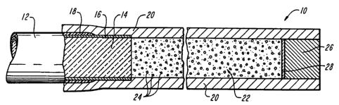
In FIG.1 an optical fiber diffusive tip assembly 10 is shown including an
optical
fiber 12 having a light-transmissive core 14, a cladding 16, and an outer
buffer coating 18.
The end face of fiber core 14 is inserted into a housing 20 which contains
scattering
medium 22 with individual scatterer particles 24. Preferably, the medium 22
has a greater
refractive index then the housing 20. At the distal end of the housing 20, an
end plug 26 is
disposed with a mirror reflector 28
Light propagating through optical fiber core 14 is transmitted into the
scatterer
medium 22 and scattered in a cylindrical pattern along the length of the
assembly 10. Each
time the light encounters a scatterer particles, it is deflected and, at some
point, the net
PCT/US95111246
w0 96/07451
-11-
deflection exceeds the critical angle for internal reflection at the interface
between the
housing 20 and medium 22. When this happens, the light will exit. Light which
does not
exit during this initial pass through the tip is reflected by the minor 28 and
returned through
the tip assembly. During the second pass, the remaining radiation (or at least
a major
portion of this returning radiation) again encounters the scatterers 22 which
provide further
circumferential diffusion of the light.
In FIGS. 2 and 2A, another diffusive tip assembly 40 is shown having
essentially
identical elements to those shown in FIG. 1, except for the disposition of a
bundle of optical
fibers 12A-12E. The individual cores of the fibers are exposed and transmit
light into the
scatterer medium 22.
FIG. 2A is a cross-sectional view of the device of FIG. 2 showing the
placement of
the bundle of optical fibers 12A-12E and the surrounding tube 20, scatterer
medium 22 and
reflector 28.
In FIGS. 3 and 3A, another diffusive tip assembly 40A is shown again having
essentially identical elements to those shown in FIG. 1, except for the
disposition of a
bundle of optical fibers 12A-12E. The individual cores of the fibers are
exposed and
transmit light into the scatterer medium 22, but the individual fibers
terminate at different
locations within housing 20, thereby permitting extended axial diffusion.
FIG. 3A is a perspective view of the fiber bundle of FIG. 3 showing the
placement
of the bundle of optical fibers 12A-12E within the housing.
In FIG. 4, an alternative diffuser tip assembly 50 is shown in which a
laminate of
multiple layers is used for the scatterer tube 20. Thus, innermost layer 20A
encases the
scatterer medium 22. Surrounding this innermost layer 20A is an intermediate
layer 20B.
A third optional layer 20C is then formed about the first two layers 20A, 20B.
Such a
configuration permits the use of different polymeric tubing materials and/or
allows the
introduction of pigmented or etched structures as part of tubing 20.
In FIG. 5, another embodiment of a diffusing tip assembly 60 is shown
incorporating a longitudinal reflector strip 62. As further illustrated in the
cross-sectional
section of FIG. 5A, the longitudinal reflector 62 can be formed as a partial
layer or foil
element within a laminate structure, e.g., between layer 20 and layer 30. The
longitudinal
reflector 62 illustrated in FIGS. 5 and 5A cooperates with the scatterer
medium 22 to create
an azimuthal exposure pattern of approximately 180°, although it should
be clear that other
angles of exposure can be simply achieved by widening (or narrowing) the
circumferential
extent of the reflector element 62. Various alternative co~gurations of the
reflector can be
constructed. For example, the reflector can be disposed on the outside of the
housing or can
be formed as a coating rather than a foil element. Moreover the longitudinal
reflector can
be used without reflective end surface 28, if enhanced axial uniformity is not
needed.
In FIG. 6A, an alternative design is shown for the end reflector. As shown,
end
reflector 28A presents a convex surface to the scattering medium and, thereby,
varies the
w0 96/07451 PCT/US95/11246
-12-
exposure pattern. In FIG. 6B, yet another alternative design for the end
reflector is shown,
wherein the reflective surface is disposed at the distal rather than proximal
end face of the
plug 26. In this embodiment, plug 26 is optically transmissive and the
reflecting surface
28B is formed as a concave surface. In this embodiment, a filler element 29
may also be
disposed at the end of the tube 20.
In FIGS. 7A-7C, the effects of different scatterer concentrations on the
diffusion
pattern of the tip assembly is illustrated. The optimal concentration of
scatterer particles
incorporated into the scatterer medium will, of course, vary with the diameter
of the tube,
the length of the tube and the wavelength as well as other factors.
Nonetheless, a optimal
concentration can be readily determined empirically. FIG. 7A illustrates the
situation where
too many scatterers have been loaded. Most of the light is diffused
immediately upon entry
into the scatterer tube. FIG. 7B illustrates the situation where the scatterer
medium is too
dilute and a bright spot occurs in the vicinity of the reflector. FIG. 7C
illustrates a preferred
embodiment of the present invention in which the scatterer concentration and
mirror
location are chosen such that the light is diffused in a substantially uniform
axial pattern.
It should also be appreciated that the length of the scatterer tube (e.g., the
distance
between the fiber end face and the reflector) will also affect the uniformity
of the diffused
radiation. FIG. 8 illustrates how the mirror placement changes the exposure
pattern for a
given light source, tube diameter and scatterer concentration. As the tube is
extended and the
distance between the fiber and mirror increases, a drop-off in uniformity is
observed. Again,
optimal dimensions for a particular application can be determined empirically.
FIG. 9 is graph of intensity for one preferred embodiment of the invention, a
fiber tip
assembly similar to that shown in FIG. 1 have a Teflon~ FEP tubular housing
(O.D. of about
0.5 millimeters and LD. of about 0.25 millimeters) filled with a silicone and
titania scatterer
composition and capped with an aluminum-coated reflective mirror. The
scatterer medium
was formulated by mixing 70 parts of clear silicone, MastersilTM Formula 151-
Clear
(available from Masterbond, Inc. of Hackensack, New Jersey) with one part of
titania filled
silicone, MastersilTM Formula 151-White (also available from Masterbond). The
result was a
diffusive tip assembly which uniformly transmitted red light at about 633
nanometers over
its entire length of 25 millimeters.
FIG. 10 illustrated the azimuthal exposure patterns for two embodiments of the
present invention. The pattern formed by the squares represents intensity of
light diffused
outwardly with a fiber tip assembly similar to that shown in FIG. 1. This
azimuthal exposure
pattern is essentially isotropic. The pattern formed by the diamonds
represents intensity of
light diffused outwardly with a fiber tip assembly similar to that shown in
FIG. 5. This
azimuthal exposure pattern is essentially semi-cylindrical.
An exemplary manufacturing process suitable for joining a diffuser assembly to
a
glass-clad or polymer-clad optical fiber having an outer diameter of about SO
to about 1000
micrometers can begin by stripping off the buffer from the end of the optical
fiber, e.g.,
w0 96/07451 PCT/US95/11246
-13-
exposing about two or three millimeters of the inner fiber core and its
cladding. (It is not
necessary to strip the cladding away from the core.) Prior to stripping, the
fiber end face
preferably should be prepared and polished as known in the art to minimize
boundary or
interface losses. A transparent tubular structure which will form the housing
for the scatterer
medium is then slipped over the prepared fiber end and, preferably slid beyond
the fiber end.
For example, if a tip assembly of about 20 millimeters is desired, the tubing
can be about 100
millimeters long and slid over about 75 millimeters of the fiber, leaving an
empty lumen of
about 25 millimeters in front of the fiber end face. In one preferred
embodiment, the housing
is Teflon~ FEP tubing, available, for example, from Zeus Industries (Raritan,
New Jersey).
FIG. 11 illustrates the transmission spectrum of Teflon~ FEP, showing that
this
material is well suited for use as a scatterer-encasing material across a
spectrum of light from
infrared to ultraviolet.
The assembly is then injected with a scatterer-loaded material, such as a
silicone,
epoxy or other polymeric material(if a solid diffuser is desired) or a
suitable liquid, such as
water or a deuterium oxide solution, containing colloidal scatterer particles,
such as silica,
alumina, or titania, (if a liquid diffuser is desired). As mentioned above,
one exemplary
scatterer medium can be formulated by mixing 70 parts of clear silicone,
MastersilTM
Formula 151-Clear (available from Masterbond, Inc. of Hackensack, New Jersey)
with one
part of titania filled silicone, MastersilTM Formula 151-White (also available
from
Masterbond), and a conventional silicone curing or hardening agent. The tube
lumen should
be completely filled with the silicone, epoxy or other carrier mixture to
avoid entrapment of
air bubbles. The reflector (e.g., an aluminum, gold or other reflector-coated
plug) is inserted
into the distal end of the tube. The reflector at the distal end of the
scatterer tube can be a
deposited metal or dielectric coating. In one preferred embodiment, a room
temperature
hardening agent is used and the diffuser assembly is simply allowed to
solidify overnight.
Optionally, as a final step, an outer Teflon~ jacket can be disposed about the
apparatus to encase and protect the entire tip assembly including the inner
scatterer tube and
fiber end. The outer jacket is particularly useful in constructing large
azimuthal angle, non-
cylindrical diffusers. In such applications, an inner scatterer assembly is
constructed and then
a reflective strip is disposed along the axis of the assembly to block light
diffusion where the
housing is covered with the reflector and thereby define a non-cylindrical
exposure pattern.
The extent of the circumferential coverage by the reflector will determine the
azimuthal
exposure pattern. The use of an outer jacket also permit a wider variety of
tubing choices for
the inner component of the scatterer housing. Thus, any transparent material
can be used as
the inner tube and the outer Teflon~ jacket will still ensure that the problem
of contact
adhesion is minimized.
It should be clear that the manufacturing processes described above are merely
illustrative, and various alternative techniques can be practiced to construct
the fiber tip
assemblies of the present invention. For example, automated extrusion methods
and/or
WO 96/07451 PCT/US95/11246
-14-
injection molding approaches can be employed to mass produce fibers with
integral diffusive
tip assemblies.
The amount of scatterer incorporated into the diffusive tip assembly will vary
with the
carrier and the desired length, and can therefore be adjusted to meet
particular applications.
S Different scatterers may be more or less useful in particular applications.
Table 1 below
illustrates certain relevant characteristics of three different scatterer
compositions:
Scatterer Density Transmission Spectrum
Composition (grams/cc) (wavelength in micrometers)
Ti02 4.0 .45 - 11
Si02 2.1 .2 - 7
A1203 3.6 .2 -9
In certain applications, it may be desirable to mix two or more scatterer
compositions
together to achieve blended characteristics.
Liquid scatterer compositions can be used to extend phototherapy into the
ultraviolet (UV) and infrared (IR) regions of the spectrum. In particular,
structures
1 S employing deuterium oxide and other heavy water solutions are useful to
transmit IR light
with low losses and minimal tip heating. Distilled water suspensions of
scatterers are used
for UV light delivery.
The above-described manufacturing techniques were used to produce diffusing
tips
joined to fibers ranging from about 100 to about 600 micrometers in diameter.
When fiber
bundles are joined to the diffuser tip, the individual fibers can be even
smaller, e.g., as small
as 25 micrometers in diameter. The cylindrical light-diffusing assemblies
produced axial
exposure patterns of about 2 cm to about 4 cm in length. The azimuthal
exposure angle was
either 360° for assemblies resembling FIG.1 or about 180° for
those resembling FIG. 5.
Other azimuthal exposure patterns can be obtained by modifying the
circumferential extent of
the longitudinal reflector strip 62 of FIG. 5. The solid tubes were clear
Teflon~ and were
injected with the above-described mixture of silicone and micron-sized
titania. The liquid-
filled tubes were similarly constructed but contained a water or D20 solution
loaded with
colloidal alumina or silica. A exemplary liquid scatterer composition of
colloidal alumina is
available as Formulation 12733 from the Johnson Matthey Co. (Seabrook, New
Hampshire).
In use, it is preferably diluted with water by a factor of about 100:1 and pH-
balanced with
acetic acid.
In FIG. 12 another phototherapeutic apparatus 80 according to the invention is
shown having two chambers filled with different scattering media to effect an
increasing
diffusion pattern. Apparatus 80 includes an optical fiber 12 having a light-
transmissive core
14. The end face of fiber core 14 is inserted into a housing 20 which contains
a first
w0 9610'7451 PCT/US95/11246
-15-
chamber with a first scattering medium 21 with individual scatterer particles
22A. The
housing further includes a second chamber which can have a transparent core 23
(e.g., an
FEP rod or beading) surrounded by a toroidal space filled with a second medium
having
scatterers 22B of a different loading density or composition. At the distal
end of the
housing 20, an end plug 26 is disposed with a mirror reflector 28.
Light propagating through optical fiber core 14 is transmitted into the
scatterer
medium 22A and scattered in a cylindrical pattern along the length of the
assembly 10. Each
time the light encounters a scatterer particles, it is deflected and, at some
point, the net
deflection exceeds the critical angle for internal reflection at the interface
between the
housing 20 and medium 21. When this happens, the light will exit. Similarly,
light which
passes through this first chamber is transmitted to the second chamber 23
where it encounters
the scatterers 22B, causing more of the light to be reflected. Light which
does not exit during
this initial pass through the tip is reflected by the mirror 28 and returned
through the tip
assembly. During the second pass, the remaining radiation (or at least a major
portion of this
returning radiation) again encounters the scatterers 22A and 22B which provide
further
circumferential diffusion of the light.
In FIG. 13, another phototherapeutic apparatus 100 is shown including a jacket
112
having a plurality of light diffusing loops 114A,114B which can be expanded
out of, or
retracted back into, the instrument housing 112 by control wire 116. As shown,
the apparatus
100 can further include a radio opaque region 118 which facilitates location
of the instrument
by radiographic means. Although the apparatus is illustrated with only two
loops, in some
applications it can be desirable to have a greater (or lesser) number of
loops.
In FIGS. 14A-14D, the deployment of loop elements 114A and 114B is shown
schematically. FIG. 14A illustrates a fully retracted mode in which most of
the loop elements
are withdrawn into the housing 112. In FIG. 14B, a control wire 116 has been
moved
partially forward and a larger portion of diffusive loop elements 114A and
114B projects
outward from the housing 112. In FIG. 14C, the control wire has been slid
forward even
further and the loop elements 114A,114B now are nearly fully deployed. In FIG.
14D, the
control wire 116 is partially retracted after extension to effect a "heart-
shaped" diffuser.
In FIG. 15A, a truncated, cross-sectional view of a diffusive loop element 114
is
shown connected to two optical fibers, each having a light transmissive core
120A,120B and
a cladding/buffer coating 129. The end face of each fiber core 120A,120B is
inserted into a
housing 128 which contains a scattering medium 124 with optional individual
scatterer
particles 125. Preferably, the medium 124 has a greater refractive index than
the housing
128.
FIG.15B is a graph of intensity vs. radial distance for two fibers as shown in
FIG.
15A. The curve 121A illustrates the intensity of diffused radiation vs. axial
length of one
fiber while curve 121B represents a similar intensity distribution for a
second fiber which has
been deployed in an opposite configuration. The cumulative intensity
distribution of these
WO 96/07451 ~ ~ PCT/US95/11246
-16-
two fibers are shown by curve 123. By employing pairs of fibers that are
joined in opposite
directions, one can thus achieve nearly uniform distribution of the diffusive
radiation.
A similar radiation distribution pattern can be achieved by employing a
reflective end
cap on each loop, as shown in FIG. lb. In this figure, a truncated, cross-
sectional view of a
diffusive loop element 114 is shown having an optical fiber with a light
transmissive core 120
and a cladding/buffer coating 129. The end face of fiber core 120 is inserted
into a housing
128 which contains a scattering medium 124 with optional individual scatterer
particles 125.
Preferably, the medium 124 has a greater refractive index than the housing
128. At the distal
end of the housing 128 an end plug 126 is disposed. Optionally, the end plug
may also be
fitted with a mirror reflector 140 to create a distribution pattern like that
shown in FIG. 15B.
Light propagating through the optical fiber core 120 is transmitted into the
scatterer
medium and scattered in an cylindrical pattern along the length of the
assembly 114. Each
time the light encounters a scatterer particle, it is deflected and, at some
point, the net
deflection exceeds the critical angle for internal reflection at the interface
between the
housing 128 and the medium 124. When this happens the light will exit. The
housing can
either be made suff=iciently long to ensure that virtually all of the light
entering it is eventually
scattered and diffused in a single path, or as noted above, a reflective
mirror can be fitted to
the distal end of each diffuser assembly. When a mirror is employed, light
propagating
through the medium 124 will be at least partially scattered before it reaches
mirror 140.
Light which does not exit during this initial pass through the tip will be
reflected by mirror
140 and returned through the tip assembly. During the second pass, the
remaining radiation
(or at least a major portion of this returning radiation) again encounters the
scatterers which
provide further circumferential diffusion of the light.
In FIG.17, the loop diffuser apparatus of the present invention 10 is shown
schematically in operation. The diffuser apparatus 100 is coupled to a source
of
phototherapeutic radiation 136 (e.g., a laser) and positioned within a
patient's body to provide
phototherapy. As shown in FIG. 17, the diffuser assembly can be designed to
fit within the
instrument channel of an endoscope 132. The endoscope can further include
viewing means
134 and/or at least one additional channel 138 for the introduction of
irrigation saline or
therapeutic solutions. Alternatively, the diffusing assemblies of the present
invention can be
incorporated into catheter-type instruments that are introduced into the
patient's body without
the assistance of an endoscopic channel.
In FIG. 18, an outer jacket (e.g., of Teflon~ material) is shown disposed
about the
apparatus to encase the fiber 112 and loop diffuser assembly 114. The outer
sheath surrounds
the entire optical transmission apparatus and ensures that the radiation-
generating
components do not come into direct contact with the patient's body and,
thereby, permits
reuse of the instrument. Only the outer sheath 150 needs to be disposed after
each use.
The devices of the present invention can be used for various therapeutic
purposes.
One application is photodynamic therapy (PDT), a form of light-activated
chemotherapy. In
WO 96/07451 PCT/US95/11246
-17-
this approach, photosensitive dyes are delivered by injection or other
vehicles such that the
dye is preferentially accumulated in cancer cells. When the cells which have
taken up the dye
are irradiated at an appropriate wavelength (e.g., with red light), a
photochemical reaction
occurs that yields radicals (usually singlet oxygen) which poison the cell.
Thus, the present
invention further encompasses the use of diffused radiation to activate
photosensitive dyes.
One advantage of the present invention is that it permits PDT at remote
treatment sites via a
catheter, trocar, hollow needle or other hand held instrument in a minimally
invasive manner
because diffusive fiber tip assemblies can now be constructed with outer
diameters on the
order of only a few hundred micrometers.
The present invention also encompasses the use of diffuse radiation in
photocoagulation and/or hypodermic therapy of tumors and hyperplasia. For
example, the
phototherapy apparatuses described above can be used to treat liver,
pancreatic or prostate
tumors, or benign prostate hyperplasia. The application of diffuse radiation
to heat prostate
tissue can be used in lieu of transurethral resection of the prostate, balloon
dilatation of the
prostate or ultrasonic hyperthermia. In particular, the directional probes
described above can
be especially useful in improving the outcome of prostate treatment by heating
more tissue
directly in less time, and in distributing irradiation over a larger volume of
prostatic-tissue,
thus increasing the therapeutic heating effects while reducing the risk of
overheating damage
to surrounding tissue structures such as the sphincter. The invention further
permits
interstitial laser coagulation of hepatic and pancreatic tumors. The desired
effects are
achieved by thermal destruction of cancerous tissue by depositing laser
radiation via a
diffusive fiber tip carrier by a hypodermic needle or similar instrument
inserted
percutaneously into the tumor. In each of these procedures, therapy can be
delivered while
the patient is awake; general anesthesia as well as open surgery are avoided.
In heat-based phototherapy techniques, the diffusive fiber tip assemblies of
the
present invention allow for the formation of large distributed heat sources
within the target
tissue. The invention significantly alters the rate of heat deposition in
tissue, especially in the
regions immediately surrounding the fiber tip, where tissue overheating and/or
carbonization
would limit the effectiveness and inhibit efficient heat transfer. Since the
radiation is
distributed by the diffuser assembly over a larger volume of tissue, more
tissue is heated
directly and there is less need to rely on connective or conductive heat
transfer through
nearby tissue to reach the periphery of the tumor.
Moreover, the materials disclosed herein for the diffusive tips and jackets
further
enhance the therapeutic effects by permitting high radiation transmission and
low absorption,
thereby ensuring the tip assembly itself does not overheat during usage. In
addition, the use
of Teflon~ tubes and/or coatings further improve the procedures by avoiding
the problem of
tip fusion or contact-adhesion between the tip assembly and biological tissue
during usage. It
has been found that Teflon~ FEP materials (polyperfluoroethylene-propylene
copolymers)
are preferable for most applications because they do not discolor if they are
etched prior to
WO 96/07451 ~ ~ PCT/US95/11246
-18-
loading with the scatterer medium, although Teflon~ PFA materials
(polytetrafluoroethylene
polymers with perfluoroalkoxy side chains) and Teflon~ PTFE
(polytetrafluoroethylene) and
other fluoropolmers may also be useful.
The non-cylindrical, large azimuthal angle diffusers of the present invention
are also
particularly useful in therapeutic applications. By directionalizing the
diffused radiation, the
devices disclosed herein can provide therapeutic radiation to large volumes of
tissue while
also protecting sensitive tissue or biological structures. For example, in
prostate treatment, a
semi-cylindrical or other large azimuthal angle diffuser can disposed within
the urethra and
rotated into a position such that the prostate is subjected to phototherapy
while the patient's
sphincter muscles and/or other tissue regions are largely shielded from
irradiation. In
addition the non-cylindrical diffusive tip assemblies can be used to deliver a
greater dose of
radiation to tissue and rotated, if necessary during use to effect a
circumferential (or partially-
circumferential scan of the target tissue at the higher intensity level.
The diffusive tip assemblies of the present invention can be used in various
other
medical applications, such as, for example, heat-setting of stents, activation
of photoreactive
suturing materials, curing of prosthetic devices, activation of adhesives for
implants and the
like.
In Fig. 19, yet another phototherapeutic apparatus 200 according to the
invention is
illustrated having a tubular sheath 212 and an inner optically-transmissive
fiber element 214.
The distal end of the sheath 212 is fluted such that axial compression of the
sheath results in
expansion of strut elements 218 in the fluted region 216.
Fig 20 is a more detailed cross-sectional view of the distal end of the
apparatus of Fig.
19. The optically-transmissive element is shown having an optical fiber 220
with an optically
transmissive core 222 surrounded by a cladding, and buffer coating. The end
face of fiber
core 222 is inserted into a housing 228 which contains a scattering medium 224
with optional
individual scatterer particles 225. Preferably, the medium 224 has a greater
refractive index
than the housing 228. At the distal end of the housing 228, and end cap 226
can be disposed.
Optionally, the end cap may also be fitted with a reflective mirror 240. The
end cap 226 can
further be ground or polished to a point 230 to facilitate penetration of body
tissue.
Light propagating through the optical fiber core 222 is transmitted into the
scatterer
medium and scattered in an cylindrical pattern along the length of the
assembly 214. Again,
each time the light encounters a scatterer particle, it is deflected and, at
some point, the net
deflection exceeds the critical angle for internal reflection at the interface
between the
housing 228 and the medium 224. When this happens the light will exit. The
housing can
either be made sufficiently long to ensure that virtually all of the light
entering it is eventually
scattered and diffused in a single path, or as noted above, a reflective
mirror can be fitted to
the distal end of each diffuser assembly. When a mirror is employed, light
propagating
through the medium 224 will be at least partially scattered before it reaches
mirror 240.
Light which does not exit during this initial pass through the tip will be
reflected by mirror
WO 96/07451 PCT/US95/11246
-19-
240 and returned through the tip assembly. During the second pass, the
remaining radiation
(or at least a major portion of this returning radiation) again encounters the
scatterers which
provide further circumferential diffusion of the light.
In Fig. 21, the phototherapeutic apparatus in the present invention 200 is
shown
schematically in operation. The diffuser apparatus with its fluted stopper is
coupled to a
source of phototherapeutic radiation 236, (e.g., a laser) and positioned
within a patient's body
to provide phototherapy. As shown in Fig. 21, the diffuser assembly can be
designed to fit
within a standard guiding catheter 232. The catheter 232 can further include
electrical
sensing elements 234 and/or at least one additional channel 238 for
introduction of saline or
therapeutic solutions.
In Fig.22A, the use of the phototherapeutic apparatus of the present invention
is
shown schematically. As illustrated, the instrument 200 is positioned next to
a segment of a
patient's body tissue where penetration and radiation is desired. As shown,
the apparatus
includes an outer sheath 212 having a fluted region 216 and an inner optically-
transmissive
1 S fiber element 214 with tip 226. In one preferred embodiment, the fiber 214
and sheath 212
are constructed with sufficient clearance to permit saline or other
therapeutic liquids to be
released during the procedure. In particular, saline flushing of the fiber tip
214 may be
desirable to cool the tissue surface proximal to the treatment site.
In Fig. 22B, initial penetration of the apparatus 200 is shown. In this
illustration, the
optically-transmissive fiber has penetrated the patient's tissue but the end
217 of sheath 212
has not yet touched the tissue surface.
In Fig. 22C, the fiber 214 has penetrated further into the patient's tissue
and the sheath
212 has now been pushed into a position abutting the patient's tissue. As the
instrument is
advanced, the fluted region 216 begins to expand due to the compressive forces
exerted
during penetration. Struts 218 are pushed out radially from the body of the
apparatus.
In Fig. 22D, the apparatus is shown in a fully deployed position wherein a
predetermined length of the optical fiber 214 has now penetrated the patient's
body tissue and
the radially-expanded struts 218 have been fully compressed into a maximal
position creating
a large cross-sectional obstruction to further penetration.
Various materials can be used to form the outer sheath including, for example,
Teflon~ and other fluorocarbon polymers. The struts 218 can be formed by axial
slices at
various locations on the sheath. For example to construct a four strut stopper
device, one
would make four longitudinal cuts into the sheath, separated by 90°
from each other. The
length of the cuts will determine the radial extent of the stopper. In one
embodiment it may
also be desirable to fill the sheath polymer with a radio-opaque substance,
such as barium or
bismuth in order to permit visualization under angiography.
In FIG. 23 a further adaptation of the present invention is shown in a
phototherapeutic
apparatus 300 for sterilizing an inner lumen of an endoscopic medical
instrument 332 is
WO 96/07451 PCT/US95/11246
-20-
shown including a source of ultraviolet radiation 336, an optical fiber 312
and a diffusive tip
assembly 314.
In use, the apparatus 300 serves to sterilize or clean an inner lumen of the
endoscopic
instrument 332. The optical fiber 312 with its light-diffusing distal tip
assembly 314 is
inserted into the lumen requiring sterilization. In one technique, the optical
fiber tip can be
inserted through the entire instrument and then slowly retracted. The
radiation source is
activated to transmit light via the fiber 312 to the diffusive tip assembly
314. As the
apparatus is retracted through the endoscope lumen 338, cytotoxic radiation is
delivered to all
portions of the inner lumen walls. Any debris or deposits on the inner lumen
walls are
likewise irradiated to kill any microbes which may be harbored in such
deposits.
In FIG. 24 a diffusive tip assembly 314 is shown in more detail proving the
optical
fiber 312 having a light transmissive core 320 and a buffer coating or
cladding 321. The end
face of fiber core 320 is inserted into a housing 328 which contains a
scattering medium 324
with optional individual scattering particles 325. As in previous embodiments,
preferably,
the medium 324 again has a greater refractive index than the housing 328. At
the distal end
of the housing 328, an end plug 326 is disposed with a mirror reflector 340.
Light propagating through the optical fiber core 320 is transmitted into the
scatterer
medium 324 and scattered in a cylindrical pattern along the length of the
assembly 314. Each
time the light encounters a scatterer particle, it is deflected and, at some
point, the net
deflection exceeds the critical angle for internal reflectance at the
interface between the
housing 328 and the medium 324. When this happens, the light will exit. Light
which does
not exit during the initial pass through the tip is reflected by the mirror
328 and returned
through the tip assembly. During the second pass, the remaining radiation (or
at least the
major portion of this returning radiation) again encounters the scatterers 325
which provide
further circumferential diffusion of the ultraviolet light.
The optimal concentration of scatterer particles incorporated into this
scatterer
medium will, of course, vary with the diameter of the tube, the length of the
tube and the
wavelength as well as other factors. Nonetheless, an optimal concentration can
readily be
determined empirically for ultraviolet radiation in the range of about 400
nanometers to about
200 nanometers, one preferred composition for the scatterer medium is
colloidal alumina
suspended in acetic acid. It should also be appreciated that the length of the
scatterer tube
(e.g., the distance between the fiber end facing and the reflector) will also
affect the
uniformity of the diffused radiation.
Optionally, as shown in FIG. 24, an outer Teflon~ jacket 350 can be disposed
about
the apparatus as a final step to encase and protect the entire tip assembly
including the inner
scatterer tube 314 and fiber end 312.
w0 96/07451 ~ PCT/US95/11246
-21-
In use, the apparatus is slid into an endoscope lumen and connected to a UV
light
source. The light source is activated and the UV radiation is transmitted to
the diffusive tip,
where the scatterers project a cylindrical exposure pattern to the lumen
walls. The apparatus
can then be slid forward or backwards (or in both directions) to bathe the
entire lumen with
sterilizing irradiation.