Note: Descriptions are shown in the official language in which they were submitted.
CA 02200220 1997-OS-30
DRUG FOR TUMOR GROWTH SUPPRESSION
IPCS A61K 31/00
The invention relates to the field of biology and medicine, in particular
to suppression of the growth of malignant tumors.
The following drugs are known for suppression of tumor growth.
1. Cis-dichlorodiaminoplatinum. Having a wide range of action, it is
used for treatment of solid tumors of various localization [ 1, 2], but
exhibits
high nephrotoxicity [3).
2. Photodynamic therapy of tumors, in which photosensitizers - free
tetrapyrrolic macrocyclic ligands or their complexes with metals, e.g.
aluminum
complex of sulfonated phthalocyanine Al[Pc(S03H)4] - are used. Their use is
only possible in the case of surface located tumors, more exactly - accessible
for a laser probe [4] .
3. A drug consisting of a complex of copper (II) with
glycylglycylhistidine tripeptide ([Cu(GGH)]Cl) and sodium salt of ascorbic
acid in a ratio of 1:10 is the drug most similar to that proposed; it causes
an
increase in the life-span (ILS) of mice with Ehrlich ascitic carcinoma [5].
Drawbacks of the prototype are low effectiveness related to the low
selectivity of that complex in respect of tumors and its instability in
physiological conditions, and a high toxicity due to the products of
decomposition of the complex.
The object of the present invention is to find more effective and less
toxic drugs for suppression of tumor growth.
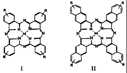
The essence of the proposed invention is that a drug consisting of a
biogenic reductant and a complex of cobalt or iron with substituted
phthalocyanines (I) or naphthalocyanines (II) is used to suppress tumor
growth.
CA 02200220 1997-OS-30
2
I II
where R = COONa, S03Na, CH2C5HSN~C1-, CH2(NH2)2S~CCl-.
The scientific foundation for the invention is literary data on the
selective accumulation of tetrapyrrolic macrocyclic compounds and their
complexes with metals in tumorous tissues [4, 6] and the facts that we have
established which show the high catalytic activity of phthalocyanine complexes
of cobalt and iron in model chemical systems, in particular:
1 ) they are homogenic catalysts of the autooxidation row of biogenic
reductants and analogues thereof, e.g. ascorbic acid, ubiquinone and cysteine;
2) the (intermediate) formation of active forms of oxygen - the anion-
radical of superoxide, hydrogen peroxide and a hydroxyl radical, the
cytostatic
and other biological activity of which is well known - takes place in this
reaction [7];
3) in the conditions of this reaction, the proposed complexes cause an
oxidative degradation of nucleic acids.
METHODS AND RESULTS OF TESTS
Tests of the proposed drug for cytotoxic and antitumor activity were
carried out on cultures of tumor cells (in vitro) and on mice with regrafted
tumors (in vivo).
CA 02200220 2003-10-23
3
Determination of the activity of the drugs
Qn cultures of tumor cells (in vitro)
Method 1.
The method of evaluation of the cytostatic effect of a combination of
phthalocyanines with ascorbic acid in a system in vitro was developed on a
culture of tumor cells of human testicular carcinoma (Cao V line).
The culture of cells was grown in a monolayer in medium 199
comprising a lOgb solution of an embryonic calf serum.
At the beginning of the experiment the cells were inoculated at a density
of 100000/ml in a total volume of 2 ml and incubated at 37°C for 24
hours.
Then the testing was carried out in the following variants:
1. Control.
The growth medium for the samples was replaced with a fresh intact
nutrient medium and incubated for 48 hours, then 3H-thymidine (3H-T) in a
final concentration of 1 ~,Curie/ml was introduced into the medium of the
samples, it was washed with a Hank's solution, a 2.Sg'o solution of perchloric
acid, and the acid=insoluble fraction was hydrolyzed in 5 ml of Sq6 perchloric
acid. The hydrolyzates in a volume of 100 ~I were transferred into flasks with
a scintillation fluid SF-8 and the level of radioactivity in the samples was
registered on a Rackbeta~ (Sweden) fluid scintillation counter. The average
values of the level of radioactivity were calculated.
2. In order to evaluate the effect of ascorbic acid on the growth of cells
of the Cao V line, the growth medium of the samples was replaced with a fresh
medium, comprising ascorbic acid in a concentration of 1x10⁻⁴ M, incubated
for 4 hours, and 'then the sample was processed according to the method
described in pare 1.
3. In order to evaluate the effect of phthalocyanine metal complexes on
the growth of cells of the Cao V line, the growth medium of the samples was
replaced with a fresh medium, comprising complexes in a predetermined
concentration, the method described in pare 1 was followed.
I ~~
CA 02200220 2003-10-23
4
4. In order to evaluate the cytostatic effect of the combination of
phthalocyanine and ascorbic acid, ascorbic acid was added into the growth
medium comprising the complex in a ratio of concentration equal to 1:10, and
the method described in para 1 was followed.
The suppression of the 3H-T inclusion in the test samples was calculated
according to the equation
average value of decomposition/min/test samples
(1 _ _______________________________________,_______________ ) x 100%
average value of decomposition/min/control samples
Statistic processing of the results was carried out using the method of
sample analysis.
Method 2.
Determination of the cytostatic activity of the drugs being tested was
carried out using a biotest based on inhibiting the proliferation of a
regrafted
culture of cells of human lung adenocazcinoma A-549, due to the action of
cytotoxic agents.
In order to cultivate cells of the A-549 line, the Eagle medium was used
with the addition of 100 pg/ml of heptamycin and a 10 qb solution of
embryonic calf'serum, preliminarily inactivated by heating. Cultivation of the
cells was carried out under standard conditions: at 37~C in a humid
atmosphere, comprising 5~ carbon dioxide.
When a cytostatic biotest was being set up, 100 Ecl of a suspension of
cells of the A-549 line at a concentration of 7.5x10⁴ cells/ml were placed in
2S each of the craters of a flat 96-crater microboard (~~g~stedt~" USA) with
addition of the drugs being tested in a volume of 100 ~,1/crater at the
beginning
of the phase of logarithmic growth.
The cytostatic activity of the drugs being tested was evaluated by the
colorimetric method, based on the capability of mitochondria) dehydrogenases
of live test-cells of restoring exogenetically introduced soluble 3-(4,5-
dimethyl
2-thiazolyl)-2,5-Biphenyl-2H-tetrazole bromide (MTT, "Sigma Chemical Co.,"
i
CA 02200220 2003-10-23
USA) into an. insoluble crystalline formazan. Wherein, 20 ~,1 of a solution of
MTT at a concentration of 5 mg/ml were put into each crater of the 96-crater
microboard, after which the cell cultures were centrifuged, the whole volume
of
the culture medium was removed from the craters. For solubilization of the
5 colored reaction products (crystals of formazan), 150 ~,1 of
dimethylsulfoxide
("Sigma") were introduced into each crater of the microboard and the
microboard was incubated at room temperature and continuously shaken. The
results were registered upon absorption at a wavelength of S50 nm on the
minireader "Dynatech~" FRG).
The level of inhibition of proliferation of the cell cultures by the drugs
being tested for cytostatic activity was calculated according to the equation:
Pt
IP(~'v) = 100 - ----- x 100,
P
where: IP is the level of fnhibition of the proliferation;
Pt is the level of proliferation in the test (with the thugs):
absorption of the dye in test samples;
P~ is the level of proliferation in the control (without the drugs):
absorption of the dye in test samples.
Data are presented in Table. 1 on the absence of cytotoxic activity of
ascorbic acid (AH2) by itself and of phthalocyanine complexes relative to
cells
of the Cao V and MCF-7 lines at a predetermined criterion of activity CEO
equal to 10'4 M for a 4$-hour period of incubation.
CA 02200220 1997-OS-30
6
Table 1. Antiproliferation activity of components of the claimed drug
(phthalocyanine and naphthalocyanine complexes of metals and ascorbic acid)
relative to tumor cells of a human in vitro
Content CESp, M
Human testicular Breast adenocarcinoma
carcinoma
CaoV* MCF-7 line
Co[Pc(S03Na)2] 2.5x10'4
Fe[Pc(S03Na)2] 2x10'4
Fe[Pc(COONa)g] 0.5x10'4
Co[Pc(CHZCSHSN~)6]CY6 5X10'4 1x10'4
Co[Pc(S03H)2] $X10'4 1X10'4
Co[Pc(CH2(NH2)2S~CCl')g] 2.5x10'4
Co[Nc(CH2CSHSN~)2]Cl'2 1x10'4
Co[Pc(COONa)g] 5x10'4 1x10'4
AH2 > ixl0'3 > 1x10'3
* AH2 - ascorbic acid
The compound was considered to be active if CESp [the concentration at
which a 50% suppression of inclusions 3H-T in the cells (see method 1)] was
1x10'4M under these conditions of the experiment. As is evident from the
presented results, the complex Co[Nc(CH2CSHSN~)2]Cl'2 and the complex
Fe[Pc(COONa)8] had limited activity, all the other complexes and AH2 were
inactive.
Data are grouped in Table 2 which were obtained during a study of the
inhibition of the proliferation of tumor cells in vitro when phthalocyanines
and
ascorbic acid were used together in noncytotoxic concentrations.
CA 02200220 1997-OS-30
7
Table 2. Inhibition of the proliferation of tumor cells in the presence of
phthalocyanine and naphthalocyanine complexes of metals and ascorbic acid
when simultaneously administered into a culture of cells
Inhibition of proliferation ( % )
Human Breast Human
testicular adenocarcinoma, lung
carcinoma carcinoma
CaoV* MCF-7* line A-549**
Co[Pc(S03Na)2] 0 0
Co[Pc(S03Na)2]+AH2 52 83
Fe[Pc(S03Na)2] 0
Fe[Pc(S03Na)2]+AHZ 12
Fe[Pc(COONa)g] 50
Fe[Pc(COONa)g]+AH2 51
CO[Pc(CH2CSHSN~)6C1'6 0 U 0
CO[Pc(CH2CSHS1V~)6Cl-6+AH2 74 50 55
Co[Pc(S03H)2] 0 0
Co(Pc(S03H)2]+AH2 76 30
Co[Pc(CH2(NH2)2S~CCl-)g] 0
Co[Pc(CH2(NH2)2S~CCl-)g]+AH2 64
CO[Nc(CH2C5HSN~)2]Cl-2 0
Co[Nc(CH2CSHSN~)2]Cl-2+AH2 64
Co[Pc(COONa)g] 0 0
Co[Pc(COONa)8]+AH2 99.5 86
* - method 1, [complex] = 5~ 10-SM, [AHZ] = 1 ~ 10-4M.
** - method 2, [complex] = 100 ~,g/ml, [AH2] = 227 ~.g/ml
CA 02200220 1997-OS-30
8
Determination of antitumor activity of the drugs on mice with
rearafted tumors !in vivo~
Tests were carried out on tumor strains: Ehrlich ascitic carcinoma
(example 1), ascitic hepatoma 22 (examples 2, 3), solid breast adenocarcinoma
Ca-755 (example 4).
The complexes are dissolved in a sterile physiological solution until a
concentration of from 0.005 to 1 % is obtained. Ascorbic acid is dissolved in
sterile distilled water or an isotonic solution of sodium chloride to a
concentration of from 0.011 to 2.2%. The complexes and ascorbic acid are
- administered intraperitoneally, intrapleurally, intravenously or into the
tumor
itself.
Example 1.
A tumor, an Ehrlich ascitic carcinoma, was grafted into mice
intrapleurally. The tests were carried out in a manner similar to that of
Example 2.
Mice of the control group without treatment died on the 9th-15th day
with development of tumorous pleurisy.
Mice, who had received treatment with a complex of Co[Pc(COONa)8]
in a single dose of 75 mg/kg with subsequent administration of 165 mg/kg of
ascorbic acid, lived for 18-40 days. Death from toxicity was not observed
(Table 3).
Example 2.
A tumor, ascitic hepatoma 22, was grafted intraperitoneally, the grafting
dose was 106 cells per mouse. Mice of both sexes were used. The weight of
each mouse was at least 18 g. Treatment was begun 48 hours after the tumor
was grafted. Mice of the control group without treatment lived 19.3+1.7 days
and died with expressed ascites. In the group of mice receiving treatment with
a complex of Co[Pc(COONa)g] in a single dose of 100 mg/kg with subsequent
administration of ascorbic acid (a course dose of S50 mg/kg), one mouse died
on the 19th day with ascites, the remaining 8 of the 9 mice lived without
symptoms of tumor for more than 70 days. Death from toxicity was not
observed (Table 3).
CA 02200220 1997-OS-30
9
Example 3.
Tests were carried out in a manner similar to that in example l, but the
tumor, ascitic hepatoma 22, was grafted to the mice intrapleurally.
Mice of the control group without treatment lived 5.7+ 1.6 days and died
with exudation in the pleural cavity in a volume of about 2.0 ml.
In the group of mice who received treatment with a complex of
Co[Pc(COONa)8] in a single dose of 75 mg/kg with subsequent administration
of 165 mg/kg of ascorbic acid, the mice lived more than 70 days without
symptoms of a tumor. Death from toxicity was not observed (Table 3).
Table 3. Effect of complex Co[Pc(COONa)$] and ascorbic acid (AHZ) on the
life-span of mice with grafted tumors, as compared with the prototype
Substance Tumor strain
Ehrlich carcinoma Hepatoma 22
ALS, Recovery, ALS, Recovery,
Co[Pc(COONa)8]+AH2 296 0 370 70
[Cu(GGH)]Cl(prototype [5]) 60 0
Example 4.
Breast adenocarcinoma Ca-755 was grafted into the mice using 50 mg of
tumorous tissue. Treatment was begun 48 hours or on the 9th day after the
tumor was grafted. The complexes and ascorbic acid were administered in
several ways: intravenously, intraperitoneally, intratumorously (Table 4).
The results obtained with mice having tumors were evaluated by means
of generally accepted indexes of antitumor activity, with mice who were not
given antitumor therapy being used for control.
Calculation of the increase of life-span was made using the equation:
Ltest ' I-control
~,g = __________________ x 100%,
I-control
where L is the life-span in days.
CA 02200220 1997-OS-30
Inhibition of tumor growth was calculated for solid tumors using the
equation:
Vaverage control - Vaverage test
TGI = ______________________-_-_-__ x 100% ,
Vaverage control
where Vaverage is the average volume of the tumor, calculated as the
product of three measurements and expressed in cubic cm.
Regression of the tumor was determined for a developed solid
adenocarcinoma Ca-755, the percentage of regression was calculated by the
10 equation:
Vo - vn
R = ---------- x 100 % ,
Vo
where R is the percentage of regression,
Vo is the initial average volume of a tumor,
Vn is the average volume of a tumor after treatment for "n" days.
Table 4. Inhibition of the growth of a solid breast adenocarcinoma Ca-755
after administration of phthalocyanine complexes of cobalt and ascorbic acid
(~2)
Complex Dose {mg/kg) TGI*,%
Co[Pc(CH2C5HSN~)6]Cl-6 25 58
AH2 27.5 38
Co[Pc(CH2CSHSN~)6]Cl-6 + AH2 25 + 27.5 91
Co(Pc(S03Na)2] 25 68
AH2 56.75 66
Co(Pc(S03Na)2] + AH2 25 + 56.75 88
CA 02200220 1997-OS-30
11
Co(Pc(COONa}g] 10 44
AH2 22 38
Co[Pc(COONa)8] + AH2 10 + 22 61
* - data in respect of Co complexes;Co[Pc(CHZCSHSN~}6]Cl-6 ~'
and Co[Pc(S03Na)2] are presented for the 14th day, data in
respect of the complex Co(Pc(COONa)8] are presented for the
16th day after transplantation of the tumors.
Thus, use of the proposed drug makes it possible to suppress effectively
the growth of a wide range of malignant tumors, in particular to achieve the
suppression of proliferation of cancer cells (in vitro) and inhibition of the
growth of tumors in mice (in vivo) and substantially increase their life-span,
including that obtained in comparison by use of the prototype.
20
CA 02200220 1997-OS-30
12
LITERATURE
1. Rose W.C. et al., Cancer Treatment Rep., 1982, 66, 135-146.
2. Gorbunova V.A., Voprosy Onkology, 1989, 35, 325-331.
3. Belgorodsky V.V. et al., Voprosy Onkology, 1975, 21, 95-105.
4. Mironov A.F., "Photosensitizers on the base of porphyrins and
related compounds for photodynamic therapy of cancer" in book "Itogi nauki i
tekhriiki," VINITI, Moscow, 1990, 3, 5-62.
5. Kimoto E., Tanaka H., Gyutoku J., Morishige F., Pauling L.,
Cancer Research, 1983, 43, 824-828.
6. Amato L, Science, 1993, 262, 32-33.
v 7. Au~t S.D., Morehouse L.A. and Thomas C.E., J. of Free Radicals in
Biology & Medicine, 1985, 1, 3-25.
20