Note: Descriptions are shown in the official language in which they were submitted.
220~4 ~1
WO 96tOgO13 PCT/US95/10931
~an~ . Tntr~ l"minal 8tent9
Field of the Invent;on
The present invention relates to an
e~r~n~hle, intraluminal stent and an apparatus and a
method for deploying the eYp~nA~hle, intraluminal
stent in a body passage. More particularly, the
present invention relates to a type of intraluminal
stent capable of ~p~o ~ing an intact, intraluminal,
venous graft providing an inner stent lining having an
endothelial layer and an apparatus and method for
deploying and mech~nically eYp~n~ing the vein-lined
stent within a body r~sr-ge through the application of
an outward force on the ~xternal surface of the stent.
Background of the Invent;on
As an alternative to vascular surgery,
balloon angioplasty has been a common method for
unblorki~g narrowed or occluded blood vessels. In
this procedure, an angioplasty balloon is inflated
within a steno~A vessel in order to dilate the vessel
to provide an enlarged lumen. Altho~lg~ balloon
angioplasty has been s~lccessful in restoring flow in
stenotic or occluded vessels, these vessels often
restenose due to elastic recoil of the diseased
tissue. Subintimal Ais~ction is also caused by
balloon induced stresses and results in geometric
irregularities at the inner wall leading to flow
disturh~nces and decreased flow.
220 0~
WO96/09013 PCT~S95/10931
Con~equently, intraluminal stenting has been
used with increasing frequency to improve the sl~cc~ss
rate of transluminal balloon angioplasty. These
tllhlll A~ stents are il.~,G~ ce~ via catheter, ~YpAn~
to a preset diameter and left in situ to resist
elastic recoil and to hold A issections against the
vessel wall. There are essentially three types of
conventional stents all of which are metallic. The
three types are balloon eYpAn~hle, self-expandable,
and memory metals (i.e., nitinol). The balloon-
eY~An~hle stents deform plastically beyond the
elastic limit of the material and are relatively rigid
at their ~YpAnAed diameter. The balloon expAn~hle
stents are mounted over a deflated angioplasty balloon
and then positioned within a vessel. The balloon is
then inflated transmitting outward radial forces
across the t~h~lAr stent that plastically deforms into
a final larger diameter against the vessel wall. The
balloon is then deflated and removed from the vessel.
Self-exrAn~hle stents rely on the potential energy
stored in a reduced diameter to spring back to some
new, larger diameter when released. Self-expandable
stents tend to be more compliant than balloon-
~YpAn~Ahle stents- Self-eYrAn~Ahle stents are
compressed into a smaller diameter and then inserted
into a sheath. The sheath is then inserted into a
vessel and removed at the desired location to expose
the stent. The compressed stent springs open against
the vessel wall exerting a constant outward force
thereby fixing the stent in place. Memory metal or
nitinol stents assume a final enlarged diameter from
an initial r~ ce~ diameter in the presence of
temperature changes. Nitinol stents, along with
resorbable polymeric stents, are not as widely used as
balloon- and self-eYpAn~Ahle stents. Memory metal
Wo96tO9013 2 2 0 3 ~ ~ S PCT~S95/10931
stents respond to temperature changes by changing from
a r~uGeA diameter to a final ~YpAn~ed configuration
at the stenotic site.
;
Although patency rates have improved when
stenting is used in conjunction with balloon
angioplasty, thrombosis and neointimal hyperplasia
within the region of the stent continue to compromise
the potential utility of these devices. Stent surface
thrombogenicity and processes regulating neointimal
lo hyperplasia are considered to be major contributors to
the long-term problems associated with conventional
stenting.
Ultimately, endothelialization of the stent
surface properly represents the best chance for
sllcceccful use of a stent since the endothelial layer
has the potential to inhibit low-flow thrombosis and
to moderate factors involved in maintaining luminal
patency. The course of events leading to
endothelialization of any metallic stent surface
begins with thrombus formation at the stent surface.
The thrombogenicity of the stent surface is dependent
on surface characteristics such as the electronegative
potential of the metal and surface roughness.
Thrombus that initially forms is eventually replaced
by fibromu~c~lAr tissue, fibrocytes and collagen.
Endothelialization is allowed to proceed across the
newly formed tissue from the endothelial cells eYrose~
between the stent latticework and from the ends of the
stent. The extent to which endothelialization occurs
~eren~-c upon the number of cells to survive the trauma
of stent deployment as well as the flow conditions set
up by the introduction of the stent. Minimization of
mech~nically induced trauma to the endothelial lining
22û04 .
Wo96/oso13 PCT~S95/10931
of the vessel certainly becomes desirable.
Accordingly, a stent design achieving a low ratio of
metal ~urface area to open surface area therefore
becomes desirable to reduce thrombogenicity while
maximizing the potential for endothelialization.
Another factor to be considered is that
blood flow is altered by the presence of a stent.
Troughs created along the stented segment of the
vessel create turbulence, boundary layer separation,
and regions of potentially low flow and low shear.
These kinds of flow conditions have been implicated as
a mechAni-cm for atherogenesis. Accordingly, a stent
having an open structural design appears to be
desirable.
lS Next, most conventional stents undergo
longitllAinAl shortening with an increase in diameter.
In the presence of arterial smooth muscle
contraction/relaxation and pulsatile flow, length
changes likely accompany diameter changes.
Endothelium may be slc~ghe~ as a result and an
additional inflammatory reaction may ensue due to
relative motion at the ctent-tissue interface. The
foreshorteni~g of conventional stents within the
target location also creates problems in deployment
accuracy and potentiates further damage to the wall of
the vein at the target location. Accordingly,
reducing or eliminating longitll~inAl shortening of the
stent during eyrAncion also becomes a desirable goal.
It is well known that the endothelial layer,
formed by the cells lining the inner wall of a vessel,
is a dynamic layer that is able to produce, secrete,
221~04~9
WO96/09013 PCT~S9~/10931
and modulate factors involved in maintaining patency
of the vessel lumen. These endothelial properties are
tho~yht to be the reason venous ron~llits have
significantly higher patency rates than synthetic
grafts when used in arterial reconstructions.
Combining the properties of endothelium and stents
would therefore be desirable to create a better
endoprosthesis. Specifically, a stent lined with an
endothelial layer would be less thrombogenic.
Additionally, the vessel wall, which is likely to be
injured by the angioplasty, would be largely shielded
from blood-borne comrQnents such as platelets which
are known to be potent instigators of neointimal
hyperplasia. Lastly, the stent itself would still
retain its ability to counteract the elastic recoil of
the vessel wall following angioplasty.
Accordingly, it would be highly desirable to
have a stent that reAllceC surface contact with the
vessel wall, that inhibits longitll~inAl shortening
during eYrAncion and that ~ ~o~s a venous lining to
provide an inner endothelial layer.
Summarv of the Invention
In accordance with the present invention, an
eYpA~hle~ intraluminal stent is provided for
2S deployment in a body passage, such as a blood vessel,
to inhibit vessel stenosis. The stent in accordance
with the present invention is easy to deploy, is made
of metal so that it can be imaged during deployment,
demonstrates a high eYrAncion ratio, counteracts
- 30 elastic recoil of the vAFclllAr wall, and has a non-
thrombogenic surface. Furthermore, because of its
unique configuration, the stent does not shorten
22504~9
WOg6/09013 PCT~S95/10931
following eYrAneion. The stent also enables vein
grafts or other biocompatible materials or surfaces to
be mounted within its lumen without compromise of
endothelial integrity or creation of vein graft
reAl~n~nGy.
A stent delivery ~ystem and method are also
provided in accordance with the present invention for
illL~Gd~cing and deploying the stent within a selected
body passage such as a constricted, diseased or
lo injured vAFclll~r site. Generally, the stent is
deployed by a me~nism that exerts an outward force
on the external surface of the stent to ~Y~AnA the
stent to an enlarged diameter, thereby leaving its
luminal environment undi&Lu~bed.
lS The stent is a generally thin-walled, mesh-
like, t~lhlll~r structure having a central lumen. The
stent includes a plurality of rigid support tabs, in
the form of end ~ ~G~ LS, which are positioned in an
annular arrangement to form a ring at each end of the
stent. The rigid support tabs are uniformly spaced
around the periphery of each end of the stent. As a
result, the rigid end ~~ 1'~~ LS are disposed in the
respective ring so that each end support is positioned
diametrically oppo~~A to another one of the end
supports in the ring. The rigid ~u~ L tabs at one
end of the stent are A i SrOFeA generally opposite
corresronAing rigid ~ -o~L tabs at the other end of
the stent. A plurality of spacer bars, in the form of
rigid struts, are used to co~nect the rigid support
tabs at the one end of the stent to the opposite rigid
.l-olL tabs at the other end of the stent. The
spacer bars span longit~l~in~lly between the support
tabs and serve as struts to maintain the length of the
223 G4 ~ .
wos6lo9ol3 PCT~S95/10931
stent. A plurality of plastically deformable
connecting links are used to interconnect adjacent
support tabs along the circumference of each end of
the stent, so that the stent is eYpAnA~hle to an
enlarged diameter through plastic deformation of the
rQnn?cting links which thereby serve to maintain the
stent in its eYr~nA~A configuration.
A further feature of the present invention
is that an endothelial layer provided by a vein
lo segment can be attached to the stent prior to
deployment of the stent. The vein segment can be
positioned within the central lumen of the stent and
then attached to the stent using sutures.
The stent delivery apparatus comprises a
lS drive unit and a catheter having a stent deployment
mech~n;sm. The stent deployment mechanism is disposed
at the distal end of the stent delivery apparatus and
is designed to ~Yr~nA the stent by applying a
radially, outwardly extenAinq force from the exterior
of the stent. The stent deployment mechAnism is
operably connected to the drive unit so that operation
of the drive unit controls operation of the stent
deployment mechAnism.
The stent deployment mechAnicm comprises a
uniform bundle of ~po~e~ attached to the distal end of
a ~GJ.L~ ol cable that interconnocts the deployment
me~h~nism with the drive unit. Free ends of the
~pokec are pA~seA through corresponding coupling tubes
att~h~A to the exterior of the stent for releasably
coupling the spokes to the stent. The free ends of
the CpQk~c are then loosely held in place by a conical
tip which is attached to the distal end of a central
2200~P
WO96/09013 PCT~S95/10931
guidewire from the a~,.LLol cable. The guidewire is
co~Yi~l with an outer flexible tube and is freely
movable within the flexible tube. The guidewire
extends from the flexible tube and passes through the
lumen of the stent along a central axis of the bundle
of nroke~. Movement of the guidewire into the
flexible tube causes the ~rokec to flex outwardly
thereby exerting an outward external force on the
stent causing the stent to dilate. Subse~uent
movement of the guidewire out of the tube causes the
free ends of the spokes to spring free of the conical
tip. Withdrawal of the stent deployment mec~nism
away from the ~Yr~nA~A stent then causes the freed
spokes to disengage from the coupling tubes of the
stent.
Brief Descri~tion of the Drawinqs
The foregoing summary~ as well as the
following detailed description of the preferred
embodiments of the present invention, will be better
underctood when read in conjunction with the
accompanying drawings, in which:
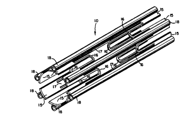
Fig. 1 is an enlarged perspective view of an
eYrAn~hle intraluminal stent in accordance with the
present invention and a delivery apparatus also in
accordance with the present invention for inserting
the stent into the lumen of a body passage and for
~Yr~ g the stent;
Fig. 2 is an enlarged, schematic end
elevational view of the stent in its uneYr~n~ed
configuration;
2200'i~
WO96/09013 PCT~S95/10931
Fig. 3 is an enlarged, schematic side
elevational view of the stent in its llne~pAn~ed
configuration;
Fig. 4a is an enlarged side elevational view
of the stent and a tool having a vein segment thereon,
depicting a method for attaching the vein segment to
the stent, using tissue adhesive, just prior to
insertion of the vein segment within the lumen of the
stent;
Fig. 4b is an enlarged side elevational view
of the stent and a tool having a vein segment thereon,
depicting a method for inserting the vein segment into
the lumen of the stent and for attaching the vein
segment to the stent using tissue adhesive, just prior
to inflation of the vein segment within the stent;
Fig. 4c is an enlarged side elevational view
of the stent with the vein segment inserted within the
lumen of the stent, depicting a method for attaching a
vein segment to the stent using tissue adhesive,
following curing of the tissue adhesive;
Fig. 5 is an enlarged cross sectional view
of the stent showing a vein segment attached to the
stent with ~L~es;
Fig. 6 is an enlarged side elevational view
of the stent deployment mech~nism~ with several wire
spokeR removed, located at the distal end of the
delivery apparatus used to deploy the stent in the
body passage;
220û4S9
W096/09013 PCT~S95/10931
Fig. 7 is an enlarged end elevational view
of the stent deployment mechA~i-em located at the
distal end of the delivery apparatus used to deploy
the stent in the body pAe~-ge;
Fig. 8 is an enlarged plan view of the drive
unit located at the proximal end of the delivery
apparatus used to deploy the stent in the body
passage;
Fig. 9a is an enlarged, schematic cross
sectional view of the stent deployment mechanism at
the distal end of the delivery apparatus with a pair
of the wire spokes shown in an unflexed position;
Fig. 9b is an enlarged, schematic cross
sectional view of the distal end of the delivery
apparatus with a pair of the wire spokes shown in a
flexed position;
Fig. lOa is an enlarged side elevational
view of the stent mounted on the stent deployment
mech~nism of the delivery apparatus in an un~Yp~n~ed
configuration;
Fig. lOb is an enlarged side elevational
view of the stent mounted on the stent deployment
mec~nism of the delivery apparatus with the wire
spokec in position for eYpAneion of the stent;
Fig. lOc is an enlarged side elevational
view of the stent mounted on the stent deployment
mechAnism of the delivery apparatus following
p~neion of the stent;
221J~i4~','
Wog6/osol3 PCT~S95/10931
Fig. lOd is an enlarged side elevational
- view of the stent and the stent deployment meçhAnism
of the delivery apparatus in position for removal of
the stent deployment mec~nism from the ~YpAn~P~ stent
while leaving the ~YrAn~ stent within the lumen of
the body passage;
Fig. 11 is an enlarged, cross-sectional view
of a hll~hing for the stent deployment merhAnism for
guiding movement of the wire spokes in accordance with
another emho~iment of the present invention;
Fig. 12 is an enlarged, exploded perspective
view of the frustaconical tip of the stent deployment
meçhAni~m for receiving the free ends of the wire
Cpok~c for use with the h-lching shown in Fig. 11;
Fig. 13 is an enlarged, cross-sectional view
of the frustaconical tip shown in Fig. 12;
Fig. 14 is an enlarged perspective view of a
stent in accordance with another preferred emho~iment
of the present invention;
Fig. 15 is an enlarged side elevational view
of a stent deployment meçh~nism having the wire spokes
removed, located at the distal end of the delivery
apparatus in accordance with another emho~iment of the
present invention;
Fig. 16 is an enlarged, exploded perspective
view of a hl~hi~g for the stent deployment mechAnism
shown in Fig. 15;
22304~,
WO96/09013 PCT~S95/10931
Fig. 17 i8 an enlarged, exploded perspective
view of the frustaconical tip of the stent deployment
me~nism shown in Fig. 15;
Fig. 18 is an enlarged side elevational view
of a swing arm for the stent deployment mechAnicm
shown in Fig. 15;
Fig. l9a is an enlarged, schematic
perspective view of a h~lC~ing in an unflexed position
for a stent deployment mechanism in accordance with
yet another emho~iment of the present invention; and
Fig. l9b is an enlarged, schematic
perspective view of the b~ching shown in Fig. l9a but
in the flexed position.
Detailed Description of the Preferred Embodiments
Referring to Fig. 1, a stent delivery
apparatus, generally designated 8, is depicted for
deploying a generally ~llh~ r, thin-walled stent 10
within a selected body passage such as a stenosed
vessel. The stent delivery apparatus 8 serves as a
catheter for inserting the stent 10 into the selected
body passage. For this purpose, the tubular stent 10
is removably mounted on a stent deployment mechanism,
generally designated 40, in the form of a wire bundle
disposed at a distal end of the stent delivery
apparatus 8.
Control of the stent deployment mech~nism 40
is effected by a manually-operable drive unit 48 in
the form of a rack and pinion microdrive. The drive
WO96/09013 2 2 0 0 Q ~~ 9 PCT~S95tl~31
unit 48 is ~i~ro~~~ at a proximate end of the stent
delivery apparatus 8 and is attached to the stent
deployment mech~n;sm 40 by a flexible control cable
49. Manual operation of the drive unit 48 controls
operation of the stent deployment mechanism 40 so that
the stent 10 can be safely deployed at a selected
target location within a body passage whereupon the
stent deployment mech-n;sm 40 and the control cable 41
are then withdrawn from the body leaving the stent 10
properly deployed at the target location.
Referring to Figs. 2 and 3, the stent 10 is
in the form of a generally tubular mesh-like
configuration providing a central lumen along its
longit~;n~l axis. The generally tubular stent 10
includes a series of tab-like rigid supports 15 which
are disposed in a generally similar arrangement at
each end of the tubular stent 10. The rigid support
tabs 15 of the stent are generally equally spaced
apart around the circular perimeter at each end of the
stent 10. In addition, the rigid support tabs 15 are
disposed so that a rigid s~rport tab 15 at one end of
the stent is substantially opposite a corresponding
rigid support tab 15 at the other end of the stent 10.
Spacer bars 17 ~O~ QCt each rigid su~o~L tab 15 at
one end of stent 10 to an opposite corresponding rigid
support tab 15 at the other end of the stent. The
spacer bars 17 serve as struts to prevent the stent
from foreshortPn;ng along its longitll~inAl axis. Each
spacer bar 17 thereby serves to maintain corresponding
pairs of ~ ~~L tabs 15 at opposite ends of the stent
in position relative to the longitl~; nA 1 axis of the
tubular stent. The spacer bars are also sufficiently
rigid to resist bowing or hpn~ing in the transverse
direction relative to the stent. As shown
2 2 0 ~; .
W096/09013 PCT~S9StlO931
schematically in Fig. 2, the ~ o~L tabs 15 at each
end of the stent are positioned adjacent to one
another in a generally ~nnll 1 Ar arrangement around the
periphery of each end of the stent so that each
r~ lG~ tab 15 has a diametrically opposing support
tab 15 at each end of the stent. A series of
deformable connecting links 16 in the form of
plastically-deformable, U-shaped, wire-like links are
used to connect adjacent rigid support tabs 15 around
the annular arrangement of the support tabs at each
end of stent 10, so that the stent 10 can be
diametrically eY~nA~A through plastic deformation of
the connecting links 16. As the stent is
diametrically eYp~nAed, the connecting links 16 bend
so that the stent maintains its generally tubular
shape. The central lumen of the stent widens but the
spacer bars 17 inhibit longitll~inAl movement of the
~ o~ tabs thereby maintaining the overall length of
the stent. The stent 10 may be expanded by applying a
radially, outwardly ext~nAing force from the exterior
of the ctent. For this purpose, external expansion
couplings, in the form of coupling tubes 18, are
oriented longit~lAin~lly relative to the stent and are
attached on the spacer bars 17 along the outside of
the stent 10. For example, the coupling tubes 18 can
be fabricated from 0.5" (12.7 mm) lengths of 26 gauge
hypodermic tubing and attached with laser welds 19 to
the exterior of the spacer bars 17 and the support
tabs 15, making sure that the lumen of each coupling
tube 18 remains open and intact. Alternatively, the
stent 10 may be fabricated by extruding the stent 10
as one piece. Deployment of stent 10 may then be
- effected using the delivery apparatus 8. The coupling
tubes 18 permit the stent 10 to be removably mounted
on the stent deployment mechanism 40 of the stent
14
220()4~
WO96/09013 PCT~S95/10931
delivery apparatus 8. The coupling tubes 18 also
serve the ~ ? of reinforcing the spacer bars 17 to
inhibit compression and/or eYrAncion of the stent
along its longit~i n~ 1 axis thereby functioning as a
strut and to inhibit henA i ~g or collapsing of the
stent in the transverse direction. In an alternative
arrangement, the coupling tubes 18 may be used instead
of the spacer bars 17 with the ends of the coupling
tubes being attached, by laser welding, to the
corresponding support tabs 15 at opposite ends of the
stent thereby serving the function of the spacer bars
17. In yet another alternative arrangement, the stent
may be used without the coupling tubes 18 in which
case the stent can be eYr~n~ from the inside at the
target location using a balloon-type catheter delivery
system. Since the stent is plastically deformed,
there is very little recoil and the stent resists
being recompressed.
In a particular emho~iment~ the stent 10 is
fabricated from a single piece of continuous tube with
no welds or solder points. For example, a 316L grade
seamless stainless steel tube with an outer diameter
of 0.125" (3.175 mm), a nominal wall thickness of
0.006" (0.1524 mm), and an overall length of 0.5"
(12.7 mm) may be used. Six rectangular regions of
material are removed from each end of the tube so that
material remains for six rigid support tabs 15. The
~u~G~ L tabs are thin, generally arcuate, and conform
in shape to the stent in the first direction.
Deformable ronn~cting links 16 and spacer bars 17 are
formed by removing six generally H-sh~pe~ regions 11
from the central portion of the tube thereby forming
open areas in the tubular stent. Thus, the stent 10
has a general tubular structure having a thin-walled
22 JO$~S
WO96/09013 PCT~S95/10931
lattice frame with sren;ngs in the frame. Wire
electrical A i~~h~rge mac~ini ng or wire EDM can be used
for removing sections of the tubing to extremely high
precision without creating burrs or deformations. The
wire diameter for cutting can be approximately 0.010"
(0.254 mm) resulting in corners that are radiused to
0.005" (0.127 mm). Using this method a stent 10 can
be made with rigid D~G~L tabs 15 that are 0.0625"
(1.588 mm) long by 0.0625" (1.588 mm) wide, spacer
bars 17 that are 0.374" (9.5 mm) long by 0.020"
(0.508 mm) wide and connecting links 16 that are
0.004" (0.102 mm) wide relative to the circumference
of the stent. After full ~yp~ncion~ the stent 10 can
assume a final diameter of approximately 0.315"
(8.0 mm) or 2.5 times its original diameter.
The total intimal surface area along a
stented segment of a vessel can be approximated from
the equation for the surface area of a tube, or ~dl,
where d is the stent diameter and l is the stent
length. For a stented region corresponding to the
stent 10 of the type shown schematically in Fig. 3
with the above dimensions, the estimated total intimal
surface area is ~(8.0)(12.7)= 319.20 mm2. The total
surface area of the stent, with coupling tubes made
from 26 gauge hypodermic tubing, that can be exposed
to the vessel lumen is estimated to be 99.02 mm2.
Using geometrical constraints, the area of metallic
surface in contact with intimal tissue is estimated to
be 54.43 mm2. Expressed in terms of percent open area
within the stented segment, the stent of the present
invention is therefore estimated to be 82.95~ open.
This result suggests that the stent of the present
invention is capable of preserving a large area of
endothelialized tissue. In addition, the amount of
WO96/09013 2 2 IJ O ~ '~ ', PCT~S95/10931
metallic surface ~YrQ~~~ to the blood is kept
relatively low.
Construction of the stent lO is not limited
to ~tainle~s steel. The ~tent lO can be made from any
material which is compatible with the human body and
any bodily fluids that the stent lO may contact.
However, the stent lO must be made from a material
that allows for ~YrAncion of stent lO and must be able
to maintain its eYrAnA~ shape while disposed within
the lumen of the body passage. In addition to
stainless steel, suitable materials for construction
of stent lO may include tantalum and titanium. The
stent lO can also be fabricated from a memory metal
such as nitinol. In addition, the stent lO does not
have to be fabricated from a single piece of
continuous tube. For example, the spacer bars 17
and/or the connecting links 16 can be made separately
from the rigid ~y~O~ tabs 15 and attached using, for
example, laser welding techniques. Alternatively, the
stent lO may be fabricated by extruding the stent lO
as one piece.
While the stent may be deployed as a "stand
alone" device, the stent may also be effectively used
as a vA~c~llAr endograft by attaching a segment of
vein, preferably an autologous vein or a synthetic
graft material within the central lumen of the stent.
Preferably, an autologous vein segment is utilized to
provide an endothelial layer as a lining for the lumen
of the stent. As shown in Figs. 4a - 4c, a vein
segment 25 may be attached to the inner surface of the
stent lO by, for example, using tissue adhesive 29.
As shown in Fig. 4a, the vein segment 25 is attached
to a piece of tubing 26 with suture 27, which is in
221l0~J~
WO96/09013 PCT~S95110931
turn attached to a syringe filled with isotonic
saline. The free end of the vein segment 25 is then
closed with ~ Le 28. The vein segment 25 is
deflated using the syringe and, as shown in Fig. 4b,
the vein segment is then inserted into the lumen of
the stent 10. A small amount of tissue adhesive 29 is
applied to multiple points along the inner surface of
the stent 10. As shown in Fig. 4c, the vein segment
25 is then inflated 80 that the outer surface of the
vein segment 25 contacts the inner surface of stent 10
especially at the points where tissue adhesive 29 has
been applied. Once the ti~sue adhesive 29 has cured,
the vein segment 25 is disconnected from the tubing 26
and the eYc~ss vein segment 25 is trimmed at each end
of the stent.
The use of tissue adhesive to secure the
vein segment to the stent may not always be suitable
or permitted. Accordingly, a vein segment may be
attached to the stent 10 in accordance with a
preferred method of using sutures 30. As shown in
Fig. 5, a length of vein segment 25 about twice as
long as the length of stent 10 is used. The vein
segment 25 is inserted within the lumen of stent 10 so
that the stent is generally centered about the vein
segment 25. The ends of the vein segment 25 are then
everted over the ends of stent 10 so as to completely
line the inner surface of stent 10 and to
substantially cover the outer surface of stent 10.
After slightly crimping the stent 10 onto the external
surface of the vein segment 25, the proximal and
distal ends of the vein segment 25 are secured to the
adventitia with interrupted 7.0 proline sutures
between each spacer bar 17. Accordingly, a stent-vein
2 2 3 ~J 4 ~,
WO96/0~13 PCT~S95/10931
complex is provided for deployment at the target
location by the stent delivery apparatus lO.
The stent delivery apparatus 8 includes a
~tent deployment mech~n~sm 40, as shown in Figs. 6 and
7, manually operated by drive unit 48, as shown in
Fig. 8. The drive unit 48 (Edmund Scientific,
Barrington, NJ, model ~J3650) is connected with the
stent deployment mechAnifim 40 by control cable 49.
The stent deployment mechanism 40 includes a series of
six spokes 42 each connected at one end to a bushing
43. The spokes 42 may be in the form of a symmetric
bundle of six spring steel wires of diameter .008
inches (.203 mm) bonded to the outer wall of the
htlC~ing 43 which is in the form of a 316L grade
stainless steel tube having a length of approximately
.25 inches (6.35 mm), an outer diameter of .0732
inches (l.86 mm) and an inner diameter of .05 inches
(l.28 mm). The free ends of the spokec removably nest
within conical tip 45 di~ at the end of a central
guidewire 44 in the form of stainless steel wire
having a diameter of .04 inc-h~s (l mm). When the
guidewire 44 is moved to retract the tip 45 toward the
cable 49, the free ends of the spokes can be
positioned to nest within the tip 45. When the
guidewire 44 is moved to displace the tip 45 away from
the cable 49 the free ends of the spokes 42 are
released as shown in Fig. 6.
In cable 49, the central guidewire 44 is
coaxially contained within a flexible guide tube 41 in
the form of a polymer tubing, such as flexible nylon
tubing having the same inner and outer diameters as
the h~l~hing 43. The hn~h;ng 43 is bonded to one end
of the guide tube 41 using, for example, epoxy, spot
WO96/09013 220a$~9 PCT~S95/10931
welding, or soldering t~chniques. The junction
between the h -C~ ing 43 and the guide tube 41 is
enclosed within an external junction sleeve 39 in the
form of a ~tainles~ steel tube segment. The other end
of the flexible guide tube extends with the guidewire
44 approximately 30 inr~eC to the drive unit 48 shown
in Fig. 8. The drive unit functions to displace the
guidewire 44 through the flexible guide tube 41 in a
controlled manner by manual rotation of actuator knob
38. The guidewire 44 and the flexible guide tube 41
are enclosed within an outer sheath tube 46 in the
form of a polymer tube such as a clear polyethylene or
teflon tubing that is approximately 30 inches long.
As best shown in Figs. 1 and 6, the end of the sheath
tube 46 at the stent deployment mechanism is capped by
a sheath cap 47 in the form of 316L grade stainless
steel tubing having a length of approximately 2
inches. The sheath cap 47 serves as a rigid housing
for accommodating the wire spoke bundle 42 when the
sheath cap is slid over the wire spoke bundle. The
sheath tube 46 may be manually retracted relative to
flexible tube 41 and guidewire 44 to displace the
sheath cap 47 from the wire spoke bundle 42 in order
to expose the wire spoke bundle. Flexible tube 41,
guidewire 44, and outer sheath 46 are concentric and
allowed to move relative to each other along their
axes.
The proximal ends of flexible tube 41 and
guidewire 44 are attached to the linear microdrive
unit 48, which allows for the axial movement of
guidewire 44 relative to flexible tube 41. As knob 38
is turned an internal rack and pinion drive mechanism
longitllAin~lly displaces the central guidewire 44
relative to the flexible inner tube 41. As
22uo4a~
WO 96/OgO13 PCr/USgS/10931
schematically depicted in Fig. 9a, when the tip 45 of
the guidewire 44 is positioned in its mid-position the
free ends of the spokes 42 are captured within the tip
45 but the spokes remain relatively extended, or
S unflexed, between the tip 45 and the hllching 43. As
schematically depicted in Fig. 9b, when the
guidewire 44 is deployed so the tip 45 moves toward
the h ~rhing 43 at the distal end of flexible tube 41,
croke~ 42 are caused to bend and flex outwardly.
In an alternative emhoAiment of the stent
deployment mech~ c-m~ the spokes 42 are attached to
flexible tube 41' by hlt-ch;ng 43', as depicted in Fig.
11, which serves as a hl-ching for the guidewire 44'.
The spokes 42 may be made from spring steel wires of
diameter 0.008" (0.203 mm). The h~ ing 43' is a
brass cylindrical section with à frustaconical end
having an outer diameter of 0.138" (3.5 mm), an inner
diameter of 0.042" (1.079 mm), and a length of 0.335"
(8.5 mm). An inner bore 56 is provided through the
hl~hing to permit the flexible tube 41' and the
guidewire 44' to pass therethrough. The cross section
of the inner bore 56 of h~lching 43' is not circular
but instead includes flattened sidewall sections to
prevent the hllching 43' from rotating around the
flexible tube 41'. The flexible tube 41' is made from
ABS plastic tubing with an approximate 3" (76.2 mm)
length of the distal end of the flexible tube 41'
being ~peA with flattened sidewall sections to mate
with inner bore 56. Six ~mall angled bores 50 are
drilled at 30~ relative to the longitudinal axis of
the hllching at the distal end of the bllching 43'. The
bores 50 are approximately 0.010" (0.254 mm) in
diameter and widen to approximately 0.020" (0.005 mm)
at the periphery of the hll~ching. The bores are
2200409
WO96/09013 PCT~S95/10931
substantially equally spaced around the distal end of
the kl~hi~g 43~ to accommodate the spokes 42, which
are epoxy glued in place within the bores 50. The
b~l~hing 43' is epoxy bQn~Q~ about the distal end of
the flexible tube 41'. The flexible tube 41' extends
from the hllChi~ for 30" before being connected to the
linear microdrive unit 48. In addition, the outer
sheath 46 is constructed from teflon tubing
approximately 30" (76.2 cm) in length with an inner
diameter sufficiently large to form a slip fit over
the bllching 43'. Attached to the distal end of the
outer sheath 46 is a tubular end cap 47 which is
fabricated from a 316L grade stainless steel tubing
approximately 2" (50.8 mm) in length and with similar
inner and outer diameters to the outer sheath 46. The
end cap 47 serves as a rigid housing that accommodates
spokes 42 when the spokes 42 are withdrawn
sufficiently into the end cap 47.
As depicted in Figs. 12 and 13, a
frustaconical tip 45' of the catheter is used in
conjunction with the hl)ching 43' depicted in Fig 11.
The tip 45' is fabricated from two brass sections, 60
and 61, having outer diameters of approximately 0.138"
(3.5 mm) and inner diameters of approximately 0.042"
(1.079 mm). The first section is generally
cylindrical with a frustaconical end. The second
section is generally cylindrical. A central aligned
bore 55 extends through the Fecon~ section and into
the first section. The cross cection of the inner
bores 55 of the two tubular sections 60 and 61 are not
circular but instead have flattened sidewall sections,
as shown in Fig. 12, to prevent the tip 45' from
rotating around the guidewire 44'. The guidewire 44'
is made from narrow hypodermic tubing with an
22 ~-t 8 ,
Wos6/o9ol3 PCT~S95/10931
approximately 3" (76.2 mm) length of the distal end of
the guidewire 44' being shaped with flattened exterior
sidewall sections to mate with inner bore 55. The
first section 60 of the tip 45' has a length of
approximately 0.236" (6 mm) and the second section 61
of the tip 45' has a length of 0.010" (2.5 mm). Six
small radially oriented bore holes 51, approximately
0.020" (0.508 mm) in diameter, are drilled at 30~
angles relative to the longitl~;n~l axis of the
central bore 55. The bore holes 51 are uniformly
spaced around the proximal end of the first section 60
to accommodate the free ends of the spokes 42, which
are loosely held in place. Small exterior
longitllAinal slots 52, 0.020" (0.508 mm) deep and
0.020" (0.508 mm) wide, are milled along the second
section 61 to act as guide slots for the spokes 42.
The longitllA i nA 1 slots 52 confine the movement of the
spokes 42 to a radial direction in a plane through the
longitllAinAl axis of the ceconA section 61 and inhibit
lateral movement of the spokec 42 out of the plane.
The proximal end of the first section 60 is held by
friction fit on the end of the guidewire 44' in
abutment with the distal end of the second section 61,
so that the slots 52 along the second section 61
register with the bore holes 51 drilled in the first
section 60. Alternatively, the first section 60 and
the second section 61 can be attached using epoxy or
spot welding techniques.
Yet another emhoAiment of the stent
deployment mechanism 40 is depicted in Figs. 15-18.
The hllching 143 is fabricated from two abutting brass
sections, 173 and 174. The first section 173 has a
larger cylindrical portion at the distal end that
tapers into a frustaconical section which terminates
220u~
WO96/09013 PCT~S95/10931
in a smaller cylindrical section at the proximate end
of the first section. The F~con~ ~ection 174 is
generally cylindrical. The two abutting sections 173
and 174 have outer diameters of 0.138" (3.5 mm), inner
diameters of 0.042" (1.079 mm), and a combined overall
length of 0.335~ (8.5 mm). An inner bore 156 passes
through both sections. The cross section of the inner
bore 156 of the two t~h~ r sections 173 and 174 is
sized and ~p~A to mate with the distal end of the
flexible tube 41 and to prevent the bushing 143 from
rotating around the flexible tube 41. The second
section 174 has a circular groove 182 milled into the
proximal end of the second tubular section 174. The
circular groove 182 is concentric with the inner bore
156. In addition, small external longitudinal
slots 180 are milled along the second tubular
section 174. A series of six swing arms 176, having
spoke su~olL tubes 177 attAchD~ to ball bearings 178,
serve as hinges and are disposed with the ball
bearings 178 positioned within the circular
groove 182, which serves as a bearing race, and the
spoke support tubes 177 aligned with the longitudinal
slots 180. The swing arms 176 and longitudinal
slots 180 confine the movement of the spokes 42 to a
radial direction in a plane through the longitudinal
axis of the secon~ section 174 and inhibit lateral
movement of the spokes 42 out of the plane. The
distal end of the first tubular section 173 is then
abutted against the proximal end of the second tubular
section 174 thereby capturing the ball bearings 178
within the bearing race ~Loove 182 to hold the swing
arms 176 in place. The first ~llh~ r section 173 is
attached to the second tubular section 174 by glue,
epoxy, laser welding, or any other suitable means.
Ends of the spokes 42 are then inserted into the spoke
WO96/09013 2 2 0 ~ 4 C, PCT~S95/10931
s~rpQrt tubes 177 and are held in place by epoxy,
glue, laser welding, or any other suitable means.
As depicted in Fig. 17, the frustaconical
tip 145 of the catheter for use in conjunction with
the h~lching 143 shown in Fig. 16 is fabricated from a
first section 160 of brass having a cylindrical
section terminating in a frustaconical point and an
~ abutting second generally cylindrical section 161 of
brass. The two sections 160 and 161 have outer
diameters of 0.138~ (3.5 mm), and inner diameters of
0.042H (1.079 mm) and an overall combined length of
.335~ (8. 5 mm). An inner bore 155 extends through the
second section 161 and into the first section 160.
The inner bore 155 of the tubular sections 160 and 161
is sized and chApe~ to accommodate the distal end of
the guidewire 44 and to prevent the tip 145 from
rotating around the guidewire 44. The second
section 161 of the tip 145 iS identical to the second
section 174 of the hl~C~ing 143. As with the
hll~hing 143, a series of swing arms 176, having spoke
support tubes 177 attached to ball bearings 178, serve
as hinges and are disposed with the ball bearings 178
positioned within the circular groove 182 serving as a
bearing race and the spoke ~ OI L tubes 177 aligned
with the longitl~inAl slots 180. The swing arms 176
and the longit~inAl slots 180 confine the movement of
the spokes 42 to a radial direction in a plane through
the longit~inAl axis of the second section 174 and
inhibit lateral movement of the spokes 42 out of the
plane. The proximate end of the first section 160 is
then abutted against the distal end of the second
section 161 thereby holding the swing arms 176 in
place. The first section 160 iS secured to the second
section 161 by glue, epoxy, laser welding, or any
22304~
W096/09013 PCT~S95/10931
other suitable means. The free ends of spokes 42 can
then be inserted into the spoke support tubes 177
where they are releasably held in place.
In still another embodiment, the bushing and
a proximate section of a frustaconical tip may be made
from a tl~hlllA~ section 274 of flexible plastic or
metal, as depicted in Figs. l9a and l9b, to guide the
movement of spokes 42. One end of the tubular section
274 is fluted with each flute 276 being sufficiently
wide to prevent movement of the spokes 42 out of a
plane through the longitllAin~l axis of the tubular
section 274 and the longitllAin~l axis of the
flute 276. The fixed ends of the spokes 42 are
attached to the separated flutes 276 of the tubular
section 274 used as the hllching by epoxy, glue, laser
welding, or any other suitable means. The free ends
of the spokes 42 may be releasably held in the
flutes 276 of the tllhlll ar section 274 used in the
frustaconical tip. When the hllching is moved
longitllAinAlly relative to the frustaconical tip by
the drive unit 48 so as to shorten the distance
between the h~lC~ing and the tip, the spokes 42 and the
flutes 276 flex as shown in Fig. l9b. When the
hllc~ing is returned to its starting position relative
to the tip, the spokes 42 and the flutes 276 return to
their unflexed positions as shown in Fig. l9a.
As shown in Fig. 14, a stent 10 is depicted
that is substantially similar to the stent shown in
Fig. 1 except that the component parts of the stent lO
shown in Fig. 14 have different sizes relative to one
another. For example, the coupling tubes 18 shown in
Fig. 14 are somewhat oversized relative to the stent
shown in Fig. 1. Also, the connecting links 16 shown
26
2200~oS
wog6/oso13 PCT~S95/10931
in Fig. 14 are somewhat more ro~ln~A than the
~onnecting links ~hown in Fig. l. While the stent
shown in Fig. 14 is presently a preferred
- configuration, both stents function in a similar
manner.
In operation, a method of deploying the
stent lO is shown in Figs. lOa-d. Referring to Fig.
lOa, the stent l0 is first installed on the stent
deployment mech~nicm 40. The spokes 42 are passed
through the coupling tubes 18 with the guidewire 44
passing through the lumen of stent l0. If a vein
segment is attached to the stent in the manner shown
in Fig. 5, the spokec must poke through the portions
of the vein segments that cover the openings to the
coupling tubes 18. After the stent is properly
positioned on the stent deployment mechanism 40, the
free ends of the spokes 42 are manually nested within
the tip 45 as shown in Fig. lOb. The outer sheath 46
is then slid over the stent l0 to cover stent lO. The
tip 45 at the distal end of stent deployment
mech~nism 40 is then inserted into the body and moved
to the target location within the body passage. The
outer sheath 46 is then pulled back to expose the
stent lO and the stent deployment mechAn;sm 40 at the
target location. The drive unit 48 is then actuated
to displace tip 45 in a direction toward the bushing
43 at the end of flexible tube 41 causing the
spokes 42 to flex radially outward thereby exerting an
external, radially outward force that expands the
stent lO as shown in Fig. lOc. Once the stent l0 is
pAn~e~ to the desired diameter, plastic deformation
of the connecting links causes the stent to remain in
its eYpAn~ed configuration providing a radially
enlarged central lumen. After the stent has been
2200lt89
WO96/09013 PCT~S95/10931
~Yr~n~eA, the tip 45 is displaced by the drive unit in
a direction away from the hllchin~ 43 at the distal end
of flexible tube 41 thereby releasing the free ends of
the rrokes 42 from the tip 45 as shown in Fig. lOd.
The guidewire 44, the flexible tube 41, and the outer
sheath 46 are then withdrawn while leaving the
stent lO in position within the body passage.
~Y~mples
Exam~le 1
A stent of the type shown in Fig. 2 was
tested by implantation in the left external iliac
artery of mongrel dogs. The stent was tested for
structural integrity, deformability, migration, and
patency.
Five adult male mongrel dogs were used for
the study. The animals were placed under general
inh~lational anesthesia with a halothane/oxygen
mixture, and administered an intravenous dose of a
preoperative cephalosporin. Both groins were shaved
and prepped with bet~ine and alcohol prior to being
draped in the usual sterile fashion. Incisions
measuring approximately 2-3 cm were made vertically
overlying the femoral vessels. The femoral artery was
isolated and controlled with vessel loops. At this
time, intravenous heparin was administered at a dose
of 100 u/kg. A transverse arteriotomy was made and a
12 ft. (3.66 m) sheath introduced within the artery.
A stent according to the present invention was mounted
on a 0.315" (8 mm) diameter balloon (1.57" (4 cm) in
length) which was attached to the end of a catheter.
The stent was positioned in the left external iliac
artery (approximately 1.97" (5 cm) from the aortic
WO96/09013 2 2 0 a ~ s, PCT~S95110931
bifurcation) under fluo~-coric guidance. The stent
was then eYpAn~ by inflating the balloon uniformly
to lO atmocr~es. Upon completion of the procedure,
an arteriogram was performed. The femoral artery was
ligated and the incision closed.
After a six week convalescent period, a
final arteriogram was performed via a left brachial
artery approach. The gradient of pressure across the
stent was then Assr~,s~ with measurements that were
taken just distal to the stent, within the center of
the stent and just proximal to the stent. All
measurements were also compared with a baseline
pressure value within the aorta near its bifurcation.
The aorta was then cannulated for the purpose of
pressure-perfusion fixation of the iliac vessel with a
2% paraformaldehyde mixture. The stent was
subsequently harvested within the vessel and submitted
for emhe~ing in methyl methacrylate for future
sectioning and histologic analysis.
The results indicate that the stent of the
present invention is deployable using a balloon
mounted to a catheter and that the stent was patent at
the completion of the study. Also, there was no
indication of migration or deformation of the stent by
arteriographic analysis. Further, there was no gross
evidence of exaggerated neointimal hyperplasia in any
area of the stent lumen or any pressure gradient
(defined as a change greater than or equal to 15% of
the systolic blood pressure).
Example 2
A vein-lined stent, of the type as shown in
Fig. 5, was subjected to conditions of high flow and
22'~0489
wos6/09ol3 PCT~S95/10931
high pressure in a bench top flow system to evaluate
the effectiveness of a vein-lined stent.
A superficial femoral vein was harvested
from a dog using stAn~rd sterile techniques. A
segment measuring approximately 0.79 in. (2 cm) in
length was selected without branches and immersed in
normal saline. The vein was mounted on a moistened
14Fr dilator with care being taken not to injure the
endothelial surface. The stent was then slid over the
lo vein segment. After slightly crimping the stent onto
the external surface of the vein, the ends of the vein
were everted and Se~r ed to the adventitia with
interrupted 7.0 proline sutures between each stent
spacer. The stent-vein assembly was mounted onto an
0.315" (8 mm) balloon catheter and deployed within a
transparent and compliant elastomeric tube that
simulated a vessel. The entire apparatus was then
subjected to a continuous flow of isotonic saline at
pressures between 15 and 200 mmHg.
Qualitative observation of the stent-vein
assembly in the continuous flow field showed that even
under high pressure, there was no flow around the
outside of the stent. The excellent seal made by the
stent-vein assembly against the vessel wall was due
primarily to the eversion of the ends of the vein over
the stent. The seal was evi~nce~ by micro bubbles
which remained stationary on the outer ~urface of the
stent in the flow field. The vein remained taut and
stationary over the stent and there was no indication
of stent or tissue migration.
WO96/09013 2 2 o ~ A ~ ? PCT~S95/10931
FY~m~le 3
The bulk ela~tic behavior of the stent of
the type shown in Fig. 2 was evaluated by mounting the
stent within a compliant tube and subjecting the stent
to increasing external pressures. The luminal area of
the stent was recorded at each pressure. Pressure was
then plotted against area reduction to estimate the
stiffne66, or inversely, the compliance of the stent.
The apparatus for loading the stent
essentially comprised a compliant vessel with an inner
diameter of 0.315" (8 mm), a pressure chamber for
housing the vessel and the stent, a pressure
transducer, and a video camera. The compliant tubes
were custom manufactured using Dow Corning
Sylgard 184. A very thin layer (approximately 0.016"
(0.4 mm)) of the material was applied in liquid state
to a polished 0.315" (8 mm) diameter cylinder mandrel
which was constantly rotating in an oven at 150~C.
The application of the liquid Sylgard to the mandrel
was carefully controlled to insure that the thickness
of the tubes did not vary around the circumference or
the length of the tubes. The tubes were removed from
the mandrel after curing and then mounted in the
pressure chamber. The stent was then mounted on a
balloon catheter (0.315" (8 mm) O.D.) and expanded
within the lumen of the tube. The compliant tube
section cont~ining the stent was supported from both
ends by rigid plexiglass fittings. The tube lumen was
open to atmospheric pressure while the pressure
chamber represented the external environment of the
compliant tube. This configuration ensured that only
the stented segment deformed under pressure. Two
additional ports in the pressure chamber served as
220C!l ~?
WO96/09013 PCT~S95/10931
access for the pressure transducer and the syringe for
imparting pressure to the system.
After the pressure chamber was filled with
water, a syringe was connected to the system. The
pressure within the system was controlled by the
syringe piston displacement and monitored by the
pressure trAnC~l~cer. A video camera was focused on
the segment of tube containing the stent. Pressure
within the chamber was increased from atmospheric
pressure in 500 pascal increments. At each
incremental increase in the chamber pressure, a
personal computer digitized the video frame of
interest. This image was ported to an image
processing program where the luminal area was
measured. The test was stopped when the luminal area
measurement had decreased by lO mm2. In order to
determine stent compliance, the change in the stent
cross-sectional area was plotted against the
incremental pressure increases. Compliance C was
estimated from the relation:
C= (A2 - A~)/(P2 - Pl)
where (A2 - A~) is the incremental area change and (P2 ~
P~) is the incremental pressure change. Stiffness is
defined as the inverse of compliance. It should be
noted from this relation that compliance is
in~p~n~ent of the stent length. The stent of the
present invention showed an initially linear elastic
behavior up to approximately lO.0 KPa. Between
lO.O KPa and 20.0 KPa, the stent began to deform more
for the same incremental increase in pressure. This
pressure-deformation behavior indicated that the yield
point of the material had been reached and that the
32
WO96/09013 2 2 0 û 4 ~ ~ PCT~S95/10931
stent was deforming plastically rather than
elastically. A linear regression was applied to the
data points up to lO.0 KPa in order to determine the
compliance of the stent in the elastic region of
deformation. The slope of the regression fit
represents the bulk stiffness and the inverse of this
slope represents the compliance. This analysis
yielded a ~tiffne~s of 5221.65 and a compliance of
0.0002. The significance of such a low compliance is
that increased rigidity is considered desirable in a
stent.
In summary, the above Examples indicate that
a stent according to the present invention can be
~Yp~n~ed within the lumen of a body passage and can be
lS used to sup~o~ an endothelial layer. As such, the
stent should improve vascular patency rates in current
applications for stents, such as obliterative disease,
arteriovenous fistulas, intimal injuries, and
aneurysmal ~ Q. The stent should also reduce
thrombogenesis and neointimal hyperplasia. The stent
should also counteract recoil of the vessel wall
following angioplasty. In addition, the stent may be
deployed by the stent delivery apparatus that couples
to the stent and exerts an outward force on the
external surface of the stent thereby leaving the
luminal environment of the stent undisturbed.
The delivery apparatus 8 is designed
specifically for the deployment of the stent lO.
However, the wire cage or basket of the delivery
apparatus could be easily adapted for transcatheter
extraction of urinary tract and biliary tract stones
or for retrieval of intravascular foreign bodies. The
wire cage of the delivery apparatus actively expands
2~ o ~ ~ o ~ ~
WO96/09013 PCT~S95/10931
and tends to passively collapse. Additionally, the
delivery apparatus of the ~ ~-ent invention might also
be modified to incorporate a high speed rotary device
within the wire cage. The delivery apparatus could
then be used for declotting prosthetic dialysis access
grafts, central veins, or even pulmonary arteries.
It will be ~ o-~..ized by those skilled in
the art that changes or modifications may be made to
the above-described embodiments without departing from
the broad inventive concepts of the invention. It
should therefore be understood that this invention is
not limited to the particular embodiments described
herein, but is intenAe~ to include all changes and
modifications that are within the scope and spirit of
the invention as set forth in the claims.