Note: Descriptions are shown in the official language in which they were submitted.
WO 96/09548 2 2 0 0 6 2 . pyGB95/02186
APPARATUS AND METHOD FOR CARRYING OUT
ANALYSIS OF SAMPLES
The present invention relates to apparatus and to a
method for carrying out optical analysis of samples and
is applicable in particular to the analysis of
biological, chemical and biochemical samples.
Many chemical, biochemical and biological assays
rely upon inducing a change in the optical properties of
a biological sample being tested. For example, in order
to test for the presence of an antibody in a blood
sample, possibly indicating a viral infection, an
enzyme-linked immunosorbent assay (ELISA) can be carried
out ~ahich produces a visible coloured deposit if the
antibody is present. An ELISA makes use of a surface
which is coated with an antigen specific to the antibody
to be tested for. Upon exposure of the surface to the
blood sample, antibodies in the sample bind to the
antigens. Subsequent staining of the surface with
specif;c enzyme-conjugated antibodies and reaction of
the enzyme with a substrate produces a precipitate which
shows up the level of antigen binding and hence allows
the presence of antibodies in the sample to be
identi-ied. This identification is usually carried out
using a light microscope which allows an area of the
substrate to be viewed by an operator.
In addition to colour staining using an ELISh,
techniques such as fluorescence and gold labelling can
be used to alter the optical properties of biological
antigen material. Similar techniques are also used in
CA 02200562 2001-04-03
general histology to visualise specific areas of tissue, e.g. particular cell
types or cell
structures, as well as in cell culture.
A significant disadvantage of existing optical analysis techniques is that
they
are open to human error because of their subjective nature. These techniques
are also
not suited to uses where a high throughput of samples is required, for example
in
blood screening applications or cervical smear tests, and are thus relatively
expensive
to use. The cost factor is exacerbated because, more often than not, different
equipment is required for each particular technique.
An object of an aspect of the present invention is to provide a technique for
carrying out the optical analysis of samples which overcomes or at least
mitigates
certain of these disadvantages.
It is also an object of an aspect of the present invention to provide an
optical
analysis technique which allows high speed automatic analysis of biological,
biochemical and chemical samples and which is versatile enough to allow it to
be
used for a variety of different studies.
These objects are achieved by adapting the technology which has been
developed in the field of audio and video compact discs to scan surfaces, to
which a
sample has been attached, using a light beam which is substantially focused
onto that
surface. A detector is arranged to detect light reflected from, or transmitted
through
that surface, and to determine from analysis of the detected light whether the
light
beam has been interfered with by the sample material.
According to a first aspect of the present invention there is provided a
method
of conducting an electromalmetic-wave inspection of a biological, chemical, or
biochemical sample, the method comprising the steps of: supporting at least
one
sample on a substrate at a surface location; directing an incident beam of
electromagnetic radiation from a radiation source onto the substrate; scanning
the
incident beam over the substrate by rotating the substrate about an axis
substantially
perpendicular to the substrate and by moving the radiation source in a
direction
having a component radial to said axis; detecting radiation reflected from and
transmitted through the substrate and the sample and providing an output
signal from
the detected radiation corresponding to the optically inspected sample of said
surface
location on which said incident beam is currently directed; modulating at
least a part
of said incident beam by distributed electromagnetic radiation modulating
means
provided on the substrate at said surface location; and analyzing said output
signal
2
CA 02200562 2001-04-03
during the scanning step to extract, from said detected radiation, digital
position
address information at said surface location on which said incident beam is
currently
directed.
According to a second aspect of the present invention there is provided, a
method of conducting an electromagnetic-wave inspection of a biological,
chemical,
or biochemical sample, the method comprising the steps of: supporting at least
one
sample on a substrate at a surface location; directing an incident beam of
electromagnetic radiation from a radiation source onto the substrate; scanning
the
incident beam over the substrate by rotating the substrate about an axis
substantially
perpendicular to the substrate and by moving the radiation source in a
direction
having a component radial to said axis; detecting radiation reflected from or
transmitted through the substrate and the sample and providing an output
signal from
the detected radiation corresponding to the optically inspected sample at said
surface
location on which said incident beam is currently directed; modulating at
least a part
of said incident beam by distributed electromagnetic radiation modulating
means
provided on the substrate at said surface location; and analyzing said output
signal
during the scanning step to extract, from said detected radiation, digital
position
address information at said surface location on which said incident beam is
currently
directed.
According to a third aspect of the present invention there is provided, a
system
for automatically carrying out an electromagnetic-wave inspection of a sample
to
determine whether the sample comprises material which interferes with incident
electromagnetic radiation, the system comprising: a substrate having a surface
for
supporting at least one sample; a source of electromagnetic radiation for
providing an
incident beam of electromagnetic radiation; means for scanning said incident
beam
across said surface on the substrate; and detector means which in use is
arranged to
detect radiation reflected from or passing through the substrate and the
sample, the
substrate being provided with distributed electromagnetic radiation modulating
means
for modulating at least a part of said incident beam with a digitally encoded
position
address at the location of said surface on which the incident beam is
currently
directed, the detector means being arranged to decode the modulated radiation
to
determine the encoded address to determine if the incident beam radiation has
been
modulated by any of said material present in the sample.
3
CA 02200562 2001-04-03
According to a fourth aspect of the present invention there is provided an
apparatus for conducting an electromagnetic-wave inspection of a biological,
chemical, or biochemical sample supported on a substrate, the apparatus
comprising:
means for supporting a substrate and for rotating the substrate about an axis
substantially perpendicular to the substrate; a source of electromagnetic
radiation for
providing an incident beam of electromagnetic radiation; drive means for
moving the
radiation source over the mounted sample in a direction having a component
radial to
said axis so that in combination with the means for rotating the substrate the
incident
beam can be scanned over the substrate; detector means for detecting radiation
reflected from or transmitted through the substrate and sample and for
providing an
output signal corresponding to the detected radiation; and decoding means for
extracting digital address information or calibration information from said
output
signal, said information having been modulated onto the incident beam by
radiation
modulating means provided on the substrate at the location on the surface of
the
substrate on which the incident beam is currently directed, and means for
using said
information to align the scan with the substrate.
The present invention enables the rapid scanning of a surface coated with
components from a sample to determine their presence and also if necessary
their
optical properties. The system is particularly suited for carrying out the
automatic
inspection of samples with a high throughput. Moreover, provision of address
information in or on the substrate enables the precise position of the
electromagnetic
radiation beam on the surface to be determined which in turn allows the
accurate
mapping of optical data, corresponding to attached material, to the surface.
This
enables regions of interest on the surface to be easily and quickly relocated.
The present invention is suited to carrying out ELISA where the specific
antigen is coated onto the
4
CA 02200562 2001-04-03
WO 96/09548 PCTIGB95I02186
_5_
surface of the substrate. The surface is then exposed
to the analyte and subsequently the specific enzyme and
the resulting sample scanned to detect and quantify the
enzyme linked to the surface. The system is also suited
to carrying out histological analysis and to the
quantitative study of gels run using electrophoresis.
Preferably, the electromagnetic radiation is light,
e.g. infra-red, visible or ultra-violet.
According to a fifth aspect of the invention
invention there is provided a system for automatically
carrying out an optical inspection of a sample to
determine whether or not the sample comprises material
which interferes with incident electromagnetic
radiation, the system comprising:
a substantially planar substrate having a surface
foz supporting the sample;
a source of electaomagnetic radiation for providing
a beam of electromagnetic radiation;
means arranged on one side of the substrate for
scanning the beam across the surface of the substrate;
a first detector .or detecting electromagnetic
radiation reflected from the substrate and the sample;
a second detector for detecting electromagnetic
radiation passing through the substrate and the sample;
and
control means coupled to the first and second
detectors and for causing said beam to scan the surface
of the substrate in dependence upon one or both of the
CA 02200562 2001-04-03
WO 96109548 PCTlGB95102186
-6-
outputs of the detectors and for detecting the presence
of said components.
In a preferred embodiment of the above third
aspect of the present invention the control means is
arranged to determine the difference between output
signals provided by said first and second detectors,
which are representative of the signals detected, for
the purpose of detecting said material without signal
artifacts arising from, for example, dirt present on the
side of the substrate opposite the support surface. The
substrate may be provided with distributed address means
for modulating the light beam with digitally encoded
position information indicative of the area currently
being scanned by the light beam, one or other of the
15 detectors being arranged to decode the received light
signal to determine the address of the location on which
the iicht beam is incident.
According to a sixth aspect of the invention
invention there is provided a system for automatically
2o carrying out an optical inspection of a sample to
determine whether or not the sample comprises material
which '_nterferes with incident electromagnetic
radiation, the system comprising:
a disc comprising a plastic base layer on the upper
25 sur~ace of which is formed a plurality of perturbations,
for interfering wita _ZCident electromagnetic radiation,
representing digitail~~ encoded data, and a surface for
suppor=ing the sample;
CA 02200562 2001-04-03
WO 96109548 PCTIGB95/02186
disc reading apparatus including a source of
electromagnetic radiation for providing a beam of
electromagnetic radiation, scanning means for scanning
the beam across the upper surface of the disk, and an
electromagnetic radiation detector for detecting
radiation reflected from and/or transmitted through the
disk and said sample components; and
means for rotating the disc about an axis
substantially perpendicular to said beam,
wherein, in addition to being modulated by
information digitally encoded onto the disc, the beam is
additionally modulated by any of said material which is
attached to the support surface of the disc.
Preferably, said electromagnetic radiation is
visible light although infra-red or ultra-violet
radiation may be suitable.
Preferably, the disc comprises a lower layer of
transparent plastic on the surface of which is
impressed, or otherwise produced, said digital
infcrmation. This surzace is coated with a partially
reflective layer, ~or example of aluminium, which in
turn may be covered by a further layer of transparent
plastic.
In an embodiment of the above sixth aspect of the
invention the upper surface of the disc is provided, with
a 3D surface topology arranged to provide growth and
attach..-~ent cues for cells grown on the surface. For
exaaple, the surface may be provided with a rectangular
CA 02200562 2001-04-03
WO 96109548 PCTlGB95l02186
-8-
grating for causing cells to align in a chosen
direction. Alternatively, growth and attachment cues
may be provided by chemical patterning of the surface,
e.g. using fibrenectin, produced, for example, using
photolithography.
In another embodiment of the above sixth aspect of
the present invention the upper surface of the disc is
coated with a gel suitable for carrying out
electrophoresis on proteins, DNA etc. In order to run
the gels radially, a first electrode may be provided at
the centre of the disc with a second electrode being
provided around the periphery of the disc. A well may
be formed in the gel into which the analyte can be
placed.
In order to calibrate a system embodying the
present invention, the disc may be provided with a
calibration track, e.g. a series of 256 grey levels.
These levels may be printed onto the surface of the
track using an ink jet printer.
According to a seventh aspect of the present
invention there is provided a substrate for use in a
system which is arranged to carry out an optical
inspection on the substrate to determine whether or not
material which interferes with incident electromagnetic
radiation is present on a surface of the substrate, t:~e
substrate including a oreformed calibration scale which
enables calibration of said system.
Preferably, the calibration scale is a series of
CA 02200562 2001-04-03
WO 96/09548 PCT/GB95/02186
_g_
graded grey regions which reflect or transmit light to
varying degrees. This scale may be printed on a surface
of the substrate using an ink jet printer.
According to an eight aspect of the invention
invention there is provided apparatus for conducting an
optical inspection of
a biological, chemical, or biochemical sample supported
on a substrate, the apparatus comprising;
means for supporting a substrate and for rotating
the substrate about an axis substantially perpendicular
to the substrate;
a source of electromagnetic radiation for providing
a beam of electromagnetic radiation;
drive means for moving the radiation source over
the mounted sample in a direction having a component
radial to said axis so that in combination with the
means ~or rotating she substrate the radiation beaa can
be scanned over the substrate; and
detector means for detecting radiation reflected
from or transmitted through the substrate and sample and
for providing an output signal corresponding to the
detected radiation.
For a better understanding the present invention
and in order to sho:~ how the same ray be carried into
effect embodiments of ~he invention will now be
described, by way of example, with reference to the
accompanying drawings, in which:
Figure 1 shows a 3iagrammatic view of a first
.,2. ~ ~ p 6 ~ _ :;;
,,
,. .
. . ... ..
-10-
embodiment of the present invention; '.
Figure 2 shows a cross-sectional view of a part of
a disc for use with the embodiment of Figure 1, drawn to
a larger scale;
Figure 3 shows a detailed diagrammatic side
elevational view of a second embodiment of the present
invention;
Figures 4 (A) to (C) illustrate graphs of outputs
from detectors D1 and D2 shown in Figure 3;
Figure 5 shdws a schematic diagram of a control
system of the embodiment of Figure 4;
Figure 6 shows a diagrammatic view of a third
embodiment of the invention; and
Figure 7 shows a schematic diagram of a control
system of the embodiment of Figure 6.
As discussed above, it is desirable to be able to
optically scan a surface to which material is attached
for the purpose of quantitative analysis of the material,
or of a sample from which the material is derived or
extracted. Figure 1 illustrates in simplified form a
system which enables this to be achieved. The system
uses a circular disc 1 (although any other suitable shape
of support substrate maybe used) which comprises lower -'
and upper layers of transparent plastic material 2,3
which provide the disc with smooth upper and lower
surfaces. Sandwiched between the two plastic layers is a
thin layer of metal 4 which provides a light reflecting
surface. The plastic may be of any suitable
a~EN~co s;~~~
_ 22 QO J62
WO 96/09548 PCTlGB95/02186
-11-
material such that the material to be optically analysed
can be attached to the upper surface of the disc in the
same way in which it would be attached to. any
conventional substrate.
If the system is used to carry out an ~LISA, the
appropriate antibody is bound to the upper surface of
the disc (this may require some pre-preparation of the
surface). The surface is then exposed to the sample to
be tested, e.g. blood plasma, in order to bind any
antigens in the sample to the antibodies. The surface
is then washed to remove any excess, unbound, material
and exposed to the appropriate enzyme-conjugated
antibodies 5 which attach to the bound antibodies. The
bound enzymes 5 can be visualised by reacting them with
a substrate to produce a coloured precipitate. The
precipitate presents a multiplicity of minute opaque
patches over the upper surface of the disc. It will be
apparent that the system is equally applicable to other
types of assay which produce a colour, or other light
interfering, change.
The disc is mounted on a rotatable shaft 6 which is
driven by a drive means (not shown in Figure 1) causing
the 3isc to rotate. An optical block 7 is mounted above
the disc and is movable along a substantially linear
track. The optical block 7 comprises a laser diode 8
which produces a beam of coherent light 9 which is
collimated and focused on to the surface of the disc 1
by a lens arrangement 10. The optical block also
22 00 X62 ~~ :, ::,.
., . .
. . .. .
... ,., » ..
-12-
comprises a detector 11 for detecting: light reflected
from the surface of the metal layer within the disc.
The lens arrangement 10 includes a polarising prism 12
which allows only vertically polarised light to pass
therethrough and a quarter waveplate (not shown) which
causes light to be rotated by 45°.
In operation, the disc is rotated by the drive
means relative to the optical block. With the block
means stationary the light beam produced by the laser
diode travels around a circular track of the disc. By
moving the optical block along its linear track it is
possible to scan the laser beam around any selected
circular track of the disc. In areas of the disc surface
where no light absorbing material is present, light
passes through the upper plastic layer, is reflected from
the metal layer, and travels back to the optical block,
through the upper plastic layer. Light entering the
optical block encounters first the quarter wavelength
plate, which rotates the light by a further 45°, and
then the polarising prism which, because the polarization
of the reflected light has been rotated by 180°, causes
the light to be redirected at right angles towards the
detector.
When the light beam is incident upon areas of the
disc surface where light absorbing material is present,
light is absorbed by the material, both upon entry to
and exit from the upper plastic layer of the disc, and
the light received by the detector ll is substantially
r=1ir~;:J ~~
:njV.,
WO 96!09548 2 2 0 0 ~ 6 ~ PCTlGB95/02186
-13-
reduced.
The detector 11 is coupled to a data analysis and
logging system which stores the output of the detector.
This system may store the output as a continuous
5, analogue signal or as discrete digital samples which
represents a reduced resolution image of the surface.
Assuming the latter, the sampling rate may be varied to
fit the data storage capacity available.
The optical, mechanical and electrical means so far
described for implementing an analyte detector are
substantially as conventionally used for reading data
from compact discs. One such conventional system is
described in the text book 'Digital Audio and Compact
Disc Technology', 2nd edition, Edited by Luc Baert et al
(Sony Service Centre Europe), Newnes, 1994.
The system of Figure 1 is adequate for obtaining an
image of the disc surface, or a portion of that surface
when the actual location of the portion itself is no
significance. However, it may be desirable to be able
to scan a selected area of the disc surface, for example
where an ELISA has been carried out only in that region,
or when it is desired to look again at a specific region
of interest.
Conventional compact discs encode digital
information in an intermediate region of the disc by
producing a series of perturbations 13 (e.g. bumps or
pits) on the upper sur=ace of a lower plastic layer 14
and subsequently ccati:~g this surface with a reflective
WO 96109548 2 2 0 0 ~ s 2 p~pg95102186
-14-
layer 15 such as a thin layer of aluminium. The
ref lective layer is then covered with a layer of
transparent plastic 16 which provides protection for the
intermediate layer (Figure 2).
It is possible to use this same technique to
digitally encode position information into the disc of
Figure 1. Assuming that the position origin is at the
centre of the disc, the first position on the innermost
circular track or spiral can be imprinted with the
position code zero (in binary representation). Position
codes can be imprinted at discrete positions (e. g. every
2 to 3~m or at any other appropriate interval) around
that innermost track incrementing by one between each
position. Similarly, the codes are incremented from
track to track. Alternatively, address information may
be distributed according to a track/sector arrangement
in the same way in which servo-codes are encoded onto
magnetic floppy and hard disks.
Over areas of the surface of the disc which are not
covered by opaque material, light incident on the upper
surf ace of the disc is transmitted through the upper
transparent plastic protective layer and is incident
upon the reflective layer. This light is reflected from
the reflective meta_ coating except where that coating
lies over a bump which causes incident light to be
dispersed and not directly reflected back to the
detector. The output from the detector can therefore be
demodulated to determine the address of the disc surface
CA 02200562 2001-04-03
~.,
. . , .
s
' ~w ,~ ..
-15-
which is currently being scanned.
Over areas of the disc surface where opaque
material causes the incident light beam to be
substantially absorbed rather then reflected, no
position information will be present at the output of
the detector. However, if the density of the opaque
material is relatively low the gaps in the address
information may not be significant.
In situations where address information is more
critical however, a more sophisticated system can be
utilised for which the optics are shown in Figure 3 and
which makes use of discs having address information
digitally encoded and distributed over an intermediate
layer as described above. This system also makes use of
the fact that the reflective layer can be made to
transmit a significant proportion of the incident light
(e. g. 40%). As with the system of Figure 1, the system
of the second embodiment includes a shaft (not shown) on
which the disc 18 is mounted and which causes the disc to
rotate and means for moving the optics along a linear
track relative to the upper surface of the disc. The
rotation and displacement means are not shown in Figure
3 for simplicity.
The optical system .of Figure 3 comprises a light
source 19, which may be for example a semi-conductor
laser or light emitting diode, arranged beneath the
disc. The output beam 20 of the light source is
directed up an optical axis 20a to a polarising prism (a
WO 96109548 2 2 0 0 s 2 PCTIGB95I02186
-16-
beam splitter) 21 which allows only light of a given
polarisation to pass, i.e. only the light received
directly from the laser. The transmitted light is then
incident upon a first lens 22 which is arranged to focus
light onto the lower surface 23 of the reflective layer
within the disc. A fraction of the light incident upon
the compact disc is transmitted through the reflective
layer and exits from the upper surface of the disc.
Any material attached to the upper surface will
interfere with light exiting the disc.
Transmitted light which is not interfered with is
received by a collimation lens 24, focused onto the
upper surface of the disc, which directs the received
light onto a partially transparent :airror 25 which in
turn allows a fraction of the incident light to pass
therethrough whilst causing the remainder to be
reflected at right angles. Light passing directly
through the partially transparent mirror is incident
upon a further lens 26 which focuses the light onto the
detect~en surface of a detector D2. Light reflected at
right angles by the mirror 25 is incident upon a lens 27
which focuses light onto a detector D3.
As already described, a fraction of the light
incident on the ref'_eccive layer within the disc is
reflected back towards the first lens 22 which acts as a
collimation lens directing light back to the polarising
prism 21. The reflected light is now horizontally
polarised and is reflected from the polarising prism at
CA 02200562 2001-04-03
W O 96/09548
-17-
PCTIGB9i101186
right angles to the optical axis. This reflected light
is received by a fourth lens 28 which focuses received
light onto a detector D1.
Light reflected by the reflective layer will be
modulated with the information digitally encoded into
the disc so that the output from the detector D1 will be
similarly modulated. As this light does not exit from
the upper surface of the disc it will not be interfered
with by material attached to the upper sample support
surface of the disc and address information can be
determined from the output of D1 with minimal error.
Although not shown in Figure 3; the optical block
situated below the disc also incorporates tracking
optics which enables the correct tracking of the disc
tracks in a similar way to that used in conventional
compact disc players. The tracking optics comprise a
diff=action grating which splits the output from the
laser into three parallel beams which are subsequently
focused by the first lens to provide three slightly
spaced-apart spots. The spacing between these spots is
such that when the central spot is directly over the
centre of one track the other two spots lie on either
side of that track. The detector Dl actually comprises
three adjacen;: detectors which receive reflected light
and the spacing of which is equivalent to that between
the beam spots. In order to align the laser correctly,
the laser position is adjusted until the output from the
centre detector is a maximum and the outputs from the
2200 X62
WO 96109548 PCTIGB95/02186
_18_
two side detectors is a minimum. A feedback control
system is used to maintain the correct tracking.
The output provided by detector D2 is modulated
with the digital address information encoded onto the
disc and, prcvided that no light absorbing material is
attached to the upper surface of the disc, is
substantially of the fona of the output of detector D1,
i.e. the ratio of the output signals of D1 and D2 will
be constant. However, if light absorbing material is
l0 present on the upper surface of the disc this will
interfere with light transmitted through the reflective
layer and the output from detector D2 will drop whilst
that from D1 will remain constant. The ratio of the
output signals of D1 and D2 will change accordingly. If
the material attached to the surface of the disc is
reflective, e.g. gold labelled, the output of D1 will
rise whilst that of D2 will fall when the light beam
scans the material. The ratio of D1 to D2 will indicate
the presence of such material.
Figure 4 illustrates the case where the bound
material is absorbent but not reflective and shows at
(A) a cross-section taken through typical disc to the
surface of which a stained cell 29 is attached. The
reflective layer beneath the support surface is encoded
with the digital address 10101. As the beam scans along
the track the ratio between the output signals of
detectors D1 and D2 (Fig. 4B) remains constant where the
upper surface is not covered by the cell. In the
2.2 ~ ~ 6 2 -: , ~..w.
., ,
... ... .~ .,
-19-
central area, however, where the cell: is shown covering
the upper surface, the signal produced by detector D2
falls so that the ratio (Fig. 4C) of the signals produced
by D1 and D2 similarly drops.
Figure 5 shows a block diagram of a system for
controlling the embodiment of Figure 3 with the flow of
data through the system being indicated by arrows. The
analogue outputs from detectors D1 and D2 are received by
an integrated circuit 30 which determines the ratio of
the two outputs. This ratio is then converted to digital
form by an analogue to digital converter 31 and
transmitted to a bitstream generator 34 for compression
using bitstream modulation. The output from detector Dl
which represents the digitally encoded address
information, is also transmitted to an address bitstream
generator 33 for compression. The two channel bitstream
data is received by a bitstream merge and display unit
which processes the data for storage and for display.
In order to provide a more stringent measure of the
variations in the intensity of light transmitted through
the disc the detector D3 is provided (although this is
optional) which receives light from the partially
transparent mirror through an aperture 53, lens 27 and
pinhole arrangement 35. This arrangement effectively
reduces the area of the disc surface from which light is
received by the detector D3 and also reduces the depth of
focus. If the output of detector D2, or the ratio D1:
D2, exceeds a predetermined threshold the output of
-.~-~~T
CA 02200562 2001-04-03
.,. ;' ,
~ T
~ ~ ~ ) 1 1
-20-
detector D3 can be used to increase the resolution with
which the surface of the disc is viewed. The use of
detectors D2 and D3 in combination prevents the
likelihood of the detector D2 producing errors if the
system used only detector D2. D3 may alternatively
provide a second type of detector for detecting for
example fluorescent light emitted by material attached
to the surface of the disc.
Figure 6 shows a further embodiment of the
invention in which absolute position information can be
determined, although the accuracy of this information
may be somewhat less then that provided by the
embodiment of Figures 4 and 5. However, the disc
construction is considerably simplified.
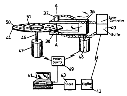
The optical inspection system has a 'U~ shaped arm
36 with a light source 52 and a detector 38 attached to
the upper and lower ends of the arm respectively. The
source and detector are connected to a laser controller
39 and a buffer 40, the latter being arranged to
transfer detected signal data to a personal computer 41
via an analogue to digital converter 42 and a data store
43.
The disc 44 upon. which the sample to be inspected __
is attached or supported is mounted on a rotatable
spindle 45 which lies parallel to the bight of 46 of the
arm 36. The spindle 45 is driven by a spindle motor 47.
The optical axes of the light source 5~ and detector 38
are aligned with one another along the axis A-A.
22 p0 562
WO 96/09548 PCTIGB95/02186
-21-
The arm 36 is coupled to a stepper motor 48 which
precisely rotates the arm in a plane parallel to the
plane of the disc 44 such that, in combination, rotation
of the arm and of the disc allows the light
source/detector arrangement to be scanned across the
entire useable surface of the disc. The stepper motor
48 is controlled by a motion controller 49, which in
turn is controlled by the computer 41, such that the
relative position of the spindle 45 can be determined to
within an accuracy of Gum.
The disc is of a completely transparent material
but is provided with a black bar 50 around a portion of
its upper peripheral surface. The bar 50 acts as an
angular calibration marking for the inspection system.
When it is required to inspect a disc, the arm 36 is
moved to an outermost "home" position, where the light
sourceidetector arrangement is situated off the disc ~4.
In this position, the laser and detector are calibrated
to ensure a constant, aaximum, output signal. The arm
36 is then rotated to move the light source/detector
arrangement towards the disc.
When the edge of the disc is detected, the arm is
held stationary until the calibration marking 50
interr::pts the beam. The leading edge of the marking 50
provides an origin to which the angular position of the
detector can be referenced whilst the edge of the disc
provides an origin for the radial position. Due to the
accuracy of the stepper motor 48 and the spindle motor
. 22 ~o X62 ; . . :w.
.. .
. . ... ,. ..
-22-
47, it is then possible to precisely determine the
position of the light source/detector arrangement
relative to the disc.
In the system of Figure 6, the disc 44 comprises
a plurality of wells or indentations 51 formed in its
upper surface. The wells contain the sample to be
inspected and are filled, for example, by microtitration.
Rather than scan the whole surface of the disc, the
personal computer may be arranged to step the light
source/detector arrangement over the disc surface from
one well to another. This is enabled by the precise
position information obtained from the calibration
marking and the disc edge. Figure 7 shows a flow diagram
of the control process for this system.
The system of Figure 6 may be modified so that the
light source 52 and the detector 38 are both arranged on
the same side of the disc, with the disc being provided
with a reflective coating on or beneath the surface on
which the sample is supported. In this arrangement the
detector detects light reflected from the reflective
coating. The two main advantages of the arrangement are
that the surface of the disc which does not support the
sample may be safely handled, as it does not lie in the _.
r.. _
light transmission path, and that the signal to noise
ratio of the optical inspection process may be increased
because light will have to pass through a sample twice
in travelling from the source to the detector.
It will be apparent that various modifications may
._ ~22 Q4 x.62
WO 96/09548 PCTIGB95/02186
-23-
be made to the above described embodiments without
departing from the scope of the invention. For
example, the support surface of the disc may be scanned
with infra-red or ultra-violet radiation rather than
visible light. It is also possible to scan the surface
with radiation which excites fluorescence in material
attached to the surface and to use the detector (D2 or
D3) arrangement to detect light at the emission
wavelength.
It is also possible to construct the disc in such a
way that the support surface is internal to the disc and
is not the upper surface of the disc. This may provide
the advantages that the sample is not damaged by
handling and that a precise volume of sample may be
analysed. To enable the system to be used for running
gels (e.g. to identify proteins, DNA etc), an
appropriate gel may be provided on the upper surface of
the disc. Electrodes ~or applying a potential across
the gel may be fonaed integrally therewith or may be
printed, or otherwise deposited, on the upper surface.
The electrodes may be spaced radially or
circumferentially. Pits may be provided in the gei into
which the material ~o be run can be placed.
Another modification to the above described
embodiaents involves replacing the light detector with a
photo-diode array, e.g, a CCD array. A preferred form
of array is a linear array extending radially with
respect to the disc. The light source would take the
WO 96109548 ~ ~ ~ PGTlGB95102186
-24-
form of a laser line generator arranged to generate a
radially extending line of light aligned with the diode
array. Some degree of optical magnification may be
incorporated between the source and the generator to
allow the resolution of the system to be varied. After
each rotation of the disc, the source/detector
arrangement would be stepped inwardly by the length of
the laser line. The advantages of this arrangement are
higher speed and higher resolution.