Note: Descriptions are shown in the official language in which they were submitted.
220 1 1 48
~IETHOD AND APPARATUS FOR MONITORING AND/OR CONTROLLING THE
~EUROMUSCULAR BLOCK, ESPECIALLY THAT PRODUCED BY MUSCLE
RELAXANT DRUGS DURING ANAESTHESIA.
The present invention pertains to a device for the monitoring and/or controlling of the
neuromuscular block, especially that produced by muscle relaxant drugs used during
anaesthesia.
Primarily, the new method and al)l)dldtus allow a quantification and control of the
neuromuscular block by means of transcutaneous stimulation directly on the muscle,
stimulating the intramuscular nerve pathways and the neural part of the motor plate without the
need of having to stimulate a peripheral motor nerve.
The invention also allows a controlled a~mini~tration of the blocker drug during the
induction of the anaesthesia, as well as control of the neuromuscular block during the operation
and control of the recovery when being awakened from the ~n~esthe~i~
ANTECEDENTS OF THE INVENTION
It is known that major advances in surgery have come about from the application of
muscle relaxant drugs during ~n~stl~ which in addition to f~f ilit~ting the work of the
surgeon also allows a less tr~lTn~tic intubation and facilitates meçh~nic~ sl.i~lion.
Likewise, muscle relaxation allows improved meçh~nic~l ventilation for intubatedpatients at intensive care wards.
It is also known that there are various factors which influence the duration and effect of
the muscle rel~Y~nts depending among other things on the body weight, the age, the physical
condition and possible pathologies of the patient to whom this type of drug is being
~mini~tered. In the event that the dose is insufficient, the intubation and the subsequent surgery
might become more difflcult; in the case of overdosage, the patient upon aw~kening from the
anaesthesia might present r~ atoly insufficiency and a need for prolonged artificial
halion, which would delay their leaving the operating theater or their a-lmi~sion to a special
care ward.
Other dangers of overdosage or poor reversal of the effects of muscle relaxants might be
serious complications such as aspiration of vomit on account of faulty reflexes or respiratory
failure, which complications may eventually cause the death of the patient.
220 1 1 48
In order to avoid these complications, at present, the status of the neuromuscular block
is evaluated by means of stimulation of a peripheral motor nerve and measurement of the degree
of motility of the muscle innervated by said nerve.
There are various methods and devices designed to stimulate peripheral nerves bymeans of transcutaneous electrodes, for example, the stim~ tors of Neuro Technology, Inc.,
Houston, Texas, USA. The most important problem lies in the evaluation or measurement and
monitoring of the neuromuscular block and the adequate a~mini~tration of the muscle relaxant
drug to m~int~in the desired level of blockage. At present, the evaluation of the block is done by
evaluation of the electrical activity, the force or the movement of'the muscle during its
contraction. The methods used up to the present date are:
1 ) The visual method. One visually evaluates the movement of a muscle after thestim~ tion of the motor nerve which innervates it. The inexactitude and difficulty of
quantification of the method are obvious, but its simplicity makes it the one which is used most
frequently today.
2) The tactile method. Similar to the prece~1in~ method, but in this case the evaluation is
done by means of the pel~e~lion through the hand of the anesthesiologist of the force of
contraction of the patient after the stimulus. As in the prece-1ing case, it is a subjective and
inexact method, offering little guarantee of dete "~ g the exact degree of neuromuscular
blockage.
3) By means of electromyography. This Co~ ls in the registration of the mllcc~ relectrical activity evoked by stim~ ng the motor nerve which innervates said muscle. There
are various devices and app~dlus on the market, which monitor the neuroml-sc--l~r block by
said technique, which is described, for example, in U.S. Patent No. 4,291,705, among others.
The primary problem which this technique entails is its difficulty of application and the
sophistication of the eq~ipmert) so that it has only been used es~nti~lly in research, without
coming to be used as a practical and routine technique.
4) By means of force tr~ncd~cers. In this case, one registers the force of the thumb when
the ulnar nerve is stimulated. Again, there are various devices on the marlcet, such as the
"Relaxograph" of Biometer Tnt~tional A/S, Denm~rk As in the preceding case, the primary
problem consists in the complexity of the technique used, which involves having to immobilize
the arm in order to perform a proper registration. For this reason, and the costly equipment, this
device is not very practical for routine use.
5) By means of accelerometry. This method is, after the visual method, the most popular
one. It is based on placing a biaxial or triaxial accelerometric sensor on the thumb which, when
2201 1 48
the ulnar nerve is stimulated, provokes a movement in same, which is picked up by the
accelerometer. This is a rather valid method, but it still has the drawback that several electrodes
separate from the sensor have to be put in place in order to stimulate the peripheral nerve. On
the other hand, the accelerometer is fragile and requires a certain immobilization of the arm.
Other placements of the accelerometer have been described, for example, on the face, in which
case one must stimulate a motor branch of the facial nerve and the stimulus electrodes must
arrive separately from the accelerometer, increasing the risk of certain of the elements becoming
detached from the skin. As an example of monitors based on accelerometry, one can mention
the "Accelograph" and "Tof-Guard" models, both of them from Biometer Intemational A/S,
Denmark, and that described in U.S. patent 4,817,628, which is another example of a facial
accelerometer.
6) By means of flexible-sheet piezoelectric sensors. The phenomenon of piezoelectricity
has been known for many years, as has been its application in the field of medicine, for
example, with the application of piezoelectric sensors which, through their deformation, capture
the plethysmographic wave ~ slllilled to the skin. Based on this principle, the monitor of U.S.
patent 5,131,401 requires, like the other methods, the stim~ tion of a peripheral motor nerve,
primarily the ulnar nerve, and it comprises some cables for the stimulus electrodes and others
for a sensor in the form of flexible piezoelectric sheets, which are placed on the palm of the
hand and register the contraction of the muscles of the hand. Like the "Tof-Guard" model, it
also has a microprocessor which, by means of a program, enables a programmed stimulation
and vi.c~ i7~tion of the quantified lc~ollse of the neuromuscular block.
The primary drawback presented by these Illolfitol~, except for the last one, is that they
require the stiml-l~tion of a peripheral motor nerve, specifically the ulnar nerve, and the
placement of the sensor on the hand or thumb. They likewise require the use of two electrodes
for separate stim--l~tion of the sensor element of the response, which increases the risk of a
detachment of the electrodes or of the sensor.
Owing to this, the placement of the electrodes may be awkward and a certain immobilization of
the limb is necessaly, limiting the placement in other positions. On the other hand, this type of
a~p~dl~ls might be more sensitive to certain intelrelc:llce or involuntary movements of the
patient.
With respect to the control of the neuromuscular block, there are various works based on
pharmacokinetic models and short-circuit models, such as those described in individual articles
of medical joumals:
"Quantitative ~cse-ccment of residual antidepolarizing block (Partil)" Ali HH et al., Br J
Anaesthesia 1971 vol. 43 pp 473-477.
3-
220 1 1 48
"~fonitoring of neuromuscular function" Ali HH et al., Anesthesiology 1976 vol. 45
pp ~16-249.
"A microcomputer based controller for neuromuscular block during surgery" Ritchie G. et al.
Ann Biomed Eng 1985 vol 13 pp 3-15.
"Microcomputer based muscle relaxation monitor and controller for clinical use" Bradlow HS et
al. Med Biol Eng Comput 1985, vol 23 pp 547-555.
"Online parameter estim~tion and control of D-Tubocurarine-in~lce.1 muscle relaxation."
Rametti LB et al., Med Biol Eng Comput 1985 vol. 23 pp. 556-564.
"Online control of Atracurium induced muscle relaxation" Bradlow HS et al, J Biomed Eng
1986 vol. 8 pp. 772-775.
"Computer-Controlled Muscle Paralysis with atracurium in the Sheep" D.G. Lampard et al,
Anesthesia and Intensive Care, vol. 41 (1986) pp 316-320.
"Clinical automatic control of neuromuscular blockade" Asbury AJ et al, ~n~esthesi~ 1986
vol. 41 pp. 316-320
"Infusion of vecuronium controlled by a closed-loop system" Br J Anaesth l986 vol. 58
pp. 1100-1103.
"Closed-loop ~rimini~tration of Atracurium" N.R. Webster et al, ~nlosthe~i~ vol. 42 (1987)
pp. 1085-1091.
"A model-based self-adjusting two-phase controller for vecu~~ -in~ ce~ muscle relaxation
during anaesthesia" Jalkist RR, EEE Transac Biomed Eng 1987 vol. 34 pp. 583-594
"Closed-loop infusion of atracurium with four different anesthetic techniques" O'Hara DA et al,
Anesthesiology 1991 vol 74 pp. 258-263.
These articles describe instances of control of the muscle relaxation by means of
computers with pharmacokinetic models, through the previously mentioned sensors, or with
closed-circuit systems.
There are correct approximations in all the instances, but even so they are still
experimental and not very safe models, which owing primarily to the need to stimulate a
peripheral nerve are impractical and complicated in use, whether because of the type of
transducer used or because of the pharmacokinetic model, which requires much data entry or
very sophisticated computers.
220 1 1 48
DESCRIPTION OF THE INVENTION
The aforesaid drawbacks can be elimin~ted with the method and apparatus of the
in~ ention.
The method for monitoring and/or controlling the neuromuscular block which is the
subject of the invention is characterized by the fact that the nerve stimulation is done on the skin
covering the muscle or muscles on which the detection of the response to said stimulation is
performed, that is, without having to stimulate any peripheral nerve.
Thanks to this method, it is not necessary to stimulate the peripheral nerve and the
stimulator-detector device can be placed on the upper or lower limb, depending on the type of
surgery being performed, without having to confine the stim~ tion to the ulnar nerve.
Optionally, the method of the invention also comprises the automatic control of the
supply of the muscle relaxant drug on the basis of a detection of the muscle response.
The invention also pe~ s to an a~p~dtus for the monitoring andlor controlling of the
neuromuscular block, which comprises means for the application of the nerve stimulation,
means for the detection of the re;,~Jonse to the stimul~tion, and means for monilu~ g the
neuromuscular block, and it is characterized by the fact that the means for the nerve stimulation
and the means for the detection of the r~l,ollse are arranged in the same body.
Thanks to this arrangement in a single body, the following advantages are achieved:
i) a single element serves to measure the arterial pressure and to measure the
neuromuscular block, achieving a more compact and handy device,
ii) the pl~r~nent becomes easier,
iii) the independent discol~e~;lion of certain of the conl?ollents is avoided,
iv) it simplifies the monito~ g in the Op~ld~ g theater.
According to another embodiment, the a~l)~dlus of the invention comprises means for
the application of the nerve stimulation, means for the detection of the response to the
stimulation, and means for the monitoling of the neuromusc.llar block, and it is characterized by
the fact that it also comprises means for supply of the drug which produces the neuromuscular
block and means of control which coordinate the generation of the stimulus, the registration of
the activity, the data processing, the computation of the drug dosage and the control of the
means for supply of the drug as a function of the computed dosage.
2201 148
The apparatus of the invention thus comprises three essential parts:
i) means for the nerve stimulation, comprising Ihe stimulator and the stimulus
electrodes,
ii) means for the detection of the muscle response, consisting of a sensor, which can be
of various types,
iii) means of monitoring and control, which can be of various types, depending on the
specific embodiment of the invention, and
iv) optionally, a closed circuit for the automatic dispensing of a drug.
The apparatus of the second embodiment can also be characterized by the fact that the
means for the application of the nerve stim~ tion and the means for the detection of the
response are arranged in a single body, whereby the advantages already described are also
obtained in this instance.
Preferably, the appal~lus is characterized by the fact that the body is a cuffof the type
used for the measurement of arterial pres~ , provided with means for the detection of the
pressure or connected to said means, whose cuff incorporates means for the application of the
nerve stimulation.
In this way, the electronics and the maneuvers involved in the pl~c~ment of the sensor
are simplified, since the same arterial ples~ul~ cuffand tr~n~d~lcer of the noninvasive arterial
pressure monitor are used. This arrangement makes it possible to simplify the technique for
measurement of the neuromuscular block.
Preferably, the means for the application of the nerve stimlll~tion comprise at least two
electrodes.
Also preferably, the means for application of the nerve stimlll~tion and the means for the
detection of the pressure are combined with the means of monitoring through a multiconductor
tube which contains on the inside an air conduit for transmitting the pressure waves and at least
one pair of electrical conductors for tr~n~mi~ion of the stimlll~tion pulses.
In a preferred embodiment, said conductors are included in the wall of the
multiconductor tube.
Due to the fact that there is only a single tube which ~ the pressure wave and the
contraction wave and which also includes the cables with transport the stimulus, the use of the
device becomes extremely simple. This solution is also more economical, since it makes use of
-- 6--
2201 148
the cuff and the electronic circuits of the arterial pressure monitor. The electrical system is more
simple and less cumbersome.
The method of the invention also represents a great advance with respect to all of the
foregoing ones, including the most modern (Tof-Guard and U.S. patent No. 5,131,401), since it
does not require the stimulation of a peripheral motor nerve in order to register the muscle
activity. The method of the invention is based on the stimulation of the intramuscular neural
pathways and the nerve en~ing~ of the muscular motor plate by the application of a stimulus
current to the skin directly over the muscle whose activity one desires to gauge.
.~,
Usually, the stimulus current does not exceed 50 mA and the time of application should
not be more than 30 ms, in order not to directly stimulate the muscle fibers, which would
produce a faulty reading of the neuromuscular block.
The means of monitoring can be any of the bloodless arterial P~5~UIe monitors existing
on the market, modified by the incol~o~lion of a pulse generator for the muscle stimulation,
with the possibility of adjusting various parameters, such as the intensity of the stimulus or the
time between re~-lin~s, and with the possibility of filrni~hin~ on-screen data co,lesl.onding to
the muscle relaxation.
The monitors can also have LEDs of light and sound alarm signals for cases of loss of
signal or contact of the electrodes (by impedance), disconnection of the sensor or any of its
parts, excessively high or low level of blockage with respect to the predetermined level, as well
as other alarms which are common in medical devices, such as current faults, loss of power
supply, etc.
The means of monitoring and control comprise an in~ tç~ amplifier, with filters for
50-60 Hz and high frequency to avoid interference from the electrical scalpel, a stimlll~tor
circuit of the type commonly used for nerve stimul~tion, capable of generating ~ up to an
intensity of 100 mA, a circuit to measure and control the intensity of the stimulus current, an air
pump to inflate the cuff, and optionally an A/D converter, a memory bus and a microprocessor
which, by means of a program in EPROM memory, controls all the functions of the device.
coor~in~ting the following closed-circuit cycle: stimulation, registration, data processing and
signal processing according to standards for ~cses~ment of the neuromuscular block (evaluation
of the TOF).
2201 148
BRJEF DESCRIPTION OF THE DRAWINGS
In order to better comprehend what has been set forth, drawings are included which
describe, schematically and only as an example, not limited hereto, the differences from the
conventional methods and a practical example of realization of the stimulus-registration device,
as well as the controller apparatus of the neuromuscular block.
Figure I is a graph of the muscular activity, demonstrating the validity of the apparatus
of the invention; figure 2 is a scheme for a conventional placement of the electrodes and the
sensor; figure 3 is a scheme in perspective view of a pressure cuf~, in which certain stimulus
electrodes have been incorporated on its inner surface, with a large-scale detail of the
multiconductor "tube"; figure 4 shows one embodiment of the app~ ls of the invention; and
figure 5 is a flow chart of a closed circuit which controls the infusion of a muscle relaxant drug.
DESCRIPTION OF A PREFERRED EMBODIMENT
Figure 2 shows a conventional placement of the electrodes 1, 2 and the sensor 3. In this
case, the electrodes 1, 2 are arranged on the ann and the sensor 3 is arranged on the finger. One
can see in the figure the two cables 4, 5 collc~ollding to the electrodes and the cable 6
corresponding to the sensor.
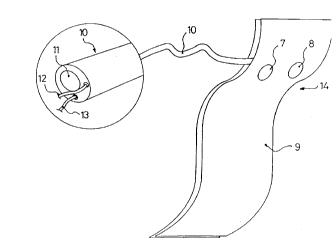
Figure 3 shows the stim~ tor-detector device of the a~p~l ls of the invention. ~n the
figure, one can see the electrodes 7, 8 arranged in a ~ Ul~ cuff9. As the large-scale detail
shows, the multiconductor tube 10 comprises an air conduit 11 and two electrical conductors 12
and 13. In the embodiment shown, the conductors 12 and 13 are included in the wall of the
multiconductortube 10.
Figure 1 shows a continuous tracing curve A which co~ onds to the method of the
invention with the pressure cuff, provided with the stimulus electrodes arranged over the muscle
in which the block is being ev~ te-l, and a continuous tracing curve B which corresponds to a
conventional method, with the stimulus electrodes over the ulnar nerve and the sensor on the
thumb.
In order to demonstrate the validity of the method of the invention and to ensure that the
direct cutaneous stim~ tion over the muscle mass in which one wishes to evaluate the
neuromuscular block does not produce direct stimulation of the muscle fibers, but instead the
stimulus follows the intramuscular nerve pathways and nerve en~lin&s of the muscle motor
plate, the following test was performed: sensors were placed on the thumb and stimulus
2201 148
electrodes over the ulnar nerve, following the instructions of the previous conventional
methods. Curve B was obtained.
In the same patient and at the same time, the pressure cuff with the electrodes on its
inner surface was put in place, following the instructions of the method of the invention. Curve
A was obtained.
Comparing the trend of both curves during the application of muscle relaxant drugs to
anaesthetized patients, results were obtained which validate the method of the invention, since
curves A and B show a similar behavior for the same method of application of the drugs.
Figure 4 shows the different components of the al)l)~dllls of the invention:
- In the first place, the stimulator-detector device 14 detailed in figure 3, which includes
the stimulus electrodes 7, 8 and the pressure cuff 9. This can be made in different sizes,
depending on the size of the limb to which it is applied, it being possible to have versions for
adults and versions for children.
The device can be disposable or reusable, and it comprises the two electrodes 7, 8,
likewise disposable or reusable, which are coupled to the cuffand have a stimulus surface
varying between 0.05 cm2 to 5 cm2 for each. The electrodes 7, 8 are placed cn the inner surface
of the arterial ~res~ur~ cuff 9, which, with the muscle contraction wave transmitted to the skin,
generates a ples~ulc wave proportional to the muscle contraction that is tr~n~mitte~ through the
tube to the prcs~u c tr~n~d~lcer, arranged inside a conventional type of arterial pressure
monitor 15.
The stim~ tion electrodes 7, 8 can be situated in various ways on the inner surface of
the cuff 9. The more far apart they are, the greater the number of nerve endings stim~ ted and
the larger will be the response.
The cycle u~sed for the measurement is as follows:
I ) Inflate the cuff slightly to ensure good contact between the electrodes and the skin
(between 10 and 300 mmHg).
2) Generation and application through cutaneous electrodes of a series of four stimuli <
50 mA and < 30 ms in duration at a frequency of 2 Hz, known as "TOF" ("Train of Four", the
standard accepted in all methods of evaluation of the neuromuscular block).
3) Registration of the pressure generated inside the cuffby the wave which is
transmitted to the surface of the skin by the contractions of the muscle.
-
- 2201 148
4) Digitization and processing of the signal, graphic representation through an LCD or
electroluminescent screen.
Figure 4 shows one embodiment of the apparatus of the invention. rn this figure, one can
see the stimulator-detector device 14 as represented in figure 3 and a conventional type of
arterial pressure monitor 15.
In the lower righthand portion of the monitor dial 15 one can see the connections 16, 17
and 18, corresponding respectively to the multiconductor tube 10, the information on oxygen
saturation, and the electrocardiogram.
Ln the upper ri~hth~nll portion one can see a pair of control buttons, one of which 19
serves to adjust the time between two readings and the other 20 is used to adjust the intensity of
the stimulus.
On the screen at left there appear two curves, the upper one corresponding to the
electrocardiograrn and the lower one to the pulse signal or plethysmograrn. The bars in the
lower part of the screen represent the res~,lses to the muscular stim~ tion over time. The
screen can also display data such as the muscular activity (Musc: 30%), heart rate (FC: 60),
arterial pressure (PA: 120/70), and oxygen saturation (Sat: 98%).
The conventional monitor 15 is provided with a pulse generator for the muscle
stimulation through the conductors 12, 13 (figure 3) and the electrodes 7, 8.
Figure 5 shows a flow chart of a closed circuit. The electrical stim~ tor 21 stim~ tes
the muscle 22. The sensor 23 provides the muscle l~onse to the means of control 24.
A program is used to control the data acquisition, A/D conversion, and analysis of the
data, filmi~hing the signals to actuate the system for supply or infusion of the muscle relaxant
25. In this way, the infusion will be automatic.