Note: Descriptions are shown in the official language in which they were submitted.
WO 96/10740 PCT/AU95100646
22C1 1 50
METHOD AND APPARATUS FOR DIAGNOSIS, DETECTION OF CELL
ABNORMALITIES AND MORPHOLOGY OF LIVING SYSTEMS
TECHNICAL FIELD
The present invention relates to an apparatus and method of
diagnostic measurement and in particular, a method and apparatus adapted
to analyse various parameters of living materials and specimens to
determine the dielectric characteristic of a specimen under test for the
purpose of diagnosis of the state of the specimen. Depending on the
specimen, a wide range of states may be susceptible to diagnosis including
such as disease in plants, ~nim~ls or humans: the revelation of residual
toxins in consumer goods from dairy products. meat products, fruit and
vegetable products, fish, grains and stock feed, oils and other liquids.
The present invention further helps identify abnormalities and
transformations in living bodies in their earliest stages, much before the
clinical appearance of a disease.
BACKGROUND ART
At the present state of technology it is well known that the dielectric
behaviour of such as plant, fruit, ~nim~l and human tissue corresponds to
broad features in their composition and structure. Recent studies have
revealed that the cell is a highly ordered dynamic entity which acts
holistically with respect to chemical and physical events within a living
body, and the existence of domains in the cytoplasm is a general rule. These
domains are electrically polarised units of ordered, packed biopolymers in
"biowater". The different organs in a living organism, with compartmental
similarity and harmonised metabolism, have basic differences in domain
arrangements which lead to a difference in dielectric responses. A disease
transformation in a living body which has a viral origin or resulting from the
action of toxins and other chemicals also changes the domain structure and
hence the polarisation and dielectric response of the tissue or cell.
A domain is herein defined as a region of a system, or a region of a
substance, comprising atoms or molecules which can be thought of as a
single entity; this single entity being responsive to electric or magnetic
fields and includes such a system having a plurality of these entities.
Examples of a domain include; a ferroelectric or ferromagnetic domain, a
cluster of atoms or molecules, an organic cell, a bacterium, a virus, a cluster
or collection of cells.
wo 96/10740 PCT/AUg5m0646
2201 1 50
A domain group is a collection of said domains having the same
response to an electric or magnetic field.
In the past, precise measurement of parameters of domains were
inconceivable due to limitations of the instruments. Measurements of
relative dielectric permittivity. energy dissipation and electrical impedance
are not possible due to very high values of electrical conductance
overshadowing real kinetic characteristics. Existing methods of
measurement are mostly based on impedance bridges, which are inadequate
at frequencies below 100Hz due to noise instability, electrode polarisation
and the time required to obtain balanced conditions. These bridges yield
relative permittivity. energy dissipation and electrical impedance values
only at discrete frequencies and therefore each frequency setting causes
disruption of sequential measurements. The dielectric properties of living
tissue from bodies vvill change when they are taken out of their natural
environment. Dead tissue will show a greater change with changes of cell
morphology. Conductivity measurement is mostly carried out by D.C.
electrometers of wide current range, often from 1o-l4 Ampere to a few
milliAmpere. This range being covered by switching to sequential decade
ranges with a mismatch of measured current values. A.C. and D.C.
measurements require different apparatus, separate sample settings and long
time switching intervals from one instrument to the next. The
morphological changes of a cell are much faster, so the obtained parameters
will refer to different intracellular structures resulting in an incorrect
correlation between these parameters. Sample size limitations sometimes
up to a few milligrams reduces electrode sensitivity and field noise
overshadows the results for fine structural studies.
DISCLOSURE OF INVENTION
In an effort to ameliorate the disadvantages of the prior art or at least
to provide a commercially viable alternative to the prior art, the present
invention proposes a dielectric diagnostic analyser (DDA) and a method of
diagnosis.
In a first aspect, the present invention consists in an apparatus
adapted to perform diagnostic analysis of a specimen having at least one
domain group as hereinbefore defined, the apparatus comprising:
excitation generating means to generate a predetermined excitation
signal;
WO 96/10740 2 2 0 1 1 5 0 PCT/AU95/00646
measuring means to measure a response signal of the specimen to
the predetermined excitation signal:
electrode means for transmitting and receiving the predetermined
excitation signal and response signal of the specimen, respectively;
analysing means arranged to analyse said response signal; and
switching means adapted to switch the electrode means between the
measuring means. and excitation generating means, in a time period less
than a polarization relaxation time period of the at least one domain group in
the specimen.
Preferably, the excitation generating means is the source of the
predetermined excitation signal and may be an electrometer or a frequency
bridge adapted to generate a predetermined signal. In one form of the
invention the measuring means compares an electrometer or a frequency
bridge arranged to measure responses received at the electrode means as the
response signals of the system.
Typically the analysing means comprises an electronic computer,
electrometer and frequency bridge arranged to analyse the response signals.
received at the electrode means and the computer has a display for
displaying a diagnostic result. Preferably the switching means is also
controlled by the computer which allows switching of the electrode means
between the excitation means and the measuring means at times less than
the smallest relaxation time, of the polarized domain group, to be measured.
In an embodiment of this invention the electrode means is in the
form of a SUCtiOl1 cup electrode, a pinch electrode, a thermocontrolled
electrode or any combination of two or more similar electrodes.
In a second aspect, the present invention provides a method of
diagnostic analysis comprising;
applying a predetermined first excitation signal to a specimen having
at least one domain group, as hereinbefore defined, so as to elicit a response
from the domain group within the specimen;
analysing the response from the domain group to determine the
maximum response of each domain group; and
comparing said maximum response to a maximum response of a
control specimen.
Preferablv the first excitation signal is a ramp function voltage sweep
or a time rate of change of voltage, and the response from the domain, in the
WO 96/10740 PCT/AU95/00646
2201 150
domain group of the specimen, is measured as a change in a current flow
through the specimen over time.
Typically the point of maximum response is at the threshold
polarization voltage of each domain group and is representative of a maxima
5 in the polarization of each domain group of the specimen.
Preferably a control specimen is any specimen, analogous to the
specimen to be diagnosed and considered to be the statistical norm of that
specimen.
In an alternative form of the second aspect of the present invention,
10 the first excitation signal is a frequency dependent applied voltage and the
response from the domains is measured so as to allow the determination of
dielectric permittivity, and dissipation energy, of each domain group. In this
form the point of maximum response of each domain group is determined by
a local maxima in the dielectric permittivity or a local minim~ in the
15 dissipation energy of that domain group.
In a third aspect, the present invention provides a method of
diagnostic analysis comprising:
all the steps of the second aspect of the present invention as well as;
applying a second excitation signal corresponding to a signal value
20 at. or near, the point of maximum response of each domain group to elicit a
further response in each domain group; and
detecting the variation and length of said further response upon
removal of the second excitation signal.
Preferably the second excitation signal is applied in the absence of
25 the first excitation signal, and the further response is measured upon
removal of the second excitation signal while each domain is relaxing to its
natural state.
Typically the detecting of the variation and length of the further
response occurs within the time in which the domains in each group relax to0 the state they were in before the second excitation signal was applied.
BRIEF DESCRIPTION OF DRAWINGS
The present invention will now be described by way of example
only, with reference to the accompanying drawings, in which:-
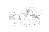
Figure 1 is a schematic diagram of a dielectric diagnostic analyser in
35 accord with an embodiment of the invention;
WO 96/10740 PCT/AU95/00646
2201 1 50
Figure 2 is a schematic diagram of a preferred embodiment of a
suction cup electrode;
Figure 3 is a schematic diagram of a preferred embodiment of apinch electrode;
5Figure 4 is a schematic diagram of a preferred embodiment of a
thermo-controlled electrode and chamber;
Figure 5 shows an hysteresis graph of the dielectric permittivity
against the applied electrode pressure on living tissue;
Figure 6 is a schematic diagram of the equivalent circuit of the
inductive, capacitive and resistive processes in the dielectric response
mechanism of domain structures analysed by the method and apparatus of
the present invention.
Figure 7 is the printout of a computer screen of two graphs of current
versus voltage each having two plotted curves, the right hand side graph
being an enlarged view of a section of the left hand side graph;
Figure 8 is a graph containing 3 curves of the relative permittivity
versus the frequency for a rat shown on a logarithmic-logarithmic scale.
Figure 9 is a graph cont~ining 3 curves of the dissipation factor
versus the frequency on logarithmic scale for a rat;
Figure 10 is an example of discharge current versus time curve for
domain structure in a Wistar rat thigh muscle;
Figure 11 is a table of relaxation time constants for four domains
(shows as r" r2, r3, r4 ) for a plurality of rat organs;
Figure 12 is a graph of a Fourier analysis of four domains in the
intracellular structure of the thigh muscle of a Wistar rat;
Figures 13 (a) and (c) are graphs of the dielectric permittivity as a
function of frequency! while Figures 13 (b) and (d) are graphs of the
dissipation factor versus frequency; all for various temperature settings of
the tongue tissue of a Wistar rat;
Figure 13(e) is a graph of current versus applied voltage for tongue
tissue at 20 degrees Celsius, before and after the tissue was heated above
42.5 degrees Celsius;
Figure 13(f) is a graph of the frequency response of the tissues versus
the inverse of the absolute temperature (temperature measured in degrees
Kelvins).
WO 96110740 PCT/AU9~100646
2201 ~ 50 6
BEST MODES
Figure 1 shows an embodiment of the first aspect of the present
invention! which comprises a switching means 97 connected to a frec~uency
bridge 95. an electrometer 96 and a computer unit 91 via appropriately
shielded cables. The computer unit 51 is also connected to a keyboard 92, a
display monitor 93 and a printer unit 94 in the usual way to provide a
computer system. The electrometer 96 and frequency bridge 95 are also
connected to the computer unit 91. such that an operator can through the
use of the kevboard 92 instruct the computer unit 91 to change the settings
on the electrometer 96 or the frequency bridge 95. Preferably, the
electrometer 96 and the frequency bridge 95 has the additional option of
changing the settings manually. The computer unit 91 can be programmed
to receive input signals, from the electrometer 96 and the frequency bridge
95, which can be analysed by means of dedicated software programmes such
as Intel's IEEE 488 and then to output the resulting analysis on the display
monitor 93 or printer 94.
The switching means 97 further having connections via a plurality of
electrically shielded conducting cables to three electrode devices. The
computer unit 91 is programmed to instruct the switch means 97 to switch
between any one of the three electrode devices. The first electrode device as
illustrated in Figure 1 and Figure 2 is a suction cup electrode 118 which
comprises an excitation electrode 114 to induce a current in a tissue
specimen 117. a measuring electrode 115 to measure the response signals of
the specimen 117 resulting from the excitation induced by the excitation
electrode 114, a guard electrode 116 to prevent unwanted surface currents
reaching the rmeasuring electrode 115, and a suction device 112 connected to
the suction cup electrode 118 by way of an airflow link 113 to the air passage
channel 125 of the suction cup 118. The suction device 112 is used to adjust
the pressure within the suction cup electrode 118, so that not only does the
cup adhere to the specimen but the contact pressure between the specimen
117 and the electrodes (i.e. the excitation electrode 114, the guard electrode
116 and measuring electrode 115) can be adjusted to an optimum pressure.
The optimum pressure between the electrodes and specimen is obtained
from a local maximum value of the dielectric permittivity in a hysteresis
plot, as shown in Figure 5. The excitation and measuring electrodes 114 and
115, respectively, are set to the optimum pressure before diagnostic
WO 96/10740 PCT/AU95/00646
220 1 ~ 50
measurements are obtained. The second electrode device illustrated in
Figure 1 and Figure 3 is hereinafter referred to as the pinch electrode 101
which comprises an excitation electrode 100, mounted on one jaw of a pair
of pincers 136. while the guard 98 and measuring electrode 99 are mounted
5 on the opposite jaw of the pair of pincers 136. At the other end of the pair of
pincers 136, a spring 132, an adjusting screw mechanism 131 and a
micrometer measuring gauge 130 are arranged to adjust and measure the
distance between the excitation electrode 100 and the measuring electrode
99 at the jaw end of the pair of pincers 136. A specimen 102 is pinched
10 between the electrodes at the jaw end and a force between the jaws is
applied by the adjustment of the screvv mechanisms 131 and spring until the
desired distance is read off the gauge reflecting the distance between the
excitation electrode 98 and the measuring electrode 99 at the jaw end
sandwiching the specimen 102 between the electrodes.
The third electrode device illustrated by Figure 1 and Figure 4 is
hereinafter referred to as the thermocontrolled electrode 105 which
comprises a first piston 141, of electrically conductive material to function
as the excitation electrode 108, and fits within a first teflon cylinder 147 so
that it protrudes from both ends. The said first teflon cylinder 147 has, a
guard electrode 107 which wraps around one end of the outer surface of the
cylinder 147 and an electromagnetic shield 144 which wraps around the
other end of the outer surface of the teflon cylinder 147.
A second teflon cylinder 149 substantially similar to the first teflon
cylinder 147! has a second piston 142 functioning as the measuring electrode
106. Piston 142 is allowed to slide in and out of the cylinder 149 by means
of an adjusting nut 145 located at the end of the piston 142 which protrudes
from second teflon cylinder 149 nearest to the electromagnetic shield 144.
A cap 146 placed over the nut 145 stops it from turning at will. The guard
electrode 107 on the second teflon cylinder 149 extends beyond the end of
the cylinder 149. The two teflon cylinders 147, 149 slide, with some
frictional force, into a third cylinder so that the guard electrodes 107 meet,
leaving a gap between the excitation electrode 108 and the measuring
electrode 106 to fit a specimen 112. The adjusting nut 145 can then be used
to change the distance between the gap. The third cylinder being a
thermocontrolled jacket 148 with two ports 150 so that fluid can be pumped
in or out. at a predetermined temperature, to thermally control the specimen
WO 96/10740 PCT/AU95/00646
220 1 1 50 8
112. The thermocontrolled jacket 148 iS connected to a thermocontrol unit
110 (seen in Figure 1) by means of tubing to the ports 150. The
thermocontrol unit 110 being capable of adjusting the flow rate and
temperature of the fluid within the jacket 148. The thermocontrol unit 110
5 further having a feedback cable 109 to the computer unit 91, so that the flow
rate and temperature of the fluid can be set or monitored.
The second aspect of the present invention comprises a method of
diagnostic analysis of the human body or specimen under test. The
following parameters can be measured directly. or indirectly by way of
10 calculations; current, voltage. specific surface conductance, specific volume conductance. domain relaxation time constants. capacitance, inductance,
relative permittivity. impedance, reactance and dissipation factor at different
frequencies and temperatures.
Figure 6 is a schematic diagram of the equivalent electric circuit for
15 the resistive, capacitive and inductive processes in the intracellular
morphology based on the known concepts of domain structures.
The embodiment of apparatus of the first aspect of this invention
hereinbefore described, enables the measurement of various parameters by
exciting the intracellular domains and measuring those parameters within
Z0 the relaxation time periods of the domains to thereby ameliorate the problem
of electrode polarisation obscuring the measurements. Values of these
parameters are therefore revealed by measuring these parameters during the
relaxation cycle after excitation.
By way of example only, we will demonstrate how the diagnostic
25 results are obtained, for the induction of cancer in a Wistar rat, using the
pinch electrode 101 hereinbefore described.
Figure 7 is a printout of two graphs for the current versus voltage
applied to a Wistar rat, the right hand side graph being an enlargement of a
section of the curves on the left hand side graph. The curve 301represents
30 the results of a test on the tongue tissue of a healthy Wistar rat and the curve
302 is a test of the same rat where the tongue was treated with a known
carcinogen and cancer was allowed to develop. The diagnosis of cancer
follows a series of steps;
In a first step, the initial rate of change of voltage "v" (hereinafter
35 called the voltage sweep rate) and the distance "d" between the excitation
electrode 100 and the measuring electrode 99 are assumed. A test run is
W O 96/10740 PCT/A U95/00646
2201 1 50
performed to obtain the current versus voltage graph similar to that of
Figure 7. Numerical data is obtained from the test run and substituted into
the following equation to obtain a new voltage sweep rate, and a new
electrode spacing amongst other parameters.
I(B)= l)rO exp( B)~ l-exp ( B )exp( B) ,
dvrO d~
where I(B) is the current of the function B and B=E-ETp". E the
electric field strength. ~TPVis the electric field at the threshold polarisation10 voltage;
"d" the distance between electrodes;
!~1,)t~ the voltage sweep rate in V/S;
"R" the total resistance of the specimen;
rO is the domain relaxation time constant and r= rO exp[U / k T],
15 where U is the activation energy and T the absolute temperature;
"A" is the constant of "softness" which is inversely proportional to the
piezomodulus of the polarising unit (domain, cell, etc.).
The test is then set up to the new voltage sweep, the nevv electrode
spacing and the other parameters, to be run again. This first step is repeated
20 mltil all of the parameters in the above equations converge to their correct
values which are determined when the values stop changing substantially
after each iteration. Finally, a test run with the correct values is performed
and the threshold polarisation voltage relating to each domain group,
indicated on the curves in Figure 7 by the local maxima, is obtained. On
25 these culves a local maxima or humps of a domain group having a threshold
polarisation voltage of less than 1 volt is indicative of some abnormality.
In a second step the relative permittivity (Figure 8) and the
dissipation factor (Figure 9) is obtained as a function of the frequency of the
applied voltage. In Figures 8 and 9 the curve marked 201 is the result of the
30 measurements of a healthy Wistar rat, the curve marked 202 is the result of aWistar rat with an ulcer and curve 203 is a Wistar rat with cancer which is
indicated by the local maxima or hump 204 in the curve.
In a third step the specimen is excited or charged to the threshold
polarisation voltage for each domain independently and allowed to
35 discharge. During this discharge cycle measurements of the discharge
WO 96110740 PCT/AU9S100646
220 1 1 50 lO
culrent versus time are obtained and analysed to reveal a relaxation time for
each domain. A computer software program designed to analyse the
relaxation times for each domain is based on the evaluation of the following
equations :-
1(~) = lo + 1, exp[-(t / r, )] + I, exp[-(l / ~2 )] + ' + 1,/ exp[-(t / ~n )]~
where In is the current amplitude and r" is the relaxation time constant for
the n~l' polarised domain group. The computer software program cross-
Q checks the results of the relaxation time constants by a Fourier analysis (as
an example of the Fourier analysis the dissipation factor D for the thigh
muscle of a Wistar rat, see Figure lZ) based on the equation:-
D( ) 1 c~)ClrL ~)C2r2 ~)Cnrn
15where Cn is the capacitance of the n~h domain group, which is related to the
current amplitude l" and the applied voltage "V'' by C" =l"r" I V . COO is the
sum of the capacitance of each domain group and ~ is the angular frequency
(2~Tf ) for " f " the frequency of the applied voltage "I~r'. Df~) is the energy
20 dissipation as a function of the angular frequency.
The experimental determination of the natural frequency of each
domain and hence the relaxation time constants. is obtained by the
computer software program IEEE 488 from Intel via the measured parameters
of the dielectric permittivity and frequency on the basis of the following
25 equations :-
(ci~)=,,(~))+ -+r"((o)+~ ))+---+i"(lv)
where
,,/ (~ rn ~ Ll+(~r /)2]
WO 96/10740 PCT/AU9S/00646
2201 150
(~) = --~ ,i" _ 2il1 ~;
L. C and R are electrical parameters of the equivalent circuit (Fig. 6)
correspond to the electromechanical coupling (piezoelectric like) within the
5 domains in living cell cytoplasm or between living cells in organisms.
In the equations above:
I ill (~il1 ((~11~ ~ oln )
~ ( 5 2 ) 1/ 2
~i" = 2L ~ ~o,n = L C
The natural frequency is referred to the inductance "Li"", capacitance
15 "C"," and resistance "R"," interrelation of the domain following the equivalent
electrical circuit in Figure 6. The resistivity "R" relates to the resistance ofeach domain group. and ~ relates to the piezoelectric constant. The relative
permittivity "f~)" is described in the above equation as a function of the
angular frequency! noting that in the equations. the subscript "11" relates to
20 the n~l' domain group.
Figure 10 is an example of a discharge current versus time curve for
the domain structure in a Wistar rat thigh muscle, however, at the top right
hand corner of the figure is a table of relaxation time constants, with
corresponding current values and "Q" or charge values for each of four
25 domain structures of the cytoplasm. If the "Q"-values of the discharge
processes sum up to give the corresponding value calculated from the input
polarisation current, then the test has been successful and the relaxation
times of each domain structure correctly reflect the dielectric characteristics
of the specimen. These relaxation times are then compared to average
30 relaxation times for a healthy specimen. similar to the table in Figure 11. If
relaxation time constants of the Wistar rat of Figure 10 are far removed from
the values indicated by the table in Figure 11 then we can surmise with very
WO 96/10740 PCT/AU95/00646
22û 1 1 50 12
good probability that there is an abnormality. The abnormality in this case
for the Wistar rat of Figure 10 was cancer.
The third step of the diagnostic procedure is performed within a
period less than or equal to the relaxation time period for the domain and
5 preferably within the time frame before any substantial change to the
intracellular morphology of the cells of the specimen under test. In the
preferred embodiment of the present invention all three steps would be
achieved in a few relaxation time cycles.
The DDA as hereinbefore described in the embodiments make
10 possible the recording of the dielectric parameters of tissue samples with
minim~l invasion. As the domains in cytoplasm are vulnerable to
spontaneous ordering. rearrangements or disruption by slight changes, for
example by temperature, the simultaneous measurements of parameters
make possible the analysis of these changes with reference to the same
15 intracellular morphology.
Figures 13(a)-(f) relate to the changes of polarisation in the tongue
tissue with a change in temperature and Figure 13(f) shows a comparison of
the minute energies required during heating below 41C.
Figure 13(e) illustrates the irreversible process that occurs to the
20 dielectric parameters and hence to living tissue (in this case tongue tissue of
a Wistar rat), before and after heating the tissue to temperatures above 42.5
degrees Celsius. The process of heating the tissue above a certain
temperature "cooks" the tissue. This "cooking" process changes the state of
the dielectric parameters of the tissue, compared to the tissue undergoing
25 chemical "fixation" (chemicals such as Kl~ofix are generally used for opticalstudies of cellular morphology) which preserves the tissue. These changes
in the dielectric parameters are shown in part in Figure 14.
Figure 14 is a table showing some dielectric pararmeters of various
tissue samples of rat organs, averaged over two rats, and a comparison of
30 these parameters for fresh or "fixated" tissue.
INDUSTRIAL APPLICABILITY
The dielectric diagnostic analyser (DDA) as described in the
embodiments of the present invention also provides a non-invasive, or at
least minim~lly invasive, technique to diagnose changes in the fine structure
35 in cell cytoplasm with respect to the complexity of chemical context,
~ w09cll0740 220 1 1 50 Pcrl~u~
13
cellular packing, disease transformation and reveal the action of preservation
(e.g., Kryofix) and st~ining (e.g., Haematoxylin) on tissue.
It will be appreciated by a person skilled in the relevant art that this
method of diagnostic testing can be applied to any specimen or substance
5 where a domain type structure within cells can be defined including any
-Y-~- Maxwell-Wagner system. To study ultrafine structure and intracellular
kinetic parameters of cells, including the cell cytoplasm, tissue, organs, the
body's metabolic processes, the detection of disease and disease
transformation at the onset of said disease including the differentiation of
10 diseases having or not having a viral origin.
-- The method herein described provides a diagnostic tool which can
- be adapted to imRging techniques. similar to medical im~gi~lg. This
diagnostic method and apparatus can be adapted to Rnim~l~ in ~nim~l
!~ husbandry, plants in agriculture, environmental diagnostics of bacteria and
-~ 15 algae in waterways and to chemical analysis of effluent amongst other fields
of use. Typically the diagnostic method hereinbefore described is well
suited to the analysis of the presence or absence of toxins and other
chemicals in specimens such as dairy products, vegetables, meat, fruit, fish,
grain, oils, seeds and stock feed products. soil, water as well as viral diseases
20 in plants, ~nim~ls or human bodies.
It will be appreciated by persons skilled in the art that numerous
variations and/or modifications may be made to the invention as shown in
the specific embodiments without departing from the spirit or scope of the
invention as broadly described. The present embodiments are, therefore, to
25 be considered in all respects as illustrative and not restrictive.
, - ~