Note: Descriptions are shown in the official language in which they were submitted.
WO 96/13612 2 2 ~ 15 9 5 PCT/US95/13847
1
DESCRIPTION
Compositions and Methods for the Simultaneous
~ Detection and Quantification of Multiple
Specific Nucleic Acid Sequences
Field of the Invention
This invention concerns compositions and methods for
simultaneously detecting and quantifying multiple nucleic
acid analytes in a single sample. Specifically, the
present invention involves the use of two or more differ-
ent chemiluminescent compounds coupled to single-stranded
nucleic acid hybridization probes. When each probe has
selectively hybridized to its target nucleic acid, the
chemiluminescent compound or "label" coupled thereto may
be distinguished from the label coupled to unhybridized
probe and from a different label hybridized to a different
target nucleic acid. Upon initiation of a chemilumine-
scent reaction, the light emitted is an indication of the
presence or amount of each hybridized probe, and thus of
the presence or amount of each target nucleic acid. The
present invention also discloses methods for separately
detecting and/or measuring the light emitted by each
chemiluminescent label in a single tube as an indication
of the presence and/or quantity of two or more nucleic
acid analytes.
Background of the Invention
Light emission as the result of a chemical reaction is
known to those skilled in the chemical arts. See Schuster
and Schmidt, Chemiluminescence of Organic Compounds, in
-Advances in Physical Organic Chemistry 18: 187-238 (V.
Gold & D. Bethel eds., Academic Press 1982). Additional-
ly, the absorbance or diffusion of light at one or more
wavelengths has been applied to the quantifying of bacte-
rial cells in suspension (see Manual of Methods for
General Bacterioloay 191 (American Society for Microbiolo-
WO 96/13612 , PCT/US95/13847
y~~~595
2
gy 1981) for the measurement of nucleic acid and protein
concentration in solution, id. at 456 and 359 respective- '
1y) and as a means of following the purification of
various compounds by chromatography and other purification
and separation techniques. However, these latter tech
niques are generally not specific with regard to the
identification of a particular compound, such as a protein
or nucleic acid species.
The use of chemiluminescent reagents as labeling
reagents in analyte-specific immunological assays is known
in the art. See e.a., W. Rudolf Seitz, Immunoassay Labels
based on Chemiluminescence and Bioluminescence, Clin.
Chem. 17:120-126 (1984). The use of acridinium
derivatives as specific labeling reagents in such assays
has been described in Weeks et al., Acridinium Esters as
Hiah Specific Activity Labels in Immunoassays, Clin. Chem.
29:1474-1478 (1983).
Assays employing chemiluminescent labels or "reporter
groups" procee-d according to a generalized mechanism. In
this mechanism, the light-emitting compound reacts with a
second compound which causes the light-emitting compound
to enter a transient high energy state. When the excited
molecule subsequently returns to a low energy state, a
photon is emitted. The reaction may or may not involve
additional cofactors or catalysts to facilitate or
accelerate the reaction. In a population of such
molecules the emitted light can be measured in a light
measuring device called a luminometer. The amount of
measured light is proportional to the concentration of
reacting luminescent compounds in the test sample.
Thus, when the compound is physically associated with .
an analyte, the amount of light generated is also
proportional to the amount of analyte in the sample, so
long as any excess or unassociated chemiluminescent
reagent has been removed from the sample before reaction
and measurement. The compound can be directly bonded to
the analyte or can be linked or bonded with a compound
2201~9~
WO 96/13612 . PCT/US95/13847
3
which itself is capable of physically associating with
the
analyte. An example of the latter would be where the
chemiluminescent reagent is bonded to an antibody specific
for the analyte of interest or to a single-stranded
nucleic acid complementary to a nucleic acid whose
presence in the test sample is suspected.
Various assay systems for the measurement of more than
one specific analyte in a single test sample have been
described. In Gorski et al., J. Histochem. and Cytochem.
25:881-887 (1977) a single label, acridine orange, was
used as a fluorescent vital dye in mixed lymphocyte
cultures. After staining the cultures were monitored at
two different wavelengths. Because the dye, which
intercalates between the bases of nucleic acids, will emit
light in the green region if associated with DNA and in
the red region if associated with RNA, it is possible to
simultaneously measure total cellular DNA and RNA by
monitoring these two wavelength regions.
Various assay systems have been devised employing two
or more different radioisotopes each incorporated in one
of a binding pair, such as a member of an antibody-antigen
pair, a receptor-substrate pair or one of two
complementary nucleic acid strands. By using
ra-dionuclides emitting different kinds of energy (such
as
y radiation and ~i particle emission) or energies of
different intensities it is possible to differentiate
between the two radionuclides, and thus between the
compounds into which they are incorporated. Scintillation
and gamma counters are commercially available which can
measure radioactive decay in more than one channel
simultaneously.
Thus, in a multi-analyte competition radioimmunoassay
(RIA) two or more populations of analyte molecules are
labeled with different radioisotopes at a known specific
activity (mCi of radioisotope/mmole of analyte). When a
test sample is mixed with the labeled analytes, the
unlabeled analyte in the test sample will compete with
the
WO 96/13612 , PCT/US9S/13847
2201595
4
labeled analyte for binding to an unlabeled specific
binding partner. The amount of unlabeled analyte in the .
test sample is proportional to the decrease in signal as
compared to the amount measured without addition of the
test sample.
Radioactive assays have obvious disadvantages. Non-
radioactive methods for detecting and measuring an analyte
in a test sample are known in the art. For example,
enzyme-linked immunoassays utilizing biotin and avidin,
carbohydrates and lectins have been described, as have
assay systems using fluorescent reporter groups such as
fluorescein and rhodamine, as well as chemiluminescent
reporter groups. Some of these systems also are
inherent-ly limited in the sensitivity with which they may
detect the analyte of interest due to inherent sensitivity
of the label, and/or by the spectral or kinetic
characteristics of the particular fluorescent or
chemiluminescent compound.
Simultaneous assays of multiple analytes using
fluorescent reporter groups having high quantum yields is
made more difficult due to the relatively broad spectra
and high backgrounds associated with these reagents.
Non-radioactive multiple labeling systems have been
reported for the measurement of proteins; Vuori et al.,
Clin. Chem. 37:2087-2092 (1991), and nucleic acids; Iitia
et al., Mol. and Cell. Probes 6:505-512 (1992), in which
chelates of fluorescent lanthanides (e-a., europium,
samarium and terbium) are coupled to one of a specific
binding pair: The unknown components are assayed either
through a competition immunoassay or by nucleic acid
hybridization, and the fluorescence is measured. The ,
fluorescent lanthanides have narrow emission peaks and the
components of the pairs Eu3+/Sm3+ -and Eu3+/Tb3+ have emission ,
maxima sufficiently far apart that they may be dis-
tinguished from each other. Moreover, the post-excitation
fluorescent decay of Eu is relatively long lived, while
that of Sm and Tb is extremely short, which provides
WO 96/13612 ~ ~ y ~ PCT/US95/13847
another way of distinguishing the signals: by measuring
the fluorescence of each chelate at different times.
A generalized multiple analyte assay system using
acridinium ester derivatives as the reporting group was
5 described in Woodhead et al., PCT Application W091/00511,
which is not admitted to be prior art and which enjoys
common ownership with the present application. Khalil et
al., PCT Application W092/12255, describe a solid phase
dual analyte immunoassay system employing an acridinium or
phenanthridinium derivative as a first chemiluminescent
reagent, and a 1,2-dioxetane, which is converted to a
chemiluminescent reaction intermediate by alkaline
phosphatase or ~i-galactosidase, as a second chemilumine-
scent reagent. The acridinium derivative yields a short-
lived photon signal upon reaction with a triggering
solution such as Hz02. The dioxetane yields a longer-lived
signal when triggered by addition of the appropriate
enzyme. Each of these reagents can be bonded to one of a
specific binding pair and is used in a solid phase
sandwich immunoassay. Each signal is measured over a
different time period.
Summary of the Invention
The present invention features the s~~::;taneous
detection and quantification of more than or.G specific
nucleic acid sequence in a sample. Specifically, each of
the labeling reagents of the present invention is linked
to a specific oligonucleotide hybridization assay probe,
the labeled probes are mixed and are allowed to hybridize
to any nucleic acid contained in the test sample having a
sequence sufficiently complementary to the probe sequence
to allow hybridization under appropriately selective
conditions. A reagent can then be added to the solution
which will specifically alter the labeling reagent
associated with unhybridized labeled probe while leaving
the labeling reagent associated with the hybridized probes
substantially unaltered. This allows each labeling
~ZU~S~J
WO 96/13612 _ PCT/US9S/13847
compound to be differentially resistant to loss of
chemiluminescent potential depending on whether the label .
is associated with a hybridized or unhybridized probe. In
a preferred embodiment, the hybridized probe-associated
label is so protected.
Usually, but not necessarily, the reaction of at least
two chemiluminescent reagents is initiated simultaneously,
and the resulting light emitted by each chemiluminescent
reagent is detected and measured essentially simul-
taneously. However in some modes of the present inven
tion, for example in the multiple pH mode discussed below,
the detection and measurement of one or more chemilumine
scent reagent is a separate temporal event from the
detection and measurement of one or more other chemi
luminescent reagents.
The emitted light may be measured differently
depending on the multiple analyte detection mode desired.
Thus, the light may be detected and measured: 1) at two or
more different wavelengths, 2) during a predeterminedtime
period, 3) over more than one set of reaction conditions
(such as different concentrations of hydrogen ion), or 4)
in a combination of these methods. Depending on the mode
and the specific chemiluminescent reagents chosen, the
data obtained from this light measurement enables the
separate detection and measurement of each chemi-
luminescent label in the test sample as an indication of
the amount of each analyte present therein.
An important feature of the present invention is
therefore the-design and selection of pairs or sets of
chemiluminescent reagents that are capable of emitting
signals sufficiently distinct from each- other or under
sufficiently different conditions to- be separately
detected and measured upon the occurrence of one or more ,
reaction-triggering events. Equally importantly, the
members of each pair or set of reagents of the present
invention are similarly susceptible to loss of their
chemiluminescent reactivity and similarly resistant to
60724-2503
CA 02201595 2000-09-14
7
the loss depending on whether coupled to hybridized or
unhybridized probe. By virtue of these latter properties,
the labeling reagents of the present invention are part-
icularly useful in, although not limited to, a homogeneous
assay system in which the presence and quantification of
the analytes of interest may be detected and measured
without the need for the analyte-bound label to be
physically separated from the unbound label prior to
detection.
However, App7.icant contemplates that the compositions
and methods oT the prese~.t invention may be used ir_
heterogeneous systems or in ccmbinations of homogeneous
and heterogenous; assay systems as well. By way cf
illustration onls~, and not as a limitation on the scope of
:.5 the present invention, such a system can involve pert orm-
ing a different hydrolysis of unhybridized probe
preferentially binding' the labeled hybrid (comprising a
labeled single-:stranded oligonucleotide probe and an
unlabeled target nucle:~.c acid) and not the unhybr idi zed
labeled oligonucleotide probe to a solid support such as
a polycationic microsphere, separating hybridized, from
unhybridize~i probe, and then measuring the chemilumine-
scence of the hyi~rid-associated label, either while still
bound to th.e suz~oort ~or after eluting from the support .
Methods for difj:erentially hydrolyzing acridinium ester
labels coupled to unhybridized probe over the same
compound coupled to a hybridized probe are described in
Arnold et al., U.S. ~latent No. 5,283,174, which enjoys
common ownership with the present invention.
Thus, the method and compositions of the present
invention make use off: the combination of the two pro-
perties mentioned above: the ability of each member of a
set of labelinc; compounds to emit a separately dis-
tinguishable signal (c:istinguishability), and the ability
of each member of a set to be susceptible to loss of
chemiluminescent activity or protected from such loss
WO 96/13612 . 2 2 U 1 ~ 9 5 pCTlUS95/13847
8
depending on whether the label is coupled to hybridized or
unhybridized probe (selectivity). Both of these pro-
perties depend not only on the structure of the labeling
compounds themselves but also on the molecular environment
in which they are placed during the course of the assay.
Additional factors can thus include, without limitation,
the type and location of attachment of the label to the
nucleic acid probe, the composition of the assay solution,
the nature and reactivity of nearby chemical moieties, the
steric properties of the labeling compound, and any
changes in molecular configuration or conformation of the
bound -label relative to the nucleic acid probe upon
hybridization of the probe to its target nucleic acid.
The exemplary labeling reagents described herein are
acridinium derivatives capable of emitting light when
reacted with a triggering reagent, for example an alkaline
solution of hydrogen peroxide. However, the Applicant
contemplates that other chemiluminescent labels or methods
(e. g. electrochemiluminescence) and triggering reagents
may be used in the multiple analyte assay of the present
disclosure, such compounds and methods being appare:~t to
one of ordinary skill in the art in light o~ the dis-
closure of this application. According?1-, the ~cliowing
examples are supplied to fully and clear::y describe the
best mode of the present invention known to the Applicant
at this time and are not intended in any way to limit the
scope of the invention.
It is an object of the present invention to provide a
rapid, cost-effective andsimple method for-simultan
eously detecting two or more distinct nucleic acid
sequences in- a test sample wherein the assay may be
conducted in a single assay tube.
It is another object of the present invention to
provide a rapid, non-radioactive assay for simultaneously
quantifying more than one different nucleic acid sequence
in a test sample, wherein at least two chemiluminescent
labeling compounds are coupled to different oligonucleo-
WO 96/13612 2 2 015 ~ .~ p~/US95/13847
9
tide hybridization probes each capable of hybridizing
to
- at least one of such sequences. After hybridizing, the
bound chemiluminescent labels are reacted, causing them
to
emit light which is measured in a luminometer. The wave-
s lengths or reaction kinetics of light emission for each
of
the labeling compounds are sufficiently unique to allow
the separate measurement of the amount of each labeling
reagent in the test sample. A luminometer may measure
emitted light over a range of wavelengths as a single
event, or may independently measure each of several narrow
wavelength ranges simultaneously. Examples of the latter
are known to those of skill in the art. For example, use
may be made of two or more photomultiplier tubes (PMT'S),
each measuring a different wavelength or range of wave-
lengths, to simultaneously measure the same sample, or
of
a diode array detector capable of measuring more than
one
wavelength of emitted light simultaneously.
It is another object of the present invention to
provide a method for the selection of sets of different
labeling compounds capable of being coupled to oligo-
nucleotide probes wherein each compound is similarly
susceptible to loss of chemiluminescent potential
depending on whether associated with a hybridized or
unhybridized probe.
It is another object of the present invention to
provide a rapid assay method for the detection of the
presence of more than one species of organism in a test
sample.
It is another object of the present invention to pro-
vide a sensitive assay system to detect or quantify the
presence of more than one type of nucleic acid in a sample
containing small numbers of each type of nucleic acid
- molecule.
It is another object to provide chemiluminescent
labeling reagents suitable for use in a multiple analyte
nucleic acid hybridization assay system wherein each such
reagent is sufficiently stable until reaction with a
X201595
WO 96/13612 . _ PCT/US95/13847
triggering reagent to be capable of use in a quantitative
assay for the presence of multiple analytes.
It is another object of the present invention to pro
vide chemiluminescent labeling reagents having
5 sufficiently different reaction kinetics to allow
differentiation of the signals of each reaction and
separate measurement of these signals. By way of example,
the "light-off" characteristics of -one member of a two
member set may cause virtually all the chemiluminescence
10 to be emitted quickly after the triggering reagent is
mixed with thebound label. The other member of the set
may have "light-off" characteristics which involve a
relatively long period of light emission following
addition of the triggering reagent. By measuring chemi-
luminescence at various times after addition of the
triggering reagent and performing an analysis of the light
emitted during this period, the signals can be effectively
differentiated and separately measured.
It is another object of the present invention to
provide chemiluminescent labeling reagents which emit
light upon "light-off" at sufficiently narrow and
different wavelengths that, by choosing the appropriate
wavelength ranges for measurement, the signals may be
sufficiently differentiated to distinguish one bound
labeling reagent from one or more other bound labeling
reagents, even when measured simultaneously.
It is another object of the present invention to
provide chemiluminescent reagents designed for use as
.reporter groups, each such reagent attached to a different
oligonucleotide hybridization probe capable of
specifically hybridizing to a target nucleic acid having
a sequence sufficiently complementary thereto to allow
detection of the target nucleic acid under hybridization ,
conditions. A feature of the preferred chemiluminescent
reagents and assay method is that, when the oligonucleo
tide hybridization probe to which each such reagent is
attached hybridizes to its target nucleic acid, each of
PG"T/US95/13847
WO 96/13612 _
11
the reagents of the present invention is similarly
protected from degradation under conditions which will
degrade that population of the reagents attached to
unhybridized oligonucleotide probe. An additional feature
of the chemiluminescent reagents of the present invention
is that they are similarly susceptible to degradation
when
coupled to unhybridized probes. Yet another feature of
the preferred method is that, although the labels are
protected from degradation when associated with a double-
stranded nucleic acid region, each such label is similarly
susceptible to reaction with an appropriate triggering
reagent causing initiation of a chemiluminescent reaction.
It is another object of the present invention to
provide a method for the detection and quantification
of
more than one analyte in a sample ina single analysis
vessel by conducting the chemiluminescent reaction at
different pH values by using acridinium ester derivatives
which have different pH optima for the chemiluminescent
reaction. This is accomplished by labelling one member
of an analyte:probe binding pair with a first acridinium
ester and labelling members of one or more other analyte:
probe binding pairs with one or more other acridinium
esters. After allowing the respective members to bind
to
their analyte pair, if present, the unbound labels are
selectively hydrolyzed to destroy the chemiluminescent
potential of the acridinium ester coupled thereto. The
remaining acridinium ester, coupled to probe:analyte
complexes, is then "lighted off" at a first pH, and the
light emission characteristics of the resulting reaction
are measured over time. The pH is adjusted to a different
pH value, and the light emission characteristics again
measured over time. This method is not limited to the
use
~
be readily apparent that three
of two pH values: it will
or more chemiluminescent compounds may be used that have
different pH optima for the chemiluminescent reaction.
Moreover, this method may be used in combination with
other discrimination methods described herein, such as
by
CA 02201595 2000-09-14
60724-2503
12
measuring the emitted light at different wavelengths or
observation of the reaction kinetics, employing
differences in wavelength or reaction kinetics to measure
or detect the presence of more than one analyte at each pH
increment.
Brief Deacrintion of the Drawincrs
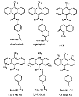
Figure 1 shows the stractures of representative
acridinium ester derivatives used as preferred embodiments
of label ing re:agent;s in the present invention. For the
structure of 1- or :3-Me-AE, 1- or 3-Me-o-F-AE, and 1- or
3-Me-m-diF-AE, a methyl group is shown near the 2 position
of the ac:ridinium ring; this indicates that the methyl
group may be attached at either the 1 or 3 position.
1:~ Figures :?a ar.~i 2b provide a chart representing
predicted pairs of acridinium ester derivatives which may
be used together ir.: the mul tiple analyte assay of the
present invention. .A "Y" indicates that two reagents ar=_
predicted to be: compatible pairs in the invention, and an
"N" indicates that the two compounds would be predicted to
be incompatibles in this assay. The numbers 1 through 3 i_~.
the left-hand column of the chart indicate the type o.
assay system: l represents a homogeneous single-phase
assay system, ~ indicates differential hydrolysis in the
aqueous phase and physical separation of hybridize'
oligonucleotide: probe:target complexes, 3 indicates an
assay sysr_em in which no differential hydrolysis takes
place, and. in which hybridized probe:target complexes are
physically separated. The numbers separated by a slash
below each named acridinium ester derivative are the time-
to-peak and reaction duration, respectively. The numbers
below this line: are the half-life of hydrolysis of each
labeling reagent while coupled to an unhybridized
oligonucleotide probe.. Finally, the selection criteria
upon which this chart is based is shown at the top of the
Figure.
WO 96!13612 . / ~ ~ ~ ~ ~ J PCT/US95/13847
13
Figure 3 is a graphical representation of an example
- using two labeling reagents in a multiple pH mode of the
present invention. In each of Figures 3A-C a triggering
reagent was added to the solution at about interval 5, and
the reaction mixtures were shifted from approximately pH
12.1 to approximately pH 13.0 at about time interval 90,
shown as the X-axis of the graph. Figure 3A shows the
emitted light of such a reaction mixture containing
standard AE only. Figure 3B shows the emitted light in a
reaction containing o-F-AE alone. Figure 3C shows the
emitted light in a reaction mixture containing both
standard AE and o-F-AE.
Figure 4 is a graphic display of the overlapping
characteristic light emission profiles from -five different
combinations of chemiluminescent labeling reagents over
time. A triggering reagent was added to each reaction
mixture at time zero. The labeling reagents used in this
figure were: o-diBr-AE, 2, 7-diMe-AE, o-Me0(cinnamyl)-AE,
o-Me-AE and o-diMe-AE.
Figure 5- is a graphic display of the overlapping
characteristic light emission profiles from five different
combinations of chemiluminescent labeling reagents over
time.- A triggering reagent was added to each reaction
mixture at time zero. The labeling reagents used in this
figure were: o-diBr-AE, a mixture of 1- and 3-Me-AE, o-AE,
o-Me-AE and o-diMe-AE.
Figure 6 is a graphic display of the overlapping
characteristic light emission profiles from seven
different chemiluminescent labeling reagents over time.
A triggering reagent was added to each reaction mixture at
time zero. The labeling reagents used in this figure were:
o-diBr-AE, 2, 7-diMe-AE, a mixture of 1- and 3-Me-AE, o
- ~ AE, o-Me0(cinnamyl)-AE, o-Me-AE and o-diMe-AE.
Figure 7 shows the superimposed characteristic light
emission profiles of four chemiluminescent labeling
reagents in a multiple pH assay mode over time. The
figure demonstrates the ability of the present assay to
220155
WO 96113612 _ PCT/US95/13847
14
detect four analytes in a multiple mode assay system.
Figures 8A through 8I graphically demonstrate the .
correlation between the expected light emission profiles
of combined chemiluminescent labeling reagents versus the
actual light emission profiles obtained. The chemilumine
scent labeling reagents used were: o-diBr-AE, o-F-AE,
standard AE and o-Me0-AE. The chemiluminescent reactions
were conducted in a multiple pH assay system under
identical conditions.
Figures 9A through 9D are chemiluminescent spectra of
two acridinium ester derivatives; Figures 9A and 9B show
the separate spectra of 2, 7 o-diMe-AE and standard AE,
respectively. Figure 9C shows a computer-generated
superimposition of each spectrum in a single plot. Figure
9D shows the -computer-generated simulation of the two
spectra.
Detailed Description of the Preferred Embodiments
The present invention comprises compositions and
methods for the specific detection of multiple different
analytes, preferably nucleic acids, in a single test
sample. Thus, -in a preferred embodiment, the invention
can be used to detect the presence of more than a single
nucleic acid sequence in a test or clinical sample. In a
particularly preferred embodiment, such nucleic acid
sequence may indicate a-particular disease state or
infection.
Definitions
Unless expressly indicated otherwise, the following
terms have the following meanings in the present
application.
By a "nucleic acid analyte" is meant at least one
nucleic acid or_ nucleotide sequence region the presence
and/or amount--of which is sought to be detected with a
single labeling reagent by the methods and compositions of
the present invention when present in a sample. The
WO 96/13612 ' ~ ~ ~ J '~ J PCT/US95/13847
analyte may be a single nucleic acid molecule having one
or more distinct target regions, or more than one
different molecule each one of which has one or more
distinct target regions. Alternatively, an analyte may
5 be a particular nucleotide sequence contained within a
single nucleic acid; hence, a single nucleic acid may
contain more than one nucleic acid analyte. However, the
Applicant contemplates that it may sometimes be desirable
that more than one target region, whether on the same or
10 different nucleic acid molecules, or both, be detected
using the same probe label. This would allow, for
example, both chromosomal r-DNA and ribosomal RNA of a
first organism to be targeted with one or more probes
bearing one label, and the chromosomal r-DNA and ribosomal
15- RNA of a second organism to be targeted by one or more
probes bearing another label. In such a case the first
analyte consists of all the targeted nucleotide sequence
regions of the nucleic acids) of the first organism, and
the second analyte consists of all the targeted nucleotide
sequence regions of the- nucleic acids) of the second
organism.
By "target region" or "target nucle-otide sequence" is
meant the portion of an analyte molecule which binds to a
given probe or class of probes. When the analyte is one
or snore -nucleic acid molecules, the target region has a
nucleotide sequence in a region of at least one of said
nucleic acids which will specifically bind a oligonucleo-
tide hybridization probe under hybridization conditions
which do not favor the hybridization of said probes to
nontargeted nucleic acids or nucleotide sequence regions.
A particular target region may be completely separate from
other target regions, whether contained on the same or
different nucleic acid molecules. Alternatively, a given
target region may, without limitation, be contained on the
satrie- nucleic acid molecule as another target region and
overlap the other target region by one or more nucleo-
tides, be overlapped by the other target region by one or
' ~2U15~~
WO 96113612 ~ _ PCT/US95/13847
16
more nucleotides, or may be contained completely within
another target nucleotide sequence. .
By "probe", "nucleic acid probe", "hybridization
probe", or "oligonucleotide probe" -is- meant an oligo
nucleotide having a nucleotide sequence sufficiently
complementary to a target nucleotide sequence comprised in
a nucleic acid analyte to permit said oligonucleotide to
hybridize therewith under highly stringent hybridization
conditions. When the word "probe" is used, it will be
understood by those of skill in the art that the term
applies to one or more oligonucleotide molecules, either
identical or non-identical, which are designed, selected,
and/or otherwise able to specifically hybridize to a
target nucleic acid region. Additionally, a probe as
defined herein may comprise a collection of different
oligonucleotide molecules targeted to one or more target
regions of the same nucleic acid analyte. Thus, the term
"probe" as used herein may mean either the singular or the
plural, such meaning being made clear by the context of
usage in the present specification. By definition, this
term preferentially applies to oligonucleotides between 10
and 100 nucleotides in length.
By "untargeted nucleic acids" is meant nucleic acids
which are not sought to be detected in a given assay using
the methods or compositions of the present invention.
By "sample" or "test sample" is meant any aqueous or
water- miscible solution, suspension, or emulsion
suspected of containing one or more nucleic acid analytes.
Such a sample may include, without limitation, purified or
unpurified nucleic acids, virus particles, and/or plant,
animal, protozoan or bacterial cells, and may be derived, ,
without limitation, from laboratory, environmental,
agricultural,--food, human,- animal, excretory or secretory
sources. A test sample may be produced as the result of
a pre-treatment of a previous sample, such as, without
limitation, by homogenizing, concentrating, suspending,
extracting, solubilizing, digesting, lysing, diluting or
22015yj
WO 96/13612 PCT/US95/13847
17
grinding the previous sample to put the suspected nucleic
acid analyte, if present, in a water-containing environ-
ment.
By "chemiluminescent label" is meant any chemical
entity or compound, capable of being coupled to another
chemical entity or compound, which can participate in a
chemically-mediated reaction that results in the emission
of light by way of a high energy chemical intermediate .
The preferred chemiluminescent labels of the present
invention are acridinium derivatives; most preferably
acridimium ester derivatives.
By "coupled" is meant that two or more chemical
entities or compounds are j oined by way of a chemical bond
or association. Thus, the term is meant to encompass
covalent bonds as well as strong non-convalent bonds such
as those formed between avidin and biotin, or a chelating
agent and one or more complexed ion.
By "targeted" is meant that a specific chemical,
physical, or biological entity is sought to be identified.
As so defined, a chemical entity may include a portion of
a larger entity, such as a nucleotide sequence region of
a nucleic acid. A biological entity under this definition
may include a grouping of organisms, such as one or more
species, genus, class, family, and so forth.
By a "light-emitting reaction" is meant a triggerable
chemical reaction that results in the detectable pro-
duction of light by one or more of the reactants.
Triggerable is intended to mean that the chemical reaction
is initiated by the addition of a reactant or energy (such
as an electrical charge) to the reaction mixture, or that
the reaction kinetics are made more favorable by adjust-
ment of one or more of the reaction conditions, such as
temperature or pH.
By "sufficiently distinct" is meant that the wave
lengths) of light emission, time-to-peak, reaction
duration or other reaction characteristics of two or more
different chemiluminescent labels can be distinguished
.._ 220595
WO 96/13612 _ PCT/US95/13847
18
when they are combined in a reaction mixture and caused to
emit light in a triggerable light-emitting reaction. ,
By "specifically hybridize" is meant that a single-
stranded nucleic acid can form a stable hydrogen-bonded ,
duplex with a targeted nucleic acid or nucleotide sequence
region under hybridization conditions which do not favor
the formation of stable double-stranded duplexes between
the same single-stranded nucleic acid and non-targeted
nucleic acids or nucleotide sequence regions.
By "similarly protected" is meant that the rates of
loss of chemiluminescent potentialof different chemi-
luminescent labels coupled to oligonucleotide
hybridization assay probes are decreased depending on
whether the probe is hybridized to a targeted nucleic acid
or nucleotide sequence region, and that the rates of said
loss are preferably within a factor of up to about 250 of
each other under the same conditions.
By "similarly susceptable" is meant that the rates of
loss of chemiluminescent potential of different chemi
luminescent labels coupled to oligonucleotide hybridi
zation assay probes by exposure to a destablizing agent
are within a factor of about 50 of each other under
identical conditions.
By "chemiluminescent potential" is meant the ab:.lity
of a given chemiluminescent label to react in a trigger
able light-emitting reaction. Loss of chemiluminescent
potential occurs when such a chemiluminescent label is
chemically degraded or transformed into a non
chemiluminescent compound.
By "reaction pH optimum" or "reaction pH optima" is
meant the pH value at which a chemiluminescent reaction
involving a given chemiluminescent label will proceed with
the highest emission of light under defined conditions. .
If more than one chemiluminescent compound is present in
the same reaction mixture there may be two or more pH
optima for the chemiluminescent reaction mixture. The
yield of light emission (as a function of pH) may rise
~20~~~~
WO 96/13612 _ PCT/US95/13847
19
steeply as the optimum pH is approached, so that a given
chemiluminescent label may emit little light at a first pH
while the same label may emit much more light at a pH
value 1.0 to 0.5 pH unit different from the first.
By "initiation" is meant the addition of energy, a
catalyst or one or more reactant to a reaction mixture
containing chemiluminescent reagents which will cause a
light-emitting reaction to commence.
By "acridinium derivative" is meant any of the family
of chemiluminescent compounds based on the acridinium
ring.
By "acridinium ester derivative" is meant any of the
family of chemiluminescent compounds based on the
acridinium ring and having an ester linkage from the C-9
position.
By "reaction kinetics" is meant the rate of a light-
emitting reaction, as determined by the amount of light
emitted by the chemiluminescent compound or compounds
participating therein in a given time interval, as a
function of time. The term "reaction kinetics" is thus
intended to include reference to the amount of time
between initiation of a chemiluminescent reaction and the
maximum extent of light emission (time-to-peak), as well
as the duration of light emission following initiation in
a given reaction mixture. The reaction kinetics of a
reaction mixture containing a given chemiluminescent label
can be plotted as amount of light emitted in a given time
period versus time, and the curve thus obtained is
reproducible and characteristic for a given chemi-
luminescent reactant under the same reaction conditions.
The reagents used in the preferred embodiments of the
present invention are acridinium derivatives, preferably
acridinium phenyl ester derivatives. Figure 1 shows
examples of representative acridinium phenyl ester
derivatives. It will be understood that other suitable
chemiluminescent reagents and acridinium ester derivatives
including other acridinium derivatives may be found
CA 02201595 2000-09-14
60724-2503
suitable for use in the present invention in light of the
present disclosure by routine screening. Acridinium
phenyl ester compounds are derivatives of acridine
possessing a quaternary nitrogen center and derivatized at
5 the 9 position to ;yield :a phenyl ester moiety. Acridinium
derivatives useful in the present invention, whether
phenyl esters or not, share the property of reacting with
hydrogen peroxide to form a transient dioxetan ring
involving the C-9 carbon. of the acridinium ring, followed
10 by the formation of an excited acridone. The radiative
relaxation of t:~.e ex;~ited acridone results in the
production of light. The synthesis of acridinium esters,
as well as a g~=nera= description of their use as
chemiluminesce nt label~._n.g reagents, is described in Weeks
15 et al . , Acridi nium Ester: as :-iiah Specific Activity Labels
in Immunoassa.vs, ~~lin. Chem. 29:1474-1478 (1984;.
In a pref err ec! embod invent , acr idinium es ten s may be
attached, using sz.andard chemical techniques, to a non-
20 nucleotide monomeric unf.t having a primary amine "linker
arm" available for bonding to the acridinium ester moiety
which is inserted between contiguous sequences of
nucleotides during the chemica'_ synthesis o~ the
oligonucleoticies, car p~.aced at a terminal position of the
oligonucleotide. See, A~rnold, et al., Non-Nucleotide
Linking Reagents f:or Nucleotide Probes, EPO Publication
No. EPO 3132'9 which e:.~_joys common ownership with the
present invention.,.
Thus, the linker arm moiety to which the label
will be attached :is placed at a predetermined position
within the oligonucleotide. It may be placed as an
insertion between or as a substitution for one or more
nucleotides comprising a nucleic acid sequence
sufficiently complementary to at least a portion of a
target nucleic: acid to bf_ able to hybridize thereto under
stringent hybridisation conditions. The solid-phase
synthesis of oligormcleot:ides is well known in the art and
CA 02201595 2000-09-14
60724-2 ~i03
2I
is described in :Brown & Brown, Modern Machine-Aided
Methods of Oliczodeoxvribonucleotide Synthesis in
0liqonucleoti des and Analocrues-A PracticAd Apz~roach
(1991).
Acridiniuvm estar derivatives may be joined to the
linker awm:hy~ridzzation probe conjugate using technicrues
well known in the art. Preferably, Applicants use the
methods described in Nelson et al., Detection of
Acrid=nium Esters by Chemiluminescence in Nor.-Isotopic
Probe Techniaues (Academic Press 1992), Arnold et al.,
Non-Nucl eotidE~ Lir_k i na Reacxents for Nucl eoti de probes, EPO
Publicat~.or. Nc. EP0 3132J.9.
Thus, in one ~~uch preferred method, ar_ N-hvd~cxv-
succinimi.de (NHS) ester ef acricinium (e.g., 4-(2-
succinimi.dylo~;ycarbonyl ethyl) phenyl-10-methyiacridinium
9-carboxylate fluorosulfonate) is synthesized as described
by Weeks et al., supra,
R~=act=or. of the primarjr amine of the linker
arm:hybridizat.ion p:_obe conjugate with the selected N'rFS
acr idinium ewer i~~ performed as fcl lows . The oligo
nucleotide h~,rbridization probe: linker arm conjugate
synthesized as described above is vacuum-dried in a Savant
Speed-Vac'"' dr5ring apparatus, then dissolved in 8 u~ of
0.125 M HEPES buffe_~ (pH 8.0) in 500 (v/v) DMSO. To this
solution is added 2 ~.1 of 25 mM of the desired NI-~S-
acridinium ester. T'he solution is mixed and incubated at
37°C for 20 minutes.
An additional 3 ~1 of 25 mM NHS-acr idinium ester in
DMSO is added to the solution and mixed gently, then 2 u1
of 0.1 M HEPE~~ buff== (pH 8.0) is added, mixed, and the
tube is allowed to :_rcubate for an additional 20 minutes
at 37°C. The :reaction is quenched with the addition of 5
~.1 0.125 M lysine in 0.1 M HEPES buffer (pH 8.0) in DMSO,
which is mixed gently into the solution.
The labeled oligonucleotide is recovered from solution
by the additic>n of 30 ~cl 3 M sodium acetate buffer (pH
~ CA 02201595 2003-O1-31
73091-28
22
5.0), 245 ~.1 water, and 5 ~1 of 40 mg/ml glycogen. Six
hundred forty microliters of chilled 100% ethanol is added
to the tube, and the tube is held on dry ice for 5 to 10
minutes. The precipitated labeled nucleic acids are
sedimented in a refrigerated microcentrifuge at 15,000 rpm
using a standard rotor head. The supernatant is aspirated
off, and the pellet is redissolved in 20 ~.1 0.1 M sodium
acetate (pH 5.0) containing 0.1% (w/v) sodium dodecyl
sulfate (SDS) .
The labeled oligomer may then be purified as necessary
and desired; methods for the purification of labeled
oligonucleotides are well known in the art. In a
preferred method described in Arnold, et al., Acridinium
Ester Labelina~ and Purification of Nucleotide Probes, U.S.
Patent No. 5,185,439 (which enjoys common ownership with
the present application),
the oligomer is purified using reverse-phase high
performance liquid chromatography (RP-HPLC). The sample
is applied to a Vydac'~C4 reverse-phase HPLC column and
eluted with a linear gradient from l0% to 15o Buffer B in
minutes where Buffer A is 0.1% (w/v) triethylammonium
acetate (pH 7.0) in HPLC grade water, and Buffer B is 100%
acetonitrile. The absorbance of the resulting effluent is
monitored at 260 nm, and 0.5 ml fractions are collected.
25 The fractions are then assayed for chemiluminescence, the,
fractions corresponding to the major active peak pre-
cipitated with ethanol, and the labeled probes resuspended
in 0.1 M sodium acetate (pH 5.0) containing O.lo SDS.
The compositions and methods of the present invention
are preferably used in conjunction with the hybridization
protection assay (HPA) described in Nelson ~t al..
Detection of Acridinium Esters by Chemiluminescence in
Non-Isotopic Frobe Techniaues (Academic Press 1992) and
Arnold et al., U.S. Patent No. 5,283,174.
In this assay format, the acridinium
ester labeling reagents are susceptible to hydrolysis when
bound to unhybridized probe but are protected from
*Trade-mark
CA 02201595 2003-O1-31
73091-28
23
hydrolysis when bound to hybridized probe. The differen-
tial hydrolysis characteristics of this system allow for
a homogeneous, single-phase assay wherein hybridization of
probe to target, discrimination between hybridized and
unhybridized probe, and detection and/or quantification of
the labeled hybridized probe can be conducted in a single
test tube. However, differential hydrolysis is not the
only method whereby hybridized and unhybridized probe can
be differentiated; other chemical modifications of the
chemiluminescent label, such as adduct formation can or
may be able to differentiate between chemiluminescent
label coupled to hybridized versus unhybridized probe.
Also, the assay format described herein is also amenable
to an assay format mixing elements of a homogeneous and a
heterogeneous assay as well.
The following examples are intended to be illustrative
only, and in no way limit the scope of the present inven-
tion, which is defined by the claims concluding this
specification.
Example 1: Initial Testing and Screenincr of Various
Acridinium Ester Derivatives
Synthesis of AE Labeling Reagents
N-hydroxysuccinimide (NHS) ester labeling reagents of
acridinium ester (AE) derivatives were synthesized
generally as described in Weeks et al., supra.
For these syntheses, materials
and reagents of highest purity available commercially were
obtained from Aldrich, Lancaster Synthesis and Fisher
Scientific. 9-Acridinecarboxylic acid (ACA), or a methyl
or dimethyl substituted derivative prepared as described
below, was converted to the corresponding acridine acid
chlorides by refuxing for 4 hours in thionyl chloride.
Commercially available hydroxyphenyl- or hydroxynaphthyl
acids -- namely, 3-(4-hydroxyphenyl)propionic acid, 3-(4-
hydroxy-3-methoxyphenyl)propionic acid, 4-hydroxy-3-
2201595
WO 96/13612 _ PCTlUS95/13847
24
methoxycinnamic acid, and 6-hydroxy-2-naphthoic acid --
were converted to benzyl (Bz) esters by treating their .
potassium salts with benzyl bromide in 95a ethanol (EtOH)
solution under- refluxing conditions for about 3 hours. .
These benzyl esters were then allowed to react with the
acridine acid chlorides in anhydrous pyridine for about 4
hours at room-temperature to give the acridine esters.
The benzyl ester protecting groups were hydrolyzed by
treating the acridine esters with 30 wt.% hydrogen bromide
(HBr) in acetic acid (HOAc) for about 4 hours at 60°C.
The resulting acid was converted to the N-hydroxy-
succinimide (NHS) ester reagent using dicyclohexyl-
carbodiimide (I7CC) catalysis in anhydrous tetrahydrofuran
(THF). Finally, transformation to the methyl acridinium
labeling reagent was accomplished by methylation of the
acridine by treatment with excess methyl trifluoro-
methanesulfonate (methyltriflate) in -anhydrous methylene
chloride for 5 to 24 hours at room temperature. The NHS-
ester labeling reagents used for the standard-AE,
naphthyl-AE, -o-Me0-AE and o-Me0-(cinnamyl)-AE were
prepared in this way. The NHS-ester labeling reagents
used for 4,5-diMe-AE, 2,7-diMe-AE, and the mixture of l-
and 3-Me-AE, required synthesis of methyl and dimethyl
substituted ACA's as described below.
The 4,5- and 2,7-dimethyl substituted derivatives of
9-acridinecarboxylic acid (ACA) were prepared through the
reactions of oxalyl chloride with dimethyl substituted
diphenylamines to provide isatin intermediates, followed
by rearrangement to produce the corresponding substituted
acridines essentially as described for 4,5-dimethy-
lacridine-9-carboxylic acid by M.S. Newman and W. H. .
Powell, J. Ora-:- Chem., 26 (1961): 812-815. First, 2,2'-
dimethyldiphenylamine and 4,4'-dimethyldiphenylamine were _
prepared by reacting 2- or 4-methylformanilide with a
slight excess of 2- or 4-bromotoluene, respectively, in
the presence of anhydrous sodium carbonate and traces of
copper in nitrobenzene at 200°C for 24 hours.- Hydrolysis
WO 96/13612 , 2 2 0 ~ 5 9 ~ PCT/US95/13847
of the resulting N,N-diphenylformamides was accomplished
by refluxing them in a 1:1 (v/v) mixture of concentrated
HC1 in acetic acid (HOAc) for 5 hours to provide the
dimethyldiphenyl amines in good yields after purification
5 over silica. Preparation of the dimethyl substituted
acridinecarboxylic acids via isatins then proceeded by
reacting the 2,2'-dimethyldiphenylamine or 4,4'-dimethyl-
diphenylamine prepared above with oxalyl chloride in
refluxing carbon disulfide for about 3 hours. After
10 evaporation of the solvent and excess reagents, the yellow
residue was taken up into fresh carbon disulfide and
treated with aluminum chloride over a period of about 30
minutes, refluxed for 4 hours and set aside at room
temperature overnight. Following evaporation of solvents,
15 the residue was partitioned between methylene chloride and
cold 10% (v/v) concentrated HCl. The orange isatins were
recovered in the organic layer. Finally, treatment of the
isatins with 10% (w/v) potassium hydroxide (KOH) for 12
hours under refluxing resulted in formation of 4,5-
20 dimethylacridine-9-carboxylic acid (4,5-diMe-ACA) and 2,7-
dimethylacridine-9-carboxylic acid (2,7-diMe-ACA), respec-
tively. In a similar manner, 3-methyldiphenylamine was
treated with oxalyl chloride and aluminum chloride to
afford a mixture of methylphenylisatins which, after
25 rearrangement by treatment with KOH, as described above,
yielded a mixture of 1- and 3-methylacridine-9-carboxylic
acid (1- and 3-Me-ACA). Esterification of 4,5-diMe-ACA,
2,7-diMe-ACA, or the mixture of 1- and 3-Me-ACA with the
benzyl ester of 3-(4-hydroxyphenyl)propionate and the
subsequent reactions described above afforded the NHS
. ester labeling reagents used for 4,5-diMe-AE, 2,7-diMe-AE,
or the~mixture of 1- and 3-Me-AE, respectively.
Several substituted hydroxyphenylpropionic acid
derivatives, not available commercially, were prepared by
conventional methods. 3-(4-Hydroxy-3,5-dibromophenyl)
propionic acid was prepared by bromination of 3-(4-
hydroxyphenyl)propionic acid with bromine in glacial
-220 ~ 59~
WO 96/13612 ~ _ .. PCTlUS95/13847
26
acetic acid (HOAc) . 3- (2-Hydroxyphenyl) propionic acid was
prepared by hydrogenation of 2-hydroxycinnamic acid over ,
palladium-on-carbon in absolute ethanol (EtOH).
Acridinium ester (AE) preparation could then proceed by
-coupling ACA with the benzyl esters of these acids as
indicated above to provide the NHS-ester labeling reagents
used for o-diBr-AE and ortho-AE, respectively.
Additionally, the propionitrile derivatives of several
substituted phenols -- namely, 2-methylphenol, 2,6
dimethylphenol, 3,5-dimethylphenol, 2-fluorophenol, and
3,5-difluorophenol -- were prepared by cyanoethylation via
aluminum chloride catalyzed condensation of acrylonitrile
with the phenol and isolation of the corresponding
substituted 4-hydroxyphenylpropionitrile. These
hydroxyphenylpropionitrile derivatives were then reacted
with the acridine acid chlorides and the resulting ester
compounds were treated with hydrogen chloride to hydrolyze
the nitrites to corresponding propionic acid derivatives
which could be processed further as described above to
afford the corresponding AE-NHS ester 1_abeling reagents.
Alternatively, the propionitriles could be first hydro-
lyzed to the corresponding propionic-acid derivative and
synthesis could proceed via the benzyl ester. The final
steps to produce the AE-NHS reagents were the same as
indicated above. The NHS-ester labeling reagents used for
o-diMe-AE, m-diMe-AE, o-Me-AE, o-F-AE, 1- or 3-Me-o-F-AE,
and 1- or 3-Me-m-diF-AE were prepared in this manner.
It -will be clear to those of skill in the art that
these synthesis schemes may be utilized more generally to
make additional and different acridinium ester derivia
tives for characterization and screening as disclosed
below.
Characterization and Screening of AE Derivatives
The chemiluminescence and hydrolysis characteristics
of these derivatives were compared to those of standard AE
(4-(2-succinimidyloxycarbonyl ethyl) phenyl-10-methyl
CA 02201595 2000-09-14
60724-2503
27
acridinium 9-carboxyl~ate fluorosulfonate).
Exemplary ~~E derivatives used to demonstrate the
present invention were naphthyl-AE, o-diBr-AE, a mixture
of I- and 3-Me-AE, 4,5-diMe-AE, 2,7-diMe-AE, o-diMe-AE, o-
Me-AE, m-diMe-.A1~, o-Me0(cinnamyl)-AE, o-Me0-AE, o-AE (an
acridinium ester derivative having the nucleic acid-
coupling linker arm attached to the phenyl ring at the
ortho position), o-F-AE, a mixture of 1- and 3-Me-o-F-AE,
standard AE:, anc? a mixture of I- and 3-Me-m-diF-AE (see
Figure I) . 1-Men-AE, 1-Me-o-F-AE, and 1-Me-m-diF-AE were
only present in a mixture with their 3-methyl isomers; as
used in this a;~plication, these nomenclatures will be
understood to mean a mixture of the corresponding 1- and
3-methyl derivatives. As s::owr. in Figure l, these
compounds were used to label various oligonucleotides to
be used as hybridization probes. It will be understood by
those of skill in the art that the present invention does
not depend on the use of any particular probe-target
combination. 'thus, to choose two or mere mutually
exclusive probe-target combinations for use with the
presently disclosed assay would be routine in light of the
present disclosure.
The ol.igonucleotides were synthesized to contain
phosphodiester bonds using standard solid-phase
~5 phosphoramidite chemistry using a Biosearch 8750 or AHI*
380A DNA synthe~~izer, and purified using polyacrylamide
gel electrc>phor~:sis; oligonucleotide synthesis and gel
purification techniques are well known in the art (see
a . Q . , Sambrook et al . , supra .
_;0 ~Tariou,s non-naturally-occurring oligo-
nucleotides, such as those having modified inter-
nucleotide :linka~~es such as phosphorothioate linkages or
those having sugar or base modifications, are also known
in the art and may have advantages such as increased
35 stability in certain applications; these nucleic acid
anal ogs are also contemplated to be used as part of the
invention of the present application.
* Trade-mark
CA 02201595 2000-09-14
60724-2503
28
As previously referred to, a linker arm terminating
in a primary amine was :incorporated into each oligonucleotide's
structure at a predetermined position in the nucleotide
sequence of the o7_igonucleotide, thus constituting an insertion
c; between nucleotides in the sequence. See ~., Arnold et al.,
Non-Nucleotide Linking_ Rea~cents for Nucleotide Probes, supra.
The AE deriva.tive:> were linked to the oligonucleotide via the
primary amine of t:he linker arm, also as detailed above. The
labelled probes were characterized and compared with regard to
their chemilu.mine~;cent, hybridization, and differential
hydrolysis properties.
Ten microliter aliquots of the labelled probes were
transferred to 12 x 75 polystyrene tubes, and chemiluminescence
was measured in a LEADEF;~ 50 luminometer (Gen-Probe
Incorporated, San Diego, CA) by the automatic injection of a
solution of 200 ,u1 of 0.1% H202 and 0.4 N HCl, a 0.1 to 2 second
delay, automatic injection of 200 ,u1 of 1 N NaOH, and
measurement of the chemi.luminescence for 5 seconds. The final
pH is approximately 13.
The particular' luminometer described herein measures
light between wavelengths 300 to 650 nm; it will be understood
that a luminometer need not detect emitted light in this range
of wavelengths in order to be useful in the methods and
compositions of the pre~;ent invention. In fact, in certain
modes of the :present method, for example, in a multiple
wavelength mode, it may be useful or necessary for a
luminometer to measure emitted light over a broader or narrower
range of wavelengths than is herein described, or over more
than one more narrow wavelength independently and
simultaneously. Thus, the breadth of wavelengths monitored in
the example described herein should be understood as being
CA 02201595 2000-09-14
60724-2503
28a
exemplary and is :zot a limitation on the scope of the present
invention.
The chemiluminescent reaction characteristics were
determined by mea;~uring the light emitted by the reacting
WO 96/13612 ~ _ L 2 O ~ ~ ~ ~ PCT/US95/13847
29
acridinium ester derivatives. The emitted light was
quantified by the luminometer using relative light units
(RLU), a unit of measurement indicating the relative
number of photons emitted by the sample at a given
wavelength or band of wavelengths. The light was detected
at multiple time points during the 5 second measurement
period. From these data the length oftime required for
each labeled oligonucleotide to reach peak light emission
("time-to-peak"), and the duration of the light emission,
were determined. "Duration" was arbitrarily defined to
mean the time required for the RLU to reach 10% of
baseline after the peak emission had occurred. These data
are presented in Table 1 below.
TABLE 1
LIGH T-OFF ISTICS
CHARACTER
Compound Standard Optimal
Conditions Conditions
Peak Dura- pH Peak Dura-
tion tion
Std-AE 0.4s 3.0s 11.9 0.75s >5s
Naphthyl-AE 0.32s 0.5s 10.2 0.5s >5s
o-diBr-AE 0.28s 0.42 10.2 0.5 >5s
1-Me-AE 0.5s 3.0s 11.9 0.75s >5s
4,5-diMe-AE 0.5s 1.8s 11.9 1.0s >5s
2,7-diMe-AE 0.5s 1.8s 11.9 1.3s >5s
o-diMe-AE 0.25s >80s 13.0 0.25s >80s
o-Me-AE 4. Os 40s 13.0 0.25s 3. Os
m-diMe-AE 0.36s 2.3s nd nd nd
o-Me0(cinnamyl)AE 0.6s 8. Os 13.0 0.5s 3.8s
CA 02201595 2000-09-14
60724-250?~
o-Me0-AE 0.35s 0.5s 11.9 0.5s >0.8s
o-p,E 0.9s S.Os 13.0 0.5s >5s
o-F-AE 0.16s 0.38s 11.2 0.6s I.2s
1-Me-o-F-AE J c).18s 0.46s 12.0 0.6s 1.2s
1
5 1-Me-m-diF-AE I t).25s 0.45s 11.3 rd rd
Additionally, the pH reauired for each labeled probe
to emit the maximum amount of light was also determined
and defined as the ":.ptimal pH". In this determination,
the reaction was initiated as described above except that
10 the first reaction solution contained 0.1 N HC1 rather
than 0.4 N HC1, and :rather than. using NaOH, the second
reaction soluticn was 0.24 M sodi um borate buffer titrated
to various pH values. The "time-to-peak" and duration
were calculated for each labeled probe at the optimum pH.
15 These data are also found in Table 1.
After detenmininc~ the cherviluminescent reaction
kinetics of: the labe:Led oligonucleotides, the hybridi-
zation ar,.d hydrolysis characteristics of each
oligonucleotide were invest=gated. The AE hydrolysis
20 characteristics fcr each hybridized and unhybridized
labeled ol~.gonuc:leotide were determined at a range of
temperatures and pH values as describe in Nelson et a'_.,
su ra, and as
briefly summarized here in the following examples.
25 Example 2: Determination of Hvdrolvsis Characteristics of
Acridinium :Ester Derivatives
This eaamplf~ demonstrates a preferred method for
screening indivi3ual chemiluminescent labels to determine
their hydrolysing characteristics, such as the rate of
30 hydrolysis, whe:~. coup:Led to hybridization assay probes.
In particular, the method is useful for a preliminary
determination of the suitability of one or more acridinium
22015g~
WO 96/13612 . PCT/US95/13847
31
ester derivatives for use in a multiple analyte assay
system. Although this method illustrates a preferred
method of chemically distinguishing hybridized from
unhybridized labeled oligonucleotide probes, other methods
of chemically or physically separating single-stranded
from wholly or partially double-stranded nucleic acids,
such as hydroxyapatite adsorption, gel filtration, or
reverse-phase chromatography are well known to those of
skill in the art.
General Procedure for Measurina Hydrolysis of Free Probe
Generally, each candidate acridinium ester is coupled
to a single-stranded oligonucleotide hybridization assay
probe and the probe: AE ester purified, as described above.
Ten microliters of each acridinium ester-labeled probe
dissolved in PSB (10 mM lithium succinate (pH 5.2), 0.1%
lithium lauryl sulfate) were added to a 12 x 75 mm
polystyrene test tube. Multiple replicate tubes are made
for each labeled probe to be tested; Applicants usually
use 13 replicate tubes for each labeled tube, three of
which are used as "time zero" (To) controls. The To
controls are placed in a test tube rack at room
temperature. To each of these tubes is added 200 ~cl 0.4
N HCl and 0.10 (v/v) H20Z, followed by addition of 100 ~cl
o f Hydrolys i s Buf f er ( 0 . 13 - 0 . 19 M Na2B40., ( pH 7 . 6 - 9 . 5 )
and
2-50 (v/v) polyoxyethylene ether (sold under the trade
name TRITON X-100 by Sigma Chemical Co., St. Louis, MO).
Applicants have found the order of addition at this step
to be important. Reagent blanks (negative controls)
contain 10 ~.1 of PSB alone and are then treated as are the
- 30 To controls.
One hundred microliters of Hydrolysis Buffer are given
to each of the 10 remaining replicates a test tube rack,
and the rack is shaken to mix. The test tube rack is
immediately placed in a circulating water bath at 60°C (or
any other desired test temperature) and timing is
initiated.
~2~1595
WO 96/13612 . PCT/US95/13847
32
At desired time points (for example 1, 2, 4, 7, 10,
20, 30, 40, and 50 minutes), 200 ~.1 of a solution of 0.4 .
N HC1, 0.1% (v/v) H2O2 are added to one tube from each set
and the tube is immediately removed from the water bath to
room temperature and mixed. The tube is allowed to stand
at room temperature for at least 1 minute.
The chemiluminescence of each sample is measured in a
luminometer, by a single injection- of a solution
containing 1 N NaOH, and measurement of the chemilumine-
scence for 5 seconds. The average RLU's of the negative
controls are subtracted from the experimental RLU's. The
net RLU's for-each sample can then be divided by the
average To RLU's and multiplied by 100_; this yields the oTo
values; the data can be plotted with log (oTo) as the y
axis and time as the x-axis.
Differential Hydrolysis (DH) Ratio Determination
The following is a generalized procedure for measuring
the ratio of the hydrolysis of the chemiluminescent label
coupled to a hybridized oligonucleotide probe as compared
to the hydrolysis of the same label -coupled in the same
manner to the same probe unhybridized to its target
nucleic acid.
Hybridization of the labeled single-stranded
oligonucleotide probe is accomplished as follows. The
following reagents are combined in a 1.5 ml
microcentrifuge tube for each acridinium ester labeled
probe to be tested: 15 ~.l of a solution of PSB containing
0.05-0.1 pmol of the AE-labeled probe (a calculated total
RLU potential of about 4-5 x 10⁶) , 0.5-1. 0 pmol equivalents
of the target nucleotide sequence (e.g., 0.25-0.5 pmol of ,
a nucleic acid having two copies of the target nucleotide
sequence), and 5-10 pmoles each. of any desired helper ,
probes to faci-litate hybridization of the probe to the
target nucleic acid. Helper probes, also called helper
oligonucleotides, are unlabeled oligonucleotides used to
increase the rate to hybridization by disrupting the
CA 02201595 2000-09-14
60724-2503
33
secondary structvire of the target nucleic acid in the area
of the target nucleotide sequence, (see Hogan et al., U.S.
Patent No. 5,030,557, which enjoys common ownership with
the present application).
However , t:he use of helper probes is not
essential to the operation of the present invention.
The microcen.trifuc~e tube is also given 15 ~C1 of 2 x
Hybridization Buffer (200 mM lithium succinate (pH 5.2),
17 ~ (w/v) 7.ithium laL:ry1 sulfat" 3 mM EDTA (ethyl ene-
diamine tet:raace:tic acid) and 3 mM EGTA ( [ethylenebis
(oxyethylen:itri lo) l -te:traacetic acid) ) . The tube is
incubated at a temperature at least about 5°C below the Tm
of the probe:ta~get duplex for a~ least 30 minutes, then
r r:r~:~ ~;or_ Buffer is added. Separate
270 ~~. of .' x c.yby_.-._..a~~
tubes should ~~e made up for each chemiluminescent
label: probe combination; one tube from each set (labeled
"Hybrid") should contain the labeled probe, the target
nucleic acid, and the reagents, another tube (labeled
"Control" ) shou:_d be made up usi__~.g the same probe and
reagents without: the target nucleic acid. Finally, for
each experiment a "Blank" set of ide::tical tubes si-:cu'_d be
made up using the hybridization reage.~.ts without label ed
probe or target nucle:_c acid.
Ten microliter aliquots of each tube are pipettes into
12 x 75 mm polystyrene: tubes; the number of such tubes is
equal to the nwnber of time points to be analyzed, plus
three tubes for TO determinations, as described above.
The three TO replicate tubes are given 200 ~.1 0.4 N
HCl , 0 .1% (v/v) HZO2, followed by 100 x1 of Hydrol ysis
3 0 Buf f er . The tubes ar a then read in the luminometer , us ing
a singl a i.nj ect:ion of 1 N NaOH, over a per iod of 5
seconds. The reagent "Blank" controls, containing 10 u1
of PSB alone, are prepared in a set of 3-6 tubes and
treated the: same: way as the TO control s .
The "Hybrid" and "Control" tubes are also given 100 ~cl
of Hydrolysis Buffer, mixed, and placed in a circulating
.~ 220 i 595
WO 96/13612 , _ PCT/US9S/13847
34
water bath at the desired temperature, e.g., 60°C. The
timer is started.
At the desired time points, one tube from each set is
given 200 ~.l of 0 . 4 N HC1, 0 . 1 0 (v/v) H202, removed from
the water bath and mixed. The tubes are allowed to sit at
room temperature for at least one minute.
The chemiluminescence of the time point samples from
each set of tubes is measured in a luminometer using an
injection of 1 N NaOH. The emitted light is measured for
5 seconds.
The data is analyzed as described above. The hybrid
hydrolysis rate, expressed as the half-life (T1/2) in
minutes, is divided by the control hydrolysis rate to
obtain the differential hydrolysis (DH) ratio. These
results are summarized in Table 2 below.
TABLE 2
HYDR OLYSIS CTERISTICS
CHARA
t1/2 Rate of
(min) Hydrolysis
Compound
Temp pH Hybrid Control Ratio
Std-AE 60C 7.6 18.1 0.67 27.0
Naphthyl-AE 60C 7.6 5.32 0.52 10.2
o-di-Br 60C 9.1 12.7 1.23 10.3
1-Me 60C 7.6 215 2.0 108
4,5-di-Me-AE 60C 9.1 99.7 0.65 154
2,7-di-Me-AE 60C 9.1 77.0 0.88 87.8
o-Me-AE 60C 9.1 13.3 0.25* 53.2
2.80* 4.8
o-Me0(cinnamyl)- 60C 7.6 63.2 2.1 30.2
AE
o-Me0-AE 60C 7.6 27.8 0.92 30.2
WO 96/13612 . ~ 2 Q ~ ~ ~ J PCT/US95/13847
ortho-AE 60C 7.6 12.7 1.23 10.3
o-F-AE 55C 7.6 57.7 0.92 62.4
1-Me-o-F-AE 55C 7.6 111 3.19 34.9
2,7-diMe-o-F-AE 55C 7.6 317 8.99 35.3
5 1-Me-m-diF-AE 55C 7.6 179 2.24 79.9
II * biphasic
From the data presented in Table 1, it was found that
sets of compounds could be selected, the members of which
10 have sufficiently distinct chemiluminescent properties to
be used as labeling reagents for the simultaneous
detection of more than one analyte in the same tube.
Surprisingly, as illustrated in Table 2, Applicants found
that some of these sets also contained member compounds
15 having similar hydrolysis characteristics; i.e., the
hybridized AE label was not only preferentially protected
from hydrolysis as compared to the unhybridized label but
the rates of hydrolysis of the members within certain
potential sets were substantially similar. Figures 2a and
20 2b show a listing of examples of sets comprising pairs of
such member compounds. The examples cited therein are in
no way intended to limit the present invention to these
embodiments. Although these Figures illustrate the
potential applicability of AE derivatives as combined
25 pairs of labeling reagents it will be understood that sets
of greater than two member compounds may be designed using
the selection criteria listed in these Figures and
disclosure. Moreover, the fact that certain member
compounds are grouped in a set together should not in any
30 way be taken to mean that these are the optimal or sole
groupings of these particular compounds, or that other
compounds would not also function as indicated. The
present invention is defined solely by the claims. The
SUBSTITUTE SH~fT (flt~l~ 2~
X201595
WO 96/13612 _~, s PCT/US95/13847
36
acridinium ester sets listed in Figures 2a and 2b are
candidates for use in at least one mode of the present
invention.
Example 3: Mode 1: Constant pH Simultaneous Reaction
Initiation
There are several modes in which the chemiluminescent
signals of labeling reagents can be used to detect more
than one nucleic acid analyte in a single sample tube
according to the present invention. This and the
following examples are illustrations of such modes.
However, by- those examples Applicants do not intend to
limit the number or description of possible assay modes,
or the composition or combination of labeling reagents for
use in the present invention.
A first experiment tested the chemiluminescence
characteristics of the AE labeling reagents coupled to
single-stranded oligonucleotides in the absence of a
target nucleic acid. Single-stranded oligonucleotides
were designed to be complementary to RNA targets derived
from Escherichia coli or Chlamydia trachomatis. The
oligonucleotides were labeled as described above: the o-
diBr derivative was coupled to an oligonucleotide
specifically complementary to E. coli target RNA, while a
mixture of the 1- and 3-Me derivatives were used to label
an oligonucleotide specifically compleme::tary to C.
trachomatis target RNA. The labeled oligonucieotides were
diluted into 10 mM lithium succinate (pH 5.0) and 0.1°s
(w/v) lithium lauryl sulfate- such that 10 ~.1 of the
resulting solution contained about 200,000 RLU
(approximately 0.002 pmoles) of each oligonucleotide. Ten
microliters of each oligonucleotide were combined with 10
~.l of the same dilution buffer in separate tubes;
additionally, 10 ~.l of each labeled oligonucleotide were
combined in a single tube. Two hundred microliters of a
solution containing 0.1 N HCl, 0.1% H20z were given to the
tube, followed by 100 ~.l of a-solution containing 0.19 M
Na2B40, (pH 7. 6) and 5 0 (v/v) TRITON~ X-100 . The resulting
SUBSTITUTE SHEET (Rt~LE 2'b~
60724-2503
CA 02201595 2000-09-14
37
solution was placed :into a LEADER' 50 luminometer, and
chemiluminescenc~e wa:a measured at various intervals
following injection of: 200 ~cl of 1 N NaOH into the sample
solution. The: lum:inometer was placed in "kinetic
analysis" mode during' the experiment; this allowed the
collection of :FLU data points at predetermined time
intervals after inir_iation of the chemiluminescence
reaction.
In another expe::iment, the same labeled oligo
nucleotides were each hybridized with an excess of their
respective targE~t RNA as described in Nelson et al . ,
supra . The
hybridization was performed in a 50 ~.1 reaction volume and
incubated a.. 55° C for 60 minutes. The f=nal solution for
hybridization contained 100 mM lithium succinate (pH 5.2) ,
8.5% (w/v) :Lithium lauryl sulfate, 1.5 mM EDTA and 1.5 mM
EGTA. Tubes containi::g 50 u1 of each individual probe
target hybridization mixture alone, or a combination of
both hybridization mi~:tures, were given 150 ~.1 of 0.19 M
sodium tetraborate (pFi 7. 6) in 5 . 0 0 (v/v) TRITON' X-100 .
The final amount of each labeled oligonucleotide was about
0.002 pmoles for each experimental tube. The samples were
placed into a LEADERa 50 luminometer , and chemil uminescence
was initiated with the. addition of 200 ~1 of 0.1°s (v/v)
2 S HzOz in 1 mM HNC~3 and, after a 0 . 1 to 2 second delay, an
automatic iwjection of 200 ~.1 of 1 N NaOH. Chemilumine-
scence was measured for various times. Again, the
luminometer was placec'. in "kinetic analysis" mode during
the experiment; this allowed the collection of RLU data
:30 points at predete:rmine<i time intervals after initiation of
the chemilumines~~ence reaction.
The data gather=_:d for the unhybridized labeled
oligonucleotides is shown in Table 3 below.
22015~~
WO 96/13612 _ PC"T/US95/13847
38
TABLE 3
Interval o-diBr-AE 1 or 3-Me-AE o-diBr-AE +
Number , 1 or 3-Me-AE
1 244 35 704
2 19152 293 21995
3 41101 1882 40563
4 44573 4056 45306
5 33485 6496 34719
6 17325 8004 23182
7 12622 8648 20631
8 6118 9346 16696
9 2345 9300 12626
10 956 8769 10243
11 521 8376 9392
12 360 7727 8498
13 262 7314 7860
14 225 6822 7334
15 187 6358 6858
16 161 5950 6463
17 142 5457 5935
18 133 5149 5489
1g 116 4776 5071
20 106 4454 4840
21 93 4111 4448
22 86 3840 4155
23 83 3575 3915
~20~595
WO 96/13612 PCT/US95/13847
39
24 73 3320 3615
25 62 3089 3370
26 58 2852 . 3157
27 57 2675 2927
28 51 2477 2781
29 52 2296 2533
30 53 2139 2410
31 ' 46 1996 2230
32 43 1855 2063
33 39 1702 1921
34 37 1592 1790
35 33 1468 1669
36 37 1381 1563
37 35 1289 1456
38 33 1189 1358
39 29 1101 1278
40 30 1035 1191
41 26 962 1119
42 26 891 1031
43 26 846 974
44 25 765 905
45 25 730 841
46 20 689 797
47 21 638 742
48 18 581 700
~~a~~~~
WO 96/13612 , PCT/US9S/13847
I
49 19 539 655
20 503 . 597
In this experiment the chemiluminescence was measured
for a total of 2 seconds with readings at intervals of
5 0.04 seconds. The results show that the o-diBr-AE label
has a sharp peak of light emission which occurs very
quickly after -the initiation of the chemiluminescence
reaction. The peak of light emission under these
conditions is at interval 4; about 0.16 seconds after
10 initiation, and then the signal decays rapidly. By
contrast, the light emitted by the mixture of 1- and 3-Me-
AE derivative peaks at about interval 8 (0.32 seconds
after initiation) and decays slowly thereafter. Moreover,
in the later intervals (intervals 14-50, and particularly
15 intervals 41-50) the signal from the o-diBr-AE derivative
is almost zero while the signal from the 1- and 3-Me
mixture is still significantly greater than background.
The data from the sample containing both labels
approximates the sum of the two individual data sets at
20 each point .
The data obtained from the experiments involving the
hybridized samples of the two labeled oligonucleotides is
shown in Table 4 below.
TABLE 4
25 Interval o-diBr-AE 1 or 3-Me-AE o-diBr-AE
Number +
1 or 3-Me-AE
1 1082 0 464
2 21715 208 19608
3 46504 1242 49854
30 4 53824 3104 55250
5 49288 5474 50275
220~5~5
WO 96/13612 . PCT/US95/13847
i
41
6 35382 7902 39082
7 . 28444 10110 36476
8 26001 12063 35488
9 20463 13638 30522
10 13750 14681 24878
11 8712 15476 21894
' 12 5460 15892 20156
13 3566 16138 19170
14 2438 16180 18450
15 1802 16048 17786
16 1402 15756 17146
17 1106 15416 16362
18 899 15044 15840
19 765 14470 15062
20 648 14001 14426
21 560 13444 13754
22 484 12986 12128
23 430 12438 12516
24 380 11923 11950
25 334 11444 11337
26 308 10931 10856
27 274 10382 10311
28 250 9926 9816
29 230 9598 9346
30 200 9093 8838
WO 96/13612 _ 2 2 0 I 5 9 5 pCT~s95/13847
42
31 185 8692 8480
32 172 8306 8040
33 154 7916 7680 '
34 141 7587 7291
35 144 7232 6896
36 128 6912 6614
37 128 6567 6295
38 114 6238 5960
39 103 5980 5658
40 100 5695 5450
41 96 5384 5165
42 96 5136 4900
43 88 4932 4688
44 79 4697 4430
45 81 4522 4230
46 75 4280 4037
47 75 4043 3850
48 71 3901 3683
49 65 3680 3486
50 66 3532 3318
The time intervals were the same as in Table 3. In
this case, the difference between the emission
characteristics of the two labels was even more clearly
distinguishable than in the previous experiment: the peak
220155
WO 96/13612 PCT/US95/13847
i
43
of E. coli-hybridized o-diBr-AE occurs again in about
interval 4; however, the peak for C. trachomatis-
hybridized 1-Me-AE is at approximately interval 14.
Again, during the later intervals (particularly intervals
41-50) the signal obtained from the o-diBr-AE derivative
is almost gone, while the signal from the 1-Me-AE
derivative is still significant. And again the profile of
the sample containing a mixture of the 2 hybridized
labeled oligonucleotides approximates the sum of the two
individual sample profiles at each point.
These data demonstrate the applicability and utility
of one mode of the method and compositions of the present
invention. The signal obtained in a single test tube
containing two different oligonucleotides labeled with
specific AE derivatives as set forth in the present
example is clearly made up of two components, one
contributed by a quickly reacting, quickly decaying
species (for example, the o-diBr-AE), and the other
contributed by a species which is slower to react and
decay (for example, the 1- and 3-Me-AE mixture). By
designing two detection time periods for analysis, an
early one (for example intervals 1-6; 0.04 - 0.24
seconds) for the detection of analyte associated with the
quickly-reacting species and a late one (for example,
intervals 41-50; 1.64 - 2.0 seconds) for the detection of
analyte associated with the slowly-reacting species, the
method and reagents of the present invention permit
virtually simultaneous detection of different nucleic acid
sequences in a single sample.
The raw data obtained from this experiment was treated
further using a reiterative data analysis method as
follows. Samples containing only one labeled probe were
used as standards for data analysis. For each standard
the ratio between the sum of the RLU values obtained in
intervals 1-10 and the sum of the RLU values obtained in
intervals 41-50 was determined (E RLU 41-50/E RLU 1-10);
for o-diBr-AE this ratio was 0.00267 and for 1-Me-AE the
WO 96/13612 ' _ J PCT/US95/13847 ,
44
ratio was 0.645. The chemiluminescent signals measured in
intervals 41-50 (in RLU) were added together and then .
divided by 0.645, the ratio obtained for the 1- and 3-Me
standard. The resulting figure is the amount of RLU
contributed in intervals 1-10 by 1- and 3-Me-AE-labeled
probe. This amount, subtracted from the total RLU in
intervals 1-10, gives the amount of RLU contributed in
these intervals by o-diBr-AE. The latter number, when
multiplied by 0.00267 (the ratio for o-diBr-AE), yields
the RLU within the intervals 41-50 which were contributed
by o-diBr-AE-labeled probe. When this figure is
subtracted from the total RLU in intervals 41-50, a
corrected value for the RLU contributed by 1-Me-AE in this
interval is yielded. This number was used to repeat the
calculation described above until the RLU contribution by
o-diBr-AE in intervals 41-50 did not change within the
chosen number of significant figures. An illustration of
the method, as applied to the raw data of Table 5, is
indicated below.
TABLE 5
Observed Initial First Second
RLU Correction
Calculation Correction
Inter- Sum 1-Me diBr 1-Me diBr 1-Me diBr
vals
1-10 341,89664,788 277,10863,639 278,25763,631278,26
5
2 41-50 41,788 41,788 741 41,047 746 41,042746
5
A personal computer was programmed to perform these
calculations. The raw data was fed directly from the
luminometer into the computer using the machine's RS-232
port, and the data processed as described above. The
intervals used in the above analysis may differ depending
on the labeling reagent chosen and it is not mandatory
that the specific intervals illustrated above or in the
following examples be used. Moreover, while the data
22(~ ~ ~~~
WO 96/13612 _ PCT/US95/13847
analysis method disclosed in this example was performed on
, data obtained in experiments using two chemiluminescent
compounds, it will be clear to one of skill in the art
that these same methods can be used to process data
5 obtained from more than two such compounds provided that
the reaction characteristics of each compound are
sufficiently different from the others.
Example 4: Mode 2: Seauential Reaction at Different pH
Values
10 In another mode the reagents and method of the present
invention may be used to detect and measure more than one
analyte in a sample by initiating the chemiluminescence
reaction at different pH values. At the first pH,
chemiluminescent reactions may be initiated, for example
15 by the addition of sodium peroxide and base in an
appropriate buffer, causing one or more of the labeling
reagents to emit measurable light while one or more
additional labeling reagents will not react to an
appreciable extent at that pH. The amount of light
20 emitted at the first pH may be measured in a luminometer.
Additionally, the emitted light may be measured over a
period of time, and the time period may be divided into
intervals as detailed above for a kinetic analysis of the
reaction. After measurement at a given pH, the pH of the
25 test solution may be adjusted to a value at which one or
more other chemiluminescent labeling reagents may react.
While the data presented herein illustrates this mode
of the invention using two AE derivatives, it will be
clear to one skilled in the art in light of this dis
30 closure that more than two pH values may be used in the
method of this invention. Moreover, in light of this
_ disclosure it will also be clear to one skilled in the art
that aspects of the various modes described herein may be
combined in a single assay system. For,example, at each
35 pH value of the "multiple pH" mode described in this
example, a set of kinetically distinct labels may be
. . ~~ ~ ~ ~ ,_
WO 96/13612 _ ~ ~ PCT/US95/1384'7
46
detected in a manner according to the previous example.
Such a system would thus allow for the detection of three
or more analytes in the same sample tube. Other com-
binations, not expressly mentioned, will also be clear to
one of ordinary skill in the art. All such combinations,
whether expressly mentioned herein or not, are intended to
fall within the scope of the present invention.
Two oligonucleotides, each labeled with an AE
derivative having a different optimum pH for the
chemiluminescent reaction, were singly diluted into a
solution containing 10 mM lithium succinate (pH 5.0) and
0.1% (w/v) lithium lauryl sulfate. The first
oligonucleotide, specific to Chlamydia trachomatis 16S
rRNA, was coupled via a linker arm to standard AE . The
second oligonucleotide, specific to Chlam~dia trachomatis
23S rRNA, was coupled via a linker arm to o-F-AE. Ten
microliters (about 0.002 pmoles) of each oligonucleotide
was combined with 10 ~.l of the same dilution buffer in
separate tubes; additionally, 10 ~1 of each labeled
oligonucleotide were combined in a single tube. Each tube
was given 40 ~.1 of 0.4 N HC1, 60 ~1 water and 200 u1 of a
solution containing 1 mM HN03 and 0.1s HBO.. The tubes
were placed into a LEADER6 50 luminometer (Gen-F~robe
Incorporated, San Diego, CA), and chemiluminessence was
measured with the automatic injection of 200 SCI of 0.24 M
boric acid (adjusted to pH 12.9 with NaOH). The
approximate pH of the solution at this--point was 12.-1.
The chemiluminescence was measured for 8 seconds followed
by another automatic injection of 200 ~,1 of 0.75 N NaOH to
an approximate--final pH of 13Ø The resulting chemi-
luminescence was measured for 10 seconds. During the ,
measurement of chemiluminescence data was collected in 0.1
second intervals and immediately downloaded into a IBM
compatible PC computer. The data was then plotted as RLU
versus time (interval number).
The results are shown in Figure 3. These data show
that more than one chemiluminescent labeling reagent
WO 96/13612
PCT/US95/13847
47
coupled to an oligonucleotide can be detected as a member
of a set of such labeling reagents chosen on the basis of
their optimal pH for reaction.
Example 5: Simultaneous Detection of Chlamydia
trachomatis and Neisseria cronorrhoeae Nucleic Acids in a
Homogeneous Assay Format
The method and compositions of the present invention
were used to simultaneously detect the presence of nucleic
acids derived from Chlamydia trachomatis (Ctr) and
Neisseria aonorrhoeae (Ngo) in a single test sample spiked
with known amounts of the target nucleic acids. In this
example the formation, selection, and detection of labeled
analyte/probe conjugates was carried out solely in the
liquid phase.
For convenience's sake, the Ctr and Ngo-specific
probes were those used in Gen-Probe's commercially
available PACE° 2 assay (Gen-Probe Incorporated, San Diego,
CA). In the commercially available assay the Ctr and Ngo
probes are each labeled with standard AE (see Figure 1)
and assayed- separately. By contrast, in the example
described herein the commercially available Ctr probes
were replaced with the identical probes labeled with l-
and 3-Me-AE, and the standard Ngo probes were replaced
with the identical probes labeled with a mixture of 1- and
3-Me-m-diF-AE. Moreover, as in the commercially available
PACE° 2 assay, the labeled probes were used together in a
"probe mix" with other non-labeled helper probes designed
to accelerate the rate of hybridization and the stability
of the formed hybrid nucleic acid. The use of helper.
probes is described above and in U.S. Patent No.
5,030,557. As stated above, helper probes are not
necessary for the practice of the present invention
although helper probes may be necessary in conjunction
with the use of particular labeled hybridization probes.
Also, as mentioned above, the present inventiondoes not
depend on the particular nucleotide sequences of the
. ~2Ui595
WO 96/13612 . PCT/US95/13847
48
nucleic acid analyte or the hybridization assay probe;
thus the specific oligonucleotide probes used in these
examples are not an essential feature of the present
invention. The present methods and compositions can be
used with any set of two or more nucleic acid analyte/
hybridization probe pairs which form mutually exclusive
stable double-stranded hybrids.
Samples were prepared for the assay by adding
different amounts of Ctr and Ngo ribosomal RNA targets to
a solution containing 3a (w/v) lithium lauryl sulfate, 30
mM sodium phosphate buffer (pH 6.8), 1 mM EDTA, 1 mM EGTA
to a final volume of 50 ~,1. The probe reagent was
prepared by combining the 1-Me-AE-labeled Ctr probe with
1-Me-m-diF-AE-labeled Ngo probe in a solution containing
200 mM lithium succinate (pH 5.1), 17a (w/v) lithium
lauryl sulfate, 3 mM EDTA, 3 mM EGTA. The total-amount of
the probes labeled with each of the AE derivatives was
about 0.2 pmoles.
Each hybridization reaction mixture contained 50 ~.l of
the target nucleic acids (or in control experiments, no
target) and 50= ~1 of the probe reagent. Each reaction
mixture was incubated at 55°C for one hour. Three hundred
microliters of 0.15 M sodium tetraborate (pH 8.5), 20
(v/v) TRITON~ X-100 were added to each sample, and the
samples incubated at 55°C for 20 minutes. Chemilumine-
scence of each sample was measured in a LEADER~ 50
luminometer with the automatic injection of 200 u1 of a
solution containing 0.1% H202, 1 mM HN03, followed after a
2 second delay by another injection of 1 N NaOH, 20 (w/v)
Zwittergent~ 3-14 (N-tetradecyl-N,N-dimethyl-3-ammonio-1-
propane-sulfonate; Zwittergent° is a registered trademark .
of CalBiochem, La Jolla, CA). The chemiluminescence of
each sample was monitored for 2 seconds using the
luminometer's kineticmode and intervals of 0.04 seconds.
The data were transferred directly from the luminometer
into a personal computer and analyzedusing calculation
methods similar to those described in Example 3 above.
-- 220159
WO 96/13612 _ PCT/US95/13847
49
The time intervals used for these calculations were
intervals 1-6 and 41-50. Two standards for each different
label were averaged, as were two blank samples, which
contained no probe or target nucleic acids. The RLU value
obtained in each time interval for the averaged blank
standards was subtracted from the RLU values for the
corresponding interval for all other samples prior to
calculation. Each sample was run in duplicate; values
shown are the average of the duplicate reaction mixtures.
The results are shown in Table 6 below.
TABLE 6
Calculate d Values
[Ngo] [Ctr] Total 1-Me-m-diF- 1-Me-AE-
( fmol ( fmol measured AE- [Ngo] [ Ctr]
) ) RLU
10 0 102330 102857 243
2.5 0 28872 27769 948
0.5 0 7624 5533 2078
0 3.0 103193 0 110774
0 0.75 29865 640 30419
0 0.15 7446 266 7510
10 3.0 202729 98729 116462
10 0.75 124013 89825 36738
10 0.15 102883 102238 4411
2.5 3.0 134348 28441 115172
2.5 0.75 56553' 23564 37321
2.5 0.15 35038 27282 8531
0.5 3.0 109582 2317 118248
220155
WO 96/13612 ' ~ . PCTIUS95/1384'7
0.5 0.75 33385 4223 33129
0.5 0.15 12490 5959 7449
0 0 2089 355 1811
These data indicate that at least two analytes (Ctr
5 and Ngo ribosomal RNA in this example)-can be-identified,
either alone or in a sample together, using the multiple
analyte method and reagents of the present invention.
Example 6: Simultaneous Detection of-Ch1amxdia
trachomatis and Neisseria cronorrhoeae Nucleic Acids in a
10 Mixed Homoaeneous/Heteroaeneous Assay Format
The method of the present invention was again used to
simultaneously detect the presence of Ctr and Ngo in a
"pure system" (i.e., no clinical specimen present); this
time in a mixed homogeneous/heterogeneous assay system
15 The assay used for this example was the PACE 2 format
(commercially available from Gen-Probe Incorporated, San
Diego, CA), modified as described herein. The probes used
in this assay were identical to those used in Example 5
and were used in a probe mix with helper probes.
20 For the assay, different amounts of either Ngo or Ctr
ribosomal RNA, or both, were combined in each tube; the
amounts of target varied between 0 and 12.5 fmoles. Final
volume of each target nucleic acid dilution was 100 ~.1;
the difference-in volume was made up with a solution of-30
25 mM sodium phosphate (pH 6.8), 3% (w/v) lithium lauryl
sulfate, 1 mM EDTA, 1 mM EGTA. The probe reagent was
prepared by mixing the 1-Me-AE probe mix and the-1-Me-m-
diF-AE probe mix in equal volumes; as in the previous
experiment, the- probe reagents also contained helper
30 probes. The probe mix contained 190 mM lithium succinate
(pH 5.1), 170 (w/v) lithium lauryl sulfate, 3 mM EDTA, 3
mM EGTA and the probes; one hundred microliters of this
was added to the target nucleic acid dilutions to yield a
60724-250f, CA 02201595 2000-09-14
51
final volume of 200 ~c:L. The tubes were shaken to mix and
incubated at 60~~C for 90 minutes. The tubes were removed
from the water bath ar.~d given 1 ml of a solution of 190 mM
sodium tetrabora.te (pF~ 7.6) , 6.89% (w/v) TRITON° X-102 and
0.01% (w/v) gelatin containing 50 ~C1 of a 1.25% (w/v)
suspension of Biomag~" 4100 magnetic particles (PerSeptive
Biosystems, Cambridge, MA) in 0.02% (w/v) sodium azide and
I mM EDTA. The tubes were incubated further at 60°C for
l0 minutes then removed from the water bath, and the rack
was immediately placed on a magnetic separation base and
allowed to stand at room temperature for 5 minutes, then
the unbound probe was separated from the magnetic bead
bound hybridizes probe by decanting the solution. See,
Arnold, et ~1., =;uropean Publication No. EPO 281390, which
.L5 enjoys common ownership with the present invention.
The
beads and adsorbed hybridized probe were washed once in a
solution of 20 mM sodiL:m tetraborate (pH 10.4), 0.1% (w/v)
Zwittergent° 3-14, then resuspended in 300 ~cl of 5a (v/v)
TRITONS X-100.
Each sample was then loaded into a LEADER" 50
luminometer. The chemiluminescent reaction was initiated
with the automatic injection of 200 ~.1 of a solution
containing C . 1% v/v) :HzOz, 1 mM HNO;, followed after a 2
second delay by arothe_- injection of 200 ~cl of a solution
cor_taining 0.7 N NaOh, and 0.5% (v/v) Zwittergent' 3-14.
The chemiluminesc:ence of each sample was monitored for 2
seconds using the luminometer's kinetic mode and inte~sals
of 0.04 seconds. The data were transferred directly from
the luminometer into a personal computer and analyzed
using calculation; methods similar to those described in
Example 3 above. The time windows used for these
calculations were. from intervals 1-7 and 34-50. Two
standards for each different label were averaged (using 5
3.~ fmoles ribosomal RNA for the Ngo control and 1.5 fmoles
ribosomal RNA fo:: the Ctr control), as were two blank
samples, which contained no probe or target nucl eic acids .
2201~9~
WO 96/13612 . PCTIUS95/13847
52 '
The RLU value obtained for the averaged blank standards in
each time interval was subtracted from the RLU values for
all other samples for the corresponding interval prior to
calculation. Each sample was run in duplicate; values
shown are the average of the duplicate reaction mixtures.
The results are shown in Table 7 below.
TABLE 7
Calculate d Values
[Ngo] [Ctr] Total RLU 1-Me-m- 1-Me-AE-
( fmol ( fmol diF-AE- [Ctr]
) ) [Ngo]
12.5 0 254925 257767 175
1.25 0 36362 38501 101
0.125 0 4197 4055 95
0 3.5 273939 0 291493
0 0.35 32857 67 32608
0 0.035 3785 0 4424
12.5 0.35 288682 248593 32160
12.5 0.035 256085 242548 3663
1.25 3.5 307178 31796 286008
1.25 0.35 66359 34839 32301
1.25 0.035 38674 36768 3273
0.125 3.5 288682 4785 287861
0.125 0.35 36157 3522 33555
0.125 0.035 7601 3906 3605 '
0 0 317 0 92
220~59~
WO 96/13612 ' ' PCT/US95/13847
53
These data demonstrate that by using the compositions
and methods of the present invention more than one analyte
(Ctr and Ngo ribosomal RNA in this case) can be clearly
identified alone or combined in the same sample tube.
Moreover, when the probes containing the labeling reagents
of the present invention are combined in the same sample
tube, the same sample volume is sufficient for the nearly
simultaneous identification of more than one analyte using
the present invention. This permits saving any remaining
sample for other purposes (such as other assays) thereby
increasing the number of assays that can be done using
samples of a given volume. Additionally, the data listed
above show that an assay conducted in accordance with this
embodiment of the present invention has high sensitivity,
with a sensitivity limit in this experiment of at least
0.125 fmole for Ngo and -0.035 fmole for Ctr. By
presenting the data in this Example Applicant does not
intend to imply, however, that this is the lower limit of
sensitivity obtainable under any set of experimental
conditions.
Example 7: Simultaneous Detection of Chlamydia
trachomatis and Neisseria aonorrhoeae ribosomal RNA in a
Clinical Specimen __
The method and reagents of the present invention were
used to simultaneously detect the presence of Ctr and Ngo
ribosomal RNA in a clinical specimen. The assay format
was the same as used in Example 6 with the following
differences.
Each sample was prepared by adding the desired amount
of ribosomal RNA to 100 ~.1 of a pool of endocervical swab
clinical specimens; each swab had been originally
suspended in a volume of 1 ml of Gen-Probe PACE° 2
transport medium (obtainable as a component of the STD
Transport Kit from Gen-Probe Incorporated, San Diego, CA) .
These specimens had previously tested negative for Ctr and
Ngo. Had the original clinical samples contained Ctr or
WO 96/13612 J ~ PCT/US95/13847
54
Ngo cells, these cells would have been lysed and their
nucleic acids (including ribosomal RNA) released into
solution by the action of components of the transport
medium.
Hybridization was conducted as in Example 5, but was
lengthened to 2.5 hours. Following hybridization,
chemiluminescence was measured as described in Example 6,
except for the following changes . ' A solution of 0 . 5 N
NaOH and 0.5o Zwittergent° was substituted for 0.7 N NaOH
and 0.5o Zwittergent°; the Ngo and Ctr ribosomal RNA
standards were 1.25 fmoles and 0.35 fmoles, respectively;
and the intervals chosen for the time windows were
intervals 1-5 and 41-50. Assay results are shown in Table
8 below.
TABLE 8
Calculate d Values
[Ngo] [Ctr] Total RLU 1-Me-m- 1-Me-AE-
( fmol ( fmol diF-AE- [Ctr]
) ) INgo]
5 0 87204 79613 186
0.5 0 9909 7825 62
0.05 0 1403 1019 62
0 5 179368 2 180452
0 0.5 20892 12 20222
0 0.05 2285 2 1985
5 5 258886 69936 174931
5 0.5 92174 57232 20408
5 0.05 81031 70362 2109
0.5 5 189936 9776 188578
0.5 0.5 26500 6887 17865
_ 220155
WO 96/13612 . PCT/US95/13847
0.5 0.05 10625 6887 1736
0.05 0.5 19758 590 18609
' 0.05 0.05 3208 939 1985
0 0 366 23 93
5 These data demonstrate that the ability of the method
and reagents of the present invention to permit the
detection and quantification of more than one analyte in
a single sample is not defeated by substances present in
a pool of clinical samples.
10 Example 8: Detection of the aaa and pol regions of HIV
DNA
The method of the present invention was used to
simultaneously detect the gag and pol regions of the human
immunodeficiency virus (HIV) genome. An advantage of the
15 multiple analyte detection feature of the present
invention for the identification of HIV is that detection
of the presence of the second region (either gag or pol)
of the HIV genome can be used to confirm the presence of
the virus in a diagnostic assay by virtue of the reduced
20 likelihood of two simultaneous false positive assay
indications in the same assay. Moreover, the detection of
more than one distinct nucleotide sequence of the same
nucleic acid analyte can help to ensure detection of a
virus or cell in cases where one target nucleotide
25 sequence has varied or mutated.
In the present example, a region of the HIV genome
. containing both the gag and pol regions was first
amplified as described below. The gag and pol nucleotide
sequence regions were then simultaneously detected using
30 the method and reagents of the present invention in a
hybridization protection assay (HPA) format.
Probes complementary to the gag and pol regions of the
HIV-1 genome- were synthesized. The gag-specific probe
CA 02201595 2000-09-14
60724-2503
56
(SEQ ID NO: 1.1) was labelled with a mixture of 1- and 3-Me-AE
and the pol-~~pecii=is probe (SEQ ID NO: 5) was labelled with
o-diBr-AE, also a:~ desc:ribed above, using a non-nucleotide
linker arm incorporated as part of the oligonucleotide during
~~ synthesis to join the 1<~bel to the probe. A cloned HIV-1 DNA
fragment containing both target sequence regions was amplified
in a 100 ,u1 volume as described in Kacian, et al., PCT
Publication nfo. WO 91/0:L384 which enjoys common ownership with
the present a.pplic:ation. Amplification primers used to amplify
the pol regic~n had nucleotide sequences SEQ ID NOs: 1 through
4. Probes and primers which are used to amplify the gag region
have nucleotide sequencE=_s SEQ ID NOs: 5 through 10.
Following amplification of the HIV-1 DNA, the probes
were hybridized tc> the gag and pol. target nucleic acid regions
by adding 100 ,u1 of a solution containing 5.5 fmol of the 1-Me-
AE labelled gag probe and 16 fmol of the o-diBr-AE labelled pol
probe in 0.2 M lithium :~uccinate (pH 5.0), 17% (w/v) lithium
lauryl sulfate, 3 mM EDTA and 3 mM EGTA to the amplification
reaction mixture. An unlabelled helper oligonucleotide of SEQ
ID NO: 6 was also used t:o assist in the amplification of the
pol target region. The reaction mixture was then incubated for
minutes at 60°C. Hydrolysis of the acridinium derivatives on
the unhybridized aerobe was accomplished by adding 300 ,u1 of a
solution of 0. 13 Nf NazB40~ (pH 9.3) , 2 % (v/v) TRITON~ X-100, and
25 13 mM iodoacetic acid and incubating the mixture for 20 minutes
at 60°C. At i,.his :pH iodoacetic acid is added to the reaction
mixture to prevent formation of acridinium ester adducts which
are unreactive in the chemiluminescent assay.
Chemilurrcinescence was measured in a LEADER~ 50
30 luminometer. Each. sample was placed in the luminometer, and
CA 02201595 2000-09-14
60724-2503
56a
the chemiluminescent reaction initiated by the automatic
injection of 200 ,u1 of 0.1% (v/v) H20z in 1 mM HN03, followed by
a 2 second delay and automatic injection
WO 96/13612 , ,~!', PCT/US95/13847
57
of 1.5 N NaOH. Chemiluminescence was measured for 2
seconds using the luminometer's kinetic mode and intervals
of 0.04 seconds. The data was collected using a personal
computer, and the raw data was analyzed using the
calculation methods described in Example 3. The time
windows used for the calculations corresponded to
intervals 1-10 and 41-50. In this case, 2 standards for
each label were used (these consisted of purified nucleic
acids containing the nucleotide sequence of each target;
either gag alone or pol alone) as well as 2 negative
controls which were treated the same as the other samples
but contained no target nucleic acids or probe. Data
obtained from duplicate standard samples were averaged,
and all samples were corrected for background as described
above. The results are shown in Table 9 below. In this
experiment, a negative assay result yields an RLU value of
less than 10,000. In all experiments using gag and pol
target nucleic acid sequences (except the controls
mentioned above), the gag and pol targets are contained
once in each target nucleic acid molecule.
TABLE 9
Calculated
Values (RLU)
Input Template Total pol gag
Nucleic Acid Observed
Sequence RLU
(before
amplification)
(Average #
Copies)
20 424149 163116 275832
20 474982 181555 288683
20 502688 175009 326109
20 487885 167060 343985
220159
WO 96/13612 _ PCT/US95/I3847
58
5 168892 72321 78129
5 275045 84425 116487
5 262052 102490 137456
5 290219 121739 140858
2.~5 181562 31211 121724
2.5 221174 53702 146548
2.5 242543 115704 116987
2.5 12327 4205 8943
1.25 214078 86608 117385
1.25 7036 4738 1637
1.25 403548 112971 277915
1.25 3246 2265 497
0 4035 3019 508
0 4119 2634 2255
0 4396 3079 730
0 4340 2954 1542
The data show that the method of the present invention
can simultaneously detect the presence of nucleic acids
having sequences corresponding to the gag and pol regions
of HIV-1. The listed number of copies of template nucleic
acids is an average number; clearly, the number of input
copies of template is an integer and not a fraction.
Indeed, the data indicate that some samples contain no
copies of the-template, as can be seen from RLU values
below 10,000 that occur in both the reaction sets
corresponding to 2.5 and 1.25 copies-of template. The
sensitivity of this assay, which combines nucleic acid
amplification with the compositions and methods of the
CA 02201595 2000-09-14
60724-2503
59
present invention, is approximately 1 to 2 copies of each
target nucleic acid sequence.
Example 9: Detection of aaQ and pol regions of HIV DNA in
a Sample Contair..incr a Clinical Specimen.
The method of the present invention was used to
simultaneously detect both the gag and pol regions of the
Human Immunodeficiency Virus (HIV) in a human blood
lysate. Whole blood which had been previously determined
to be negative for H:IV was Iysed, and the white blood
cells were collected, washed, and lysed as described in
Ryder, PCT Publication Nc. WO 93/25710 which a~.joys common
ownership with the present application.
Fifty microiiters of
the leukocyte lysate was used for each experimental tube.
:L5 A plasmid DNA containing the gag and pol regions of HT_V
(see Example 8 above) was added to the lysate, and the
added nucleic acic was amplified, hybridized, and
subjected t:o differential hydrolysis as described in
Example 8. Results are shown in Table 10 below.
<? 0 TABLE 10
j alculated
Values
(RLU)
Input Template Total pol gag
Nucleic Acid Observed
Sequence RLU
(before:
a5 amplification)
(Average #
Conies)
5 549945 264861 199071
5 545940 271571 210287
30 2.5 ~ 5037_59 261827 185001
2.5 513946 243812 195769
2.5 ~ 523'33 278479 184205
2.5 490E~89 265543 166766
2.5 518724 255322 199760
CA 02201595 2000-09-14
60724-2503
2.5 518377 259885 192783
0 8946 6296 4018
0 9113 5642 4161
These data :indicate that the gag and pol targets can
5 be detected simU.ltaneously when the target nucleic acid is
amplified in t:he presence of a cell lysate from blood
mononuclear cells. In such a detection system the
multiple analyt« assay is capable of detecting less than
2.5 copies of more than one different target nucleic acid
10 bearing a given nucleic acid secuence.
Example 10: Po:_vmera,~e Chain Reaction (PCR)
PCR is a nucleic acid amplification technique well
known to and regularly employed by those of ordinary skill
in the art (see e.g., American Society for Microbiology,
15 Diagnostic Mo7.ecular Microbiology: Principles and
Applications 5E~-70 (1993)).
and is patent=ed technology owned and licensed by
Hoffman-LaF;oche, Inc., Nutley, NJ.
A gene:-al procedure for PCR amplification of nucl eic
2C acids is taught in Sa~mbrook et al . , s~,:ora at page 14 . 18 .
In the procedure so
provided, the fcllowing ingredients are mixed in a sterile
0.5 ml mic:rocentrifuge tube for each reaction: 30 ~1 of
sterile water, 10 ~C1 of a lOX Amplification buffer (10X
25 Amplificat:.on buffer - 500 mM KCL, 100 mM Tris-C1 (pH
8.3), 15 mM Mg::l and O.lo (w/v) gelatin), 1.25 mM each
dNTP, 100 pmoles of each primer, up to 2 ~g of template
DNA, and water t:o a f_Lnal volume of 100 ~cl. The reaction
mixture is heated at 94°C for 5 minutes. 5 ~.1 of a 5
30 unit/~cl preparation of Taq DNA polymerase (Perkin-Elmer
Corz~oration, Norwalk, CT) is added to the reaction
mixture. The reaction mixture is then given 100 ~C1 of
light minei:al o:il and the reaction mixture incubated for
5 minutes at 94°C to denature hydrogen-bonded nucleic
35 acids, then for 2 minutes at 50°C to allow annealing of
WO 96/13612 ~ PCT/US9S/13847
61
the primers to the single-stranded target nucleic acids
and 3 minutes at 72°C to allow primer extension. The
reaction mixture is then sequentially incubated for 1
minute at 94°C, 2 minutes at 50°C and 3 minutes at 72°C,
in that order, through 20 cycles. The sample is incubated
at 72°C for 10 minutes in the last step of the last cycle,
then stored at -20°C for use.
Example 11: Detection of the crag and pol Regions of HIV
DNA Following PCR Amplification
The method of the present invention was used to
simultaneously detect the presence of both the gag and pol
regions of HIV DNA. In this experiment the viral DNA was
amplified using the polymerase chain reaction (PCR) prior
to detection.
The probes used were the same as used in Example 8.
HIV-1 DNA was amplified using PCR; the primer pairs used
to amplify the pol region by PCR had nucleotide sequences
of SEQ ID NOs : 2 and 4 , and the primers used to ampl i f y
the gag region had nucleotide sequences SEQ ID NOs: ~ and
10.
After amplification, nucleic acid hybridization was
carried out by mixing 20 ~C1 of the PCR react;cr. ~:i};;.ure
with 80 u1 of water, and then adding 100 ui c; :::e probe
mixture described in Example 8. The probe and target
nucleic acids were incubated together for 30 minutes at
60°C. Differential hydrolysis, measurement of the
chemiluminescence, and calculation of the results were
performed as described in Example 8. The assay results
are shown in Table 11 below.
PCTIUS95/13847
WO 96/13612 _
62
TABLE 11 '
Calculated V alues (RLU)
Input Template Total pol gag
Nucleic Acid Observed
Sequence RLU
(before
amplification)
(Average #
Copies)
25 173882 92838 65793
25 97173 53868 35657
10 107820 51472 44106
10 67621 35681 21349
2.5 65989 31730 27207
2,5 38210 18367 15040
0 975 101 732
0 286 75 56
These data demonstrate that the multiple analyte assay
method of the present invention can simultaneously detect
the presence of different nucleic acid molecules having
sequences corresponding to the gag and pol regions of HIV-
1 when the HIV--1 sequences have been amplified using the
polymerase chain reaction.
Example 12: Simultaneous Detection of More Than Two
Analytes in a Single Test Sample
As an illustration of the feasibility of detecting
more than two analytes in a single sample the following
experiments were performed.
WO 96/13612 . PCT/US95/13847
63
The following AE derivatives were individually coupled
to separate oligonucleotide probes as disclosed above:
diBr-AE; 2,7,-diMe-AE; o-Me0-(cinnamyl)-AE, o-Me-AE, and
o-diMe-AE. Approximately 0.003 pmoles of each indicated
coupled chemiluminescent label in a volume of 1.5 ~1 per
label were added to a tube as shown in Table 12 below,
then given 200 ~,l of a solution containing 0.4 N HCl, O.lo
HZO2. Each tube was loaded into a LEADER° 50 luminometer,
given an automatic injection of 1 N NaOH, and the
resulting emitted light measured over a period of 10
seconds in intervals of 0.1 second.
TABLE 12
Tube AE-Derivatives
1 o-diBr-AE and 2,7-diMe-AE
2 Same as 1 plus o-
Me0(cinnamyl)-AE
3 Same as 2 plus o-Me-AE
4 Same as 3 plus o-diMe-AE
Plots showing the resulting light emission profiles
obtained from of these experiments are shown in Figure 4.
The units of the x-axis are given in interval number, and
the units of the y-axis are given in RLU; the emission
profiles are displayed in a single overlay plot. This
plot clearly shows that the decay of each reacting
chemiluminescent compound in the-samples is sufficiently
different from each other reacting chemiluminescent
compound that each compound can be distinguished from the
others. For example, the light emission from Tube 1 (o-
diBr-AE and 2,7-diMe-AE) reaches baseline at approximately
interval 50 (5.0 seconds). Thus, the light emitted in
intervals 46-100 can be assumed to be the sum of that
emitted by Tubes 2, 3 and 4. (Tube 1 contained both o-
PCT/US95/I3847
WO 96/13612 .
64
diBr-AE and 2,7-diMe-AE; it will be appreciated by one of
skill in the art that o-diBr-AE can be clearly
distinguished from the other AE deriviatives used in this
experiment, and from 2,7-diMe-AE in particular, in a
mixture containing all these compounds, since-its light
emission reaches baseline at approximately interval 10).
Likewise, the light emitted by the chemiluminescent
compounds contained in Tube 2 (o-diBr-AE, 2,7-diMe-AE and
o-Me0(cinnamyl)-AE) reaches baseline at about interval 80
(8.0 seconds); the light emitted in intervals 69-100 can
be assumed to be the sum of the light emitted by the
chemiluminescent compounds contained in Tubes 3 and 4.
Finally, the light emitted by the compounds in Tube 3 (o-
diBr-AE, 2,7-diMe-AE, o-Me0(cinnamyl)-AE and o-Me-AE)
reaches baseline at some point after interval- 100.
Although not shown in the Figure, at this latter time the
components of tube 4 are still emitting measurable light.
Thus, by selecting the time periods during which to
measure the light emitted by the compounds in each tube,
one can distinguish between the light emitted by each
compound using a reiterative averaging process similar to
that used inExample 3 above to distinguish two
chemiluminescent labels. Using the disclosure a: the
present example as a guide, it wou:d be reasonably
expected by those of skill in the art that o-diBr-AE, 2,7-
diMe-AE, o-Me0(cinnamyl)-AE, o-Me-AE, and o-diMe-AE
coupled to oligonucleotide probes can be distinguished
under these reaction conditions. Moreover, it would also
be reasonably expected by those of skill in the art that
this ability would not be defeated when--the--probes are
hybridized to a target nucleic acid. .
Example 13: Evaluation of Additional- Chemiluminescent _
Reagents for Use in a Multiple Analyte Assay
The evaluation of the following probe-coupled
chemiluminescent reagents was performed as described in
the previous example, except emitted light was measured
~~0159~
WO 96/13612 . PCT/US95/13847
for a total of 10 seconds at time intervals of 0.1 second,
and each chemiluminescent reagent was evaluated separately
rather than in a mixture as in Example 12. Figure 5 shows
T a overlay plot of the separately assayed light emissions
5 of 1) a combination of o-diBr-AE and a mixture of 1- and
3-Me-AE, 2) the same as 1), plus ortho-AE, 3) the same as
2), plus o-Me-AE, and 4) the same as 3), plus o-diMe-AE.
As can be seen from the plot, the o-diBr-AE/1-and 3-Me-AE
mixture reacts quickly and emits little light after
10 approximately interval 40, at which time the other AE
derivatives still emit light. The ortho-AE emits little
light after about interval 80. Although this Figure does
not show the baseline resolution of the o-Me-AE deriva-
tive, additional experimentation has confirmed that the
15 light emission decay of this derivative consistantly
proceeds more quickly than does the reaction of the
remaining AE-derivative, o-diMe-AE. Extrapolation of the
curves for these latter two compounds indicates that the
kinetic profiles of these derivatives would be
20 distinguishable in later time intervals than are shown in
this Figure.
Although the coupled o-diBr-AE and 1- and 3-Me-AE
labels were combined in this experiment, it has already
been demonstrated that o-diBr-AE and a mixture of 1- and
25 3-Me-AE can be distinguished on the basis of their
characteristic reaction kinetics,(see e.g., Example 3).
These data indicate that, using the same reiterative
averaging method used in Example 3 above to distinguish
two chemiluminescent labels, the signals for each member
30 compound in this set of coupled chemiluminescent labels
are capable of being distinguished in a single sample when
a light-emitting reaction involving all the member
compounds is simultaneously initiated, and the emitted
light is detected over an appropriate period of time.
WO 96/13612 .L 2 01 ~ ~ J PCT/US95113847
66
Example 14: Evaluation of Seven Chemiluminescent Labels
for Simultaneous Use in a Multiple Analvte Assay,
The reaction kinetics of seven different chemilumine
scent labels (o-diBr-AE, 2,7-diMe-AE, a mixture of 1- and
3-Me-AE, o-linker-AE, o-Me0(cinnamyl)-AE, o-Me-AE, and o
diMe-AE) were evaluated by separately measuring the light
emitted by each compound following initiation of a
chemiluminescent reaction. Each chemiluminescent label
was coupled to a different oligonucleotide. The
experimental conditions were the same as- in Example 12
except as indicated herein. Emitted light was measured
over a total time of seven seconds at 0.1 second
intervals, and detected and measured using a luminometer.
Figure 6 -. shows the resulting light emission
characteristics of these compounds as a computer-generated
single plot comprising the superimposed individual plots
for each chemiluminescent compound. As this Figure
clearly shows, the decay of emitted light by each reacting
compound is sufficiently different and distinct from that
of each other chemiluminescent compound that each may be
separately detected and measured in a single test sample
when reaction is initiated simultaneously. It will be
appreciated by those of skill in the art in light of the
present disclosure that while this example presents data
gathered separately for each member-compound, the reaction
kinetics and decay of emitted light would not differ
substantially when these compounds are combined in a
single sample. Thus, the person of skill in the art would
realize that the present example provides a set of seven
chemiluminescent reagents which may be used simultaneously
ina single assay for the detection of seven nucleic acid ,
analytes in accordance with the compositions and methods
of the present invention.
X20 i ~'~
WO 96/13612 PCTIUS95/13847
67
Example 15: Evaluation of Chemilumunescent Reagents for
Multit~le Mode, Multiple Analyte Assay System
The following chemilumunescent reagents were evaluated
for use in a four analyte, two-pH assay system: o-diBr-AE,
o-F-AE, standard AE, and o-Me0(cinnamyl)-AE. As in the
previous example, each chemiluminescent reagent was
coupledto a different oligonucleotide. Experimental
conditions were the same as in Example 4 except the
oligonucleotides were given 74 ~.1 0.4 N HC1 + 26 ~1 H20
prior to addition of H202. Each chemiluminescent reagent
was evaluated separately.
Figure 7 shows the results of each experiment combined
in a computer generated'single plot wherein the data
obtained for each chemiluminescent reaction is super-
imposed for greater clarity. As can be seen, the o-diBr-
AE and o-F-AE participate in a chemiluminescent reaction
at the first pH. Moreover, these two reagents are clearly
distinguishable from each other with the light emitted by
the o-diBr-AE having decayed to baseline at approximately
i-nterval 25. The light emitted between intervals 25 and
75 r-epresents the contribution of the o-F-AE reagent. It
can also be seen that standard AE and o-Me0(cinnamyl)-AE
are relatively resistant to reaction at this pH, with only
a small amount of light emitted by each compound between
intervals 0 and approximately 85.
The pH of the reaction mixtures was adjusted to 13 at
a time corresponding to approximately interval 85. As can
be seen, this pH shift allowed the largely unreacted
standard AE-and o-Me0(cinnamyl)-AE to emit light at a time
when virtually all of the o-diBr and o-F-AE derivatives
had already reacted at the previous pH. The two reagents
reacting at the new pH value can also be clearly
distinguished on the basis of the time required for each
compound to completely react; standard AE has almost
completely reacted by interval 120, while o-Me0(cinnamyl)-
AE is still emitting light between intervals 120 and
approximately 175.
~ 2 015 9 J pCT~S95/13847
WO 96/13612 .
68
This example demonstrates the versatility of the
compositions and methods of the present invention. As
demonstrated herein, more than one mode of the present
invention may be combined to allow the detection of two or
more nucleic acid analytes . It will be clear to one of
skill in the art that although the data presented herein
was gathered from compounds evaluated in separate reaction
mixtures, these compounds would be reasonably expected to
have substantially similar reaction characteristics when
combined in a single reaction mixture; see, e-a., Example
16. Such a person would also understand that the reaction
characteristics of these compounds would not be materially
altered when the oligonucleotide to which they are coupled
is hybridized to a complementary nucleic acid strand.
Example 16: Correlation between Predicted and Actual
Reaction Characteristics of Combined Chemiluminescent
Reagents __
In order to demonstrate that the reaction
characteristics of the preferred acridinium ester
derivatives exemplified in the previous examples are
accurately predicted by a computer-generated
superimposition of plots - obtained - from individually
assayed chemiluminescent reagents, the following
experiment was performed. Individual reaction mixtures
were made according to the protocol of Example 15. Each
tube contained one of the following acridinium esters: o-
diBr-AE, o-F-AE, standard AE, and o-Me0(cinnamyl)-AE. In
addition, individual tubes were made using the same
amounts of each compound combined in a single tube as
follows: o-diBr and o-F-AE, standard AE and o-Me0-AE, and .
o-diBr-AE, o-F-AE, standard AE, and o-Me0-AE. All of the
chemilumunescent reagents were coupled to separate
oligonucleotides, as in the previous examples. Reaction
was initiated and measured as in Example 15. The results
are shown in Figure 8(A-I).
_ %2~ ~ 59~
WO 96/13612 . PCT/US95113847
69
Figure 8A shows a computer-generated superimposed plot
' of the light emitted by o-diBr-AE and o-F-AE which had
been separately assayed. Figure 8B shows a computer
" generated plot of the combined light emitted by both
reagents; this plot is the sum of the individual plots of
Figure 8A, and represents a prediction of-the reaction
characteristics of a single reaction mixture containing
both reagents, Figure 8C shows the actual reaction
characteristics of a mixture of these two compounds in a
single tube. These data clearly demonstrate that not only
is the decay of light emission the same for Figure 8B
(predicted curve) and Figure 8C (actual curve), but the .
kinetic curves are substantially identical.
Figure 8D similarly shows a computer-generated
superimposed plot of the light emitted by standard AE and
o-Me0(cinnamyl)-AE which had been separately assayed.
Figure 8E displays the computer-generated sum of these
superimposed plots, and Figure 8F shows the actual light
emitted by a mixture of these two compounds following
initiation of a chemiluminescent reaction. A comparison
between Figures E and F shows that the reaction
characteristics of a mixture of standard AE and o
Me0(cinnamyl)-AE are accurately predicted by adding the
curves obtained from the two individually assayed AE
derivatives.
Finally, Figure 8G shows superimposed plots of the
light emitted by all four of these individually assayed
acridinium ester derivatives. Figure 8H is a computer-
generated sum of the plots of Figure-8G, and Figure 8I
shows the light emission characteristics of a mixture of
" all four of these compounds over time. Thus, Figure 8H
shows the predicted light emission characteristics of the
' ' four compounds and Figure 8I, the actual results. Again,
there is close to an exact correlation between the
"predicted" plot of Figure 8H and the "actual" plot of
Figure 8I.
PCTIUS95/13847
WO 96/13612 L 'J
This experiment demonstrates that the characteristic
reaction kinetics of each AE label is not significantly '
different when they are mixed with other -AE labels in a
single reaction mixture. Thus, the AE labels disclosed
5 for use in the methods and compositions of the present
invention and are demonstrably suitable in a multiple
analyte assay system.
Example 17: Mode Three: Multiple wavelengths, Simul-
taneous Initiation
10 In an additional embodiment of the present invention,
multiple analytes may be simultaneously detected in a
single sample by using different oligonucleotide probes
each labeled with a different chemiluminescent label which
emits light at a different wavelength than each other
15 label.
As an example of this mode of the invention, the assay
could be runessentially as in Example 6, with the
following modifications. After the hybridization, each
tube is given 1 ml of a solution of 60 mM sodium
20 tetraborate (pH 8.9) , 6.89% (v/v) TRITON X-102 and 0.1%
(w/v) gelatin containing 50 ~.1 of a 1.25% (w/v) suspension
of BIOMAGT"" 4100 magnetic particles in 0.02%.(w/v) sodium
azide and 1 mM EDTA. Incubation and wash steps are as in
Example 6.
25 A luminometer is equipped with 4 photomultiplier tubes
(PMT'S), one monitoring emitted light in the wavelength
range from 300 nm to 700 nm, one having a 375 to 415 nm
cut-off filter, one having a 400 nm to 435 nm cut-off
filter, and one-having a 500 nm to 575 nm cut-off filter.
30 Standards of each label are loaded into the luminometer, ~
caused to emit light, and the emitted light monitored by
each PMT. Ratios of the chemiluminescence in each
wavelength window are determined for each label as
illustrated in the calculation method of Example 3.
35 Figures 9A and 9B show the chemiluminescent spectra of
2,7-diMe-AE and standard AE; the shaded portions of these
.~'Z~ ~ 59 ~
WO 96/13612 / PCT/US95/13847
spectra represent the wavelength windows referred to
above. Figure 9C is a computer-generated overlay of the
spectra of 9A and 9B. As can be seen, the maximum
' wavelength emission is different for each label, and each
label may be distinguished in a mixture of the two.
Figure 9D is a computer-generated sum of the two
individual wavelength emission profiles.
Having determined the standard ratios of wavelength
emission for the specific chemiluminescent labels to be
used, each experimental sample is loaded into the
luminometer, a light emitting reaction is initiated, and
the resulting emitted light is monitored in exactly the
same way as for the standards. Results can then be
determined using the reiterative calculation method of_
Example 3.
The foregoing examples are intended to be illustrative
only, and in no way are intended by the Applicant to limit
the scope of the present invention. Additional
embodiments are given in the following claims.
J PCT/US95113847
WO 96/13612
72
SEQUENCE LISTING
(1) GENERAL INFORMATION:
(i) APPLICANT: GEN-PROBE INCORPORATED
9880 Campus Point Drive
San Diego, California 92121
U.S.A.
(ii) TITLE OF INVENTION: --COMPOSITIONS AND METHODS FOR
THE SIMULTANEOUS DETECTION
AND QUANTIFICATION OF
MULTIPLE SPECIFIC NUCLEIC
ACID SEQUENCES
(iii) NUMBER OF SEQUENCES: 11
(iv) CORRESPONDENCE ADDRESS:
(A) ADDRESSEE: Lyon & Lyon
(B) STREET: 633 West Fifth Street
Suite 4700
(C) CITY: Los Angeles
(D) STATE: California
(E) COUNTRY: U.S.A.
(F) ZIP: 90071-2066
(v) COMPUTER READABLE FORM:
(A) MEDIUM TYPE: 3.5" Diskette, 1.44 Mb
storage
(B) COMPUTER: IBM PC
(C) OPERATING SYSTEM: MS DOS (5.1)
(D) SOFTWARE: WordPerfect (Version 5.1)
(vi) CURRENT APPLICATION DATA:
(A) APPLICATION NUMBER:
WO 96/13612
- PCT/US95/13847
73
(B) FILING DATE:
(vii) PRIOR APPLICATION DATA:
(A) APPLICATION NUMBER: 08/331,107
(B) FILING DATE: 28 October 1994
(viii) ATTORNEY/AGENT INFORMATION:
(A) NAME: Heber, Sheldon O.
(B) REGISTRATION NUMBER: 38,179
(C) DOCKET NUMBER: 210/086-PCT
(ix) TELECOMMUNICATION INFORMATION:
(A) TELEPHONE: (213) 489-1600
(B) TELEFAX: (213) 955-0440
(C) TELEX: 67-3510
(2) INFORMATION FOR SEQ ID NO:1:
(i) SEQUENCE CHARACTERISTICS:
(A) LENGTH: 24 base pairs
(B) TYPE: nucleic acid
(C) STRANDEDNESS: single
(D) TOPOLOGY: linear
(ii) SEQUENCE DESCRIPTION: SEQ ID NO:1:
ATTCCCTACA ATCCCCAAAG TCAA 24
(2) INFORMATION FOR SEQ ID N0:2:
(i) SEQUENCE CHARACTERISTICS:
(A) LENGTH: 49 base pairs
WO 96/13612 _ PCT/US95113847
74
(B) TYPE: nucleic acid
(C) STRANDEDNESS: single
(D) TOPOLOGY: linear
(ii) SEQUENCE DESCRIPTION: SEQ ID N0:2:
AATTTAATAC GACTCACTAT AGGGAGACAA ATGGCAGTAT TCATCCACA 49
(2) INFORMATION FOR SEQ ID N0:3:
(i) SEQUENCE CHARACTERISTICS:
(A) LENGTH: 45 base pairs
(B) TYPE: nucleic acid
(C) STRANDEDNESS: single
(D) TOPOLOGY: linear
(ii) SEQUENCE DESCRIPTION: SEQ ID N0:3:
AATTTAATAC GACTCACTAT AGGGAGACCC TTCACCTTTC CAGAG 45
(2) INFORMATION FOR SEQ ID N0:4:
(i) SEQUENCE CHARACTERISTICS:
(A) LENGTH: 23 base pairs
(B) TYPE: nucleic acid
(C) STRANDEDNESS: single
(D) TOPOLOGY: linear
(ii) SEQUENCE DESCRIPTION: SEQ ID N0:4:
GTTTGTATGT CTGTTGCTAT TAT 23
(2) INFORMATION FOR SEQ ID N0:5:
(i) SEQUENCE CHARACTERISTICS:
r
?201 J'~~
WO 96/13612 , PCT/US95/13847
(A) LENGTH: 29 base pairs
(B) TYPE: nucleic acid
(C) STRANDEDNESS: single
(D) TOPOLOGY: linear
(ii) SEQUENCE DESCRIPTION: SEQ ID N0:5:
CTACTATTCT TTCCCCTGCA CTGTACCCC 2g
(2) INFORMATION FOR SEQ ID N0:6:
(i) SEQUENCE CHARACTERISTICS:
(A) LENGTH: 33 base pairs
(B) TYPE: nucleic acid
(C) STRANDEDNESS: single
(D) TOPOLOGY: linear
(ii) SEQUENCE DESCRIPTION: SEQ ID N0:6:
CCAATCCCCC CTTTTCTTTT AAAATTGTGG ATG 33
(2) INFORMATION FOR SEQ ID N0:7:
(i) SEQUENCE CHARACTERISTICS:
(A) LENGTH: 46 base pairs
(B) TYPE: nucleic acid
(C) STRANDEDNESS: single
(D) TOPOLOGY: linear
(ii) SEQUENCE DESCRIPTION: SEQ ID N0:7:
AATTTAATAC GACTCACTAT AGGGAGAAGT GACATAGCAG GAACTA 46
(2) INFORMATION FOR SEQ ID N0:8:
v.2 2 015 ~ ~ PCT/US95113847
WO 96/13612
76
(i) SEQUENCE CHARACTERISTICS:
(A) LENGTH: 25 base pairs
(B) TYPE: nucleic acid
(C) STRANDEDNESS: single
(D) TOPOLOGY: linear
(ii) SEQUENCE DESCRIPTION: SEQ ID NO: B:
TGCACCAGGC CAGATGAGAG AACCA 25
(2) INFORMATION FOR SEQ ID N0:9:
(i) SEQUENCE CHARACTERISTICS:
(A) LENGTH: 49 base pairs
(B) TYPE: nucleic acid
(C) STRANDEDNESS: single
(D) TOPOLOGY: linear
(ii) SEQUENCE DESCRIPTION: SEQ ID N0:9:
ATTTTAATAC GACTCACTAT AGGGAGATTG GACCAGCAAG GTTTCTGTC 49
(2) INFORMATION FOR SEQ ID NO:10:
(i) SEQUENCE CHARACTERISTICS:
(A) LENGTH: 23 base pairs
(B) TYPE: nucleic acid
(C) STRANDEDNESS: single -
(D) TOPOLOGY: linear
(ii) SEQUENCE DESCRIPTION: SEQ ID NO:10:
AGATTTCTCC TACTGGGATA GGT 23
~.~2U ~ ~'~
WO 96/13612 , PCTlUS95/13847
77
(2) INFORMATION FOR SEQ ID N0:11:
(i) SEQUENCE CHARACTERISTICS:
(A) LENGTH: 31 base pairs
(B) TYPE: nucleic acid
(C) STRANDEDNESS: single
(D) TOPOLOGY: linear
(ii) SEQUENCE DESCRIPTION: SEQ ID NO:11:
GTCATCCATC CTATTTGTTC CTGAAGGGTA C 31