Note: Descriptions are shown in the official language in which they were submitted.
CA 02202479 1997-04-11
W O96/11634 PCTrUS9S/12443
TRANSDUCER FOR INTRALUMINAL ULTRASOUND IMAGING CATHETE-R
BACKGROUND OF THE I~v~NllON
The invention relates to an ultrasonic transducer
for use in a catheter apparatus and more particularly to such
a transducer having an electrical connection configuration
providing for optimum performance and noise rejection.
Catheters utilizing ultrasonic transducers for
performing intraluminal ultrasonic imaging are known in the
art. Typically, the transducer generates a high-frequency
electrical signal, on the order of 30 MHz, which is utilized
to generate an image.
The quality of an image is degraded by radio
frequency noise which may be mixed with the signal. A typical
hospital environment has many sources of noise, e.g., patient
monitoring systems, intra aortic balloon pumps, x-ray systems,
and computer components. These sources of noise may be
coupled to a transducer generated signal by the conductors
utilized to transmit the signal from the transducer to the
image generating system.
Since the quality of the image generated by the
system is increased when noise is reduced there is a critical
need for improved technology to reduce the noise generated
spurious RF signals present in the operating room.
SU~MA~Y OF THE lNv~NllON
The present invention is an improved system for
transmitting an RF signal from an ultrasonic tr~c~llcer in an
intraluminal catheter to an image generating system.
According to one aspect of the invention, a balanced
transmission line, isolated from the distal housing and drive
cable, transmits high frequency electrical signals from an
CA 02202479 1997-04-11
WO96/11634 PCT~S95/12443
ultrasonic transducer utilized to perform medical ultrasonic
maglng.
According to another aspect of the invention, an
active transducer element has top and bottom major surfaces
with top and bottom electrodes formed thereon. A conductive
backing element is coupled to the bottom electrode and a
backing extension element, extending toward the top major
surface, is electrically coupled to the backing element with
an insulating member electrically isolating the backing
extension element from the top electrode.
According to another aspect of the invention, the
backing element is connected to a distal housing by a non-
conducting adhesi~re. First and second leads are coupled,
respectively, to the top electrode and backing extension
element to form a balanced transmission line to facilitate
noise rejection and optimal performance.
Other advantages and features of the invention will
become apparent in view of the following detailed description
and appended drawings.
BRIEF DESCRI~TION OF THE DRAWINGS
Figs. lA and lB are top and cross-sectional views,
respectively, of a stAn~Ard transducer;
Fig. 2 is a cross-sectional view of a standard
transducer mounted in a distal housing;
Fig. 3 is a schematic view of a standard system for
energizing the transducer;
Figs. 4A and 4B are top and cross-sectional views,
respectively, of the electrical connection to the standard
transducer mounted in the distal housing;
Fig. 5 is a perspective view of a preferred
embodiment of the invention;
Figs. 6A and 6B are top and cross-sectional views,
respectively, of the electrical connection to the transducer
depicted in Fig. 5 mounted in the distal housing;
Figs. 7A and 7B are top and cross-sectional views,
respectively, of the electrical connections to a transducer in
a second preferred embodiment of the invention; and
CA 02202479 1997-04-11
W O 96/11634 PC~rAUS95/12443
Figs. 8 A and 8 B are top and cross-sectional views,
respectively, of the electrical connections to a transducer in
a third preferred embodiment of the invention.
DESCRIPTION OF THE PREFERRED EMBODIM ENTS
Figs. lA and lB are schematic diagrams depicting an
ultrasonic transducer system for emitting and detecting pulses
of ultrasonic energy; Fig. 2 is a schematic diagram of the
transducer mounted in a distal housing of a catheter; and
Fig. 3 is a schematic diagram of a typical system for exciting
the transducer to emit an ultrasonic pulse and to detect a
received ultrasonic pulse.
Turning first to Figs. lA and lB, the ultrasonic
transducer 10 includes an active transducer element 12 which
is part of an assembly. The function of the active transducer
element 12 is to convert ultrasonic pulses to electric pulses
and electric pulses to ultrasonic pulses. The transducer
element 12 is fabricated from PZT ceramic material, has a
block-like configuration, and includes front and back opposing
major surfaces 12F and 12B. These major surfaces 12F and 12B
are covered by metallic conducting films, formed of a suitable
material such as chrome or gold, which function as top and
bottom electrodes 14 and 16. The material of the films can be
formed of a foil or can be in the form of films evaporated or
sputtered onto the opposing surfaces of the transducer element
12. The top electrode 14 has a silver epoxy dot 18 disposed
thereon to be connected to a wire. The transducer element 12
may have a l/4-wave impedance matching layer on the front
surface which is not shown in the figure.
A backing element 22 of a suitable backing material
is bonded to the back surface of the transducer element 12 to
attenuate ultrasonic energy emitted by the back face 12B of
the transducer eiement 12. The backing element 22 has a front
surface 22F bonded to the back surface 12B of the transducer
element 12. An insulating layer 23 surrounds the perimeter of
the transducer element 12 and backing 22.
CA 02202479 l997-04-ll
W O96/11634 PCT~US95/12443
Fig. 2 depicts a transducer 10 mounted in a distal
housing 24. The transducer element 12 is mounted on a bed 26
of adhesive filler material, such as silver epoxy, and the
back surface 22B of the backing element 22 is in contact with
the bed 26. Typically, the backing element 22 and the bed 26
are fabricated of electrically conductive materials and
function as a conductive path forming the electrical contact
to the bottom electrode 16.
Turning now to Fig. 3, a typical system for
energizing the transducer 10 to emit ultrasonic pulses and for
detecting received pulses is depicted. This system is not
part of the invention and will be described only briefly. A
timing and control block 30 controls a transmitter 32 to emit
a series of voltage pulses of a predetermined duration
separated by a predetermined intervals. The switch 34 couples
the transmitter 32 to the transducer 10 when the pulses are
generated and couples a receiver 36 to the transducer 10
during the intervals between pulses.
The received pulses are processed by an image
generating system 38 which is not part of the invention. The
primary information utilized to generate an image is the delay
time between the transmission of an ultrasonic pulse and the
receipt of the received pulse. Other information such as the
amplitude and phase of the received pulse can also be
processed.
As is well-known, when a voltage pulse is applied to
the electrodes 14 and 16 the transducer element 12 oscillates
to generate a pulse centered on a resonant frequency
determined by the mer~An;cal and piezoelectric properties of
the transducer 10. Thus, a series of ultrasonic pulses
separated by the predetermined interval is transmitted.
Conversely, when an ultrasonic pulse is received by
the transducer 10 an imaging signal, in the form a voltage
pulse, is generated on the electrodes 14 and 16 which is
amplified by the receiver 36 and transmitted to the image
generating system 38. The pulse is typically a very high
frequency pulse, on the order of 30 MHz, which is transmitted
CA 02202479 1997-04-11
WO96111634 PCT~S95/12443
s
through a pair of conductors routed through the catheter to
the amplifier.
The electrical connection of leads to the transducer
depicted in Fig. Z is depicted in Figs. 4A and 4B. A first
lead 60 is connected to the silver epoxy dot 18 disposed on
the upper electrode and a second lead 62 is electrically
coupled to the second electrode. The electrical coupling of
the second lead 62 to the bottom electrode follows a path from
the second lead 62 through a conductive adhesive 64 coupling
the lead to a drive cable 66, through a weld to the distal
housing 24, and from the distal housing 24 to the bottom
electrode throuah the conductive acoustic backing 22. The
electrical leads 60 and 62 run the length of the catheter
imaging core.
While the above-described electrical path from the
second lead to the bottom electrode is adequate for electrical
connection alone, it creates an impedance imbalance of the
catheter transmission line. The first lead 60 is connected to
only the active transducer element 12 while the second lead 62
is connected to not only the active transducer element 22, but
is also connected through the conductive adhesive 26 to distal
housing 24 and drive cable 66 of the imaging core. Thus, the
different terminal impedances of the first and second leads 60
and 62 result in an imbalanced trAns~;ssion line. This
imbalance results in a greatly reduced ability of the catheter
to reject RF noise from external sources.
Additional, the inventors have discovered that the
drive cable 66 acts as an antenna to receive spurious rf noise
signals generated in an operating room environment. These
received noise signals are mixed with the imaging signal
generated by the transducer 10 because the second lead 62 is
electrically coupled to the drive cable 66. Thus, the
electrical isolation of the leads 60 and 62 from the drive
cable 66 reduces the mixing of spurious rf si~lA lF with the
imaging signal.
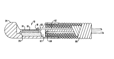
The ccnfiguration of the electrical connection
merhAni~m of a preferred embodiment of the invention will now
be described with reference to Figs. 5, 6A, and 6B. Fig. 5 is
CA 02202479 1997-04-11
W O96/11634 PCTAUS95/12443
a perspective view, Fig. 6A is a top view, and Fig. 6B is a
cross-sectional view taken along line 6B'-6B' of Fig. 5.
Referring now to those figures, the conductive backing element
22 has an extension part 70 which extends to the top surface
s of transducer assembly lo. This extension element is
electrically isolated from the top electrode by an insulating
strip 72. As can be seen from Fig. 5, the combination of the
insulating layer 23 surrounding the perimeter of the active
element and backing and insulating strip 72 electrically
isolates the top electrode 14 from the backing
element/extension 22/70. As depicted in Fig. 6B, the
insulating strip 72 does not extend through the backing
element so that a conductive path from the extension 70 to the
bottom electrode 16 of the active element 10 is formed.
Thus, as depicted in Figs. 6A and B, both leads 60
and 62 are connected to the top surface of the assembly 10.
The second lead 62 is coupled via a second silver dot 74 to
the extension 70. A non-conducting adhesive 76 is utilized as
an acoustic backing and mounting technique to connect the
transducer assembly 10 to the distal housing 24. Accordingly,
the second lead 62 is electrically isolated from the housing
24 and the drive cable 66 to provide a balanced transmission
line.
Figs. 7A and B and 8A and B depict alternative
embodiments for providing a balanced transmission line and for
isolating noise received by the drive cable from the imaging
signal.
In the embodiment depicted in Figs. 7A and B the
second lead 64 is coupled to the back of the extension 70 by
the second silver epoxy dot 74.
In the embodiment depicted in Figs. 8A and B there
is no backing extension 70 or insulating strip 72. The second
lead 62 is coupled to the backing element 22 at the bottom of
the transducer 10 and is insulated from the housing 24 and
drive cable 66 by the non-conducting adhesive 76.
The second cable 62 can be connected prior to
mounting the transducer 10 with the non-conducting adhesive 76
CA 02202479 1997-04-11
W O 96/11634 PC~rAUS95/12443
or subsequent to mounting by drilling a hole in the non-
conducting adhesive 76.
The invention has now been described with reference
to the preferred embodiments. Alternatives and substitutions
will now be apparent to persons of skill in the art.
Accordingly, it is not intended to limit the invention except
as provided by the appended claims.