Note: Descriptions are shown in the official language in which they were submitted.
CA 02202993 1997-04-17
WO 96/15255 PCTIIIS95/13790
1
ASSAY FOR PROLINE IMINOPEPTIDASE
s AND OTHER HYDROLYTIC ACTIVITIES
FIELD OF THE INVENTION
The present invention relates generally to assays for hydrolase activity (e.
g. ,
naturally occurring or artificially produced hydrolytic catalysts) in a sample
or specimen.
The present invention finds particularly useful application in the detection
of bacterial
vaginosis by assaying for the presence of proline iminopeptidase
(alternatively,
aminopeptidase) activity in a vaginal fluid sample.
BACKGROUND OF THE INVENTION
One of the most frequent reasons adult women seek medical treatment is for
abnormal vaginal discharge and related symptoms. In women who visit their
physician
is with a vaginal complaint, approximately 40% are diagnosed as having some
form of
vaginitis, and 90% of these cases are either: bacterial vaginosis (BV),
trichomoniasis or
vulvovaginal candidiasis. The most common among these is bacterial vaginosis.
BV is
associated with placental infection, premature delivery and low birth weight
babies,
increased septicemia, premature membrane rupture and episiotomy infection. As
such,
the need for rapid, accurate, cost-effective and simple point-of care
diagnostic tests for
the detection of BV is of utmost importance.
2s
The term "non-specific vaginitis" was the term initially used to distinguish
this
syndrome from the specific vaginitides caused by Trichomonas vaginalis and
yeast (i. e. ,
Candida species). Prior to 1955, the causes of non-specific vaginitis were
thought to be
a wide variety of aerobic bacteria. In l9ss, it was reported that Haemophilus
vaginalis
was the cause of this disease (Gardner and Dukes,. Am. J. Obstet. Gynecol. ,
69:962
(l9ss)). Subsequently, it was found that the specific organism had no absolute
requirement for hemin and, thus, the name was changed to Corynebacterium
vaginalis.
Also in 195s, a study was published by Gardner and Dukes which suggested that
Gardnerella vaginalis was the causative agent of BV and, thus, the organism
thought to
be responsible for BV was renamed Gardnerella vaginalis. This theory was,
however,
discredited by subsequent studies revealing that this microbe, i. e. , G.
vaginalis, is
present in the vaginal secretions of 40-s0 % of normal women, i. e. , BV-
negative women,
and in those cured of BV (Dunkelberg, et al. , Obstet. Gynecol. , 20:629
(1962)). As
3s such, the considerable overlap in the levels of G. vaginalis found in BV-
positive and BV-
CA 02202993 1997-04-17
WO 96/15255 PCT/US95/13790
2
negative women has rendered the G. vaginalis cell level inconclusive evidence
of the
disease state (Amsel, et al., Am. J. Med., 74:14 (1983).)
Since then, it has become apparent that, unlike other common infectious
diseases,
BV cannot be attributed to one specific etiologic agent, but instead results
from the
drastic alteration of the vaginal flora. The normally present aerobic
Lactobacilli (i. e. ,
the normal flora) become greatly reduced in number and there is a concomitant
overgrowth of several anaerobic bacteria and other microorganisms, including
G.
vaginalis. This alteration in vaginal flora is accompanied by an increase in
vaginal pH.
In response to these findings, the term BV was introduced to describe this
symptomatology, i. e. , to describe increased vaginal discharge without signs
of clinical
inflammation resulting from a complex change in vaginal bacterial flora.
Although BV is the most common form of vaginitis, it is the most benign in
symptomology. The primary signs and symptoms of BV are increased vaginal
discharge,
genital malodor, increased pH and the presence of clue cells. Normal vaginal
discharge
is white and floccular, with a high viscosity. With BV, the discharge is non-
adherent,
clear, thin, white or yellow grey, homogeneous, non-viscous, and watery.
Moreover,
vaginal discharge from women with BV or trichomoniasis liberates a fishy,
amine-odor
when the vaginal fluid is mixed with 10% potassium hydroxide (KOH). No such
odor is
liberated with normal vaginal discharge or, with vaginal discharge from women
with
vulvovaginal candidiasis. In addition, the vaginal pH of women with BV or
trichomoniasis is above 4.5, whereas the normal vaginal pH is less than 4.5.
Finally,
BV is often associated with the presence of "clue cells, " i. e. , vaginal
epithelial cells to
which a large number of bacteria (e. g. , G. vaginalis, Mobiluncus species,
etc. ) are
attached rendering the entire cell border obscure.
As such, the clinical "gold standard" method of diagnosing BV involves the
examination of the following four vaginal fluid criteria: the presence of clue
cells
(greater than 20 % ); vaginal secretions which are white or gray, homogenous
and with
low viscosity; a vaginal fluid pH greater than 4.5; and fishy or fish-like
amine-odor
when the vaginal fluid is mixed with 10% KOH. While these tests require
relatively.
inexpensive components to perform, they are not routinely employed in a
clinical setting.
Such tests are cumbersome, inconvenient, labor intensive, and time-consuming.
More
importantly, these tests are somewhat subjective and require the health care
professional
N
to have considerable expertise with the microscope, a tool not always
available in clinics
or offices.
In addition to the gold standard criteria, BV is sometimes diagnosed by
assessing
the shift in vaginal flora by examining Gram stained vaginal smears. This
method,
however, is difficult to perform and requires special training, thereby making
it
unsuitable for use in clinic or office settings. Alternatively, a sample of
vaginal
CA 02202993 1997-04-17
WO 96/15255 PCTIt7S95/13790
3
secretions can be sent to a laboratory for gas-liquid chromatographic analysis
for the
presence of short chain fatty acids and amines. Unfortunately, however, gas-
liquid
chromatography is time-consuming and expensive to perform.
. In 1988, a report by Thomason, et al. (Obstet. Gynecol., 71(4):607 (1988))
suggested that bacterial enzyme activity, specifically proline iminopeptidase
activity, in
vaginal fluid may be a suitable marker for BV. Thomason, et al. described a
colorimetric assay for proline iminopeptidase activity requiring saline
extraction of
vaginal fluid from a clinical swab, centrifugation, and a four hour incubation
at elevated
temperature (37° C). The enzyme catalyzes the following reaction:
z-Prolyl-/3-Naphthylamide ------------------------------ > z-Proline + j3-
Naphthylamine.
(Colorless) (proline iminopeptidase)
The beta-naphthylamine, in turn, is allowed to react spontaneously with a
solution of a
yellow dye, Fast Garnet GBC, for 5 minutes to produce a red color:
(3-Naphthylamine + Fast Garnet GBC -------------------------- > Red Color.
(Spontaneous)
Unfortunately, the proline iminopeptidase assay system described by Thomason,
et al. , supra, requires four steps to perform. The first step involves the
collection of
vaginal fluid specimens on standard clinical swabs, and the freezing of the
specimens
until a sufficient number are available to test concurrently. As the second
step, the
swabs are thawed, the vaginal fluid is eluted from the swabs with saline, and
the extracts
are centrifuged to concentrate the insoluble, particulate matter into a
pellet. If proline
iminopeptidase activity is present in the specimen, it will be present in the
particulate.
matter. As the third step, the pellet is resuspended in Tris buffered saline
at pH 7.0
containing L-prolyl-/3-naphthylamide and incubated for four hours at elevated
temperature
(37° C). During this incubation period, the substrate will hydrolyze to
release ~3-
naphthylamine if proline iminopeptidase activity is present in the sample. As
the fourth
step, a freshly prepared solution of Fast Garnet is added to the suspension,
and the
mixture incubated for five minutes. If (3-naphthylamine has be yn released by
proline
iminopeptidase or any enzyme having proline iminopeptidase activity, a red
color is
formed. In the absence of proline iminopeptidase activity, only the yellow
color of the
unreacted Fast Garnet chromogen is seen. The resulting assay as a whole is
cumbersome, labor intensive, time-consuming and not suitable for use in a
clinic or
office.
CA 02202993 1997-04-17
WO 96/15255 PCT/US95/13790
4
The association of vaginal fluid proline iminopeptidase activity with BV has
also
been documented by Livengood, et al. (Am. J. Obstet. Gynecol., 163:515 (1990))
using
an assay similar to that used by Thomason, et al. , supra. Another study by
Schoonmaker, et al. (J. Obstet. Gynecol., 165:737 (1991)) utilized a different
chromogenic substrate (i. e. , L-prolyl-para-nitroanilide) to detect proline
iminopeptidase
activity in the vaginal fluid from women with BV and normal control women. In
this
study, the procedure described by Thomason, et al. , supra, was performed
concurrently
on the specimens for purposes of comparison. The results obtained with the
second
chromogenic substrate produced diagnostic efficiencies (i. e. , sensitivity,
specificity,
positive predictive value and negative predictive value) very similar to those
seen in the
Thomason study, supra.
The Schoonmaker, et al. procedure requires the following steps:
(1) elution of vaginal fluid from clinical swabs and freezing the eluates at -
70° C until
the tests were performed; (2) thawing the specimens and concentrating the
particulate
material by centrifugation; (3) resuspending the pelleted materials and
pipetting aliquots
into microtiter wells; (4) adding the chromogenic substrate to the microtiter
wells and
incubating the mixture at 37 ° C for 4 hours; and (5) determining the
presence or absence
of a yellow color visually. Thus, as with the Thomason, et al. procedure, the
Schoonmaker, et al. procedure is cumbersome, labor intensive, time-consuming
and not
suitable for use in a clinical or office setting.
In contrast to the foregoing, the ideal BV test for point-of care use would
have
the following attributes: (1) room temperature stability to permit convenient
storage in
patient examining rooms; (2) the ability to use unprocessed or minimally
processed
vaginal fluid taken directly from the clinical swab; (3) rapid test results,
immediately
available to guide therapy or monitor therapy; (4) simple, specimen activated
format and
interpretation without multiple steps or components--ideally, the user would
only be
required to touch the unprocessed swab to the test system and check for color
formation;
(5) accuracy equal to that seen with clinical laboratory systems; and (6)
built-in,
specimen-activated positive and negative control elements to assure proper
test
performance.
To date, however, no convenient, simple, point-of care assay has been
developed
for detecting the presence of enzymatically active proline iminopeptidase, or
of any
enzyme having proline iminopeptidase activity, in an unprocessed or minimally
processed
vaginal fluid specimen. Accordingly, the present invention overcomes the
problems and
disadvantages of the prior art and which has the attributes set forth above
for the ideal
BV test. Further, the methods and test devices of the present invention are
also useful
for assaying for the presence of other known hydrolases and hydrolase
inhibitors present
in unprocessed or minimally processed samples or specimens.
CA 02202993 2001-03-07
SITMMARY OF THE INVENTION
5 The present invention provides a method for assaying for the presence or
absence of
an analyte selected from the group consisting of a catalytically active
hydrolase and an
inhibitor of a catalytically active hydrolase in a sample comprising:
(a) contacting said sample with a solid-phase conjugate consisting of a non-
enzymatic reporter group coupled to a substrate residue yet cleavable
therefrom by said
catalytically active hydrolase, said reporter group when not so coupled being
capable of
causing a detectable change in an indicator, this step being performed in an
environment in
which the condition of said reporter group as either coupled or decoupled
correlates with the
presence or absence of said analyte;
(b) during or subsequent to step (a), contacting said sample with a solid-
phase
indicator which undergoes a detectable change upon action of said reporter
group; and
(c) observing whether said indicator undergoes a. detectable change, said
detectable change being an indication of the presence or absence of said
analyte in said
sample.
The invention also provides a test device for assaying for the presence of a
catalytically active hydrolase in a sample comprising:
a receptacle defined at least in part by first and second opposing walls
having interior-
facing surfaces with a gap therebetween, said first wall, said second wall, or
both being of
light-transmitting material;
a solid-phase conjugate deposited on said interior-facing surface of one of
said
first and second walls, said conjugate consisting of a non-enzymatic reporter
group coupled to
a substrate residue yet cleavable therefrom upon contact with said
catalytically active
hydrolase, said reporter group when not so coupled being c<~pable of causing a
detectable
change in an indicator;
a solid-phase indictor deposited on said interior-facing surface of one of
said
first and second walls, said indicator being one which undergoes a detectable
change upon
action of said reporter group; and
an opening in said receptacle for introduction. of said sample.
CA 02202993 2001-03-07
Sa
The term "non-enzymatic" as used herein denotes a reporter that is not an
enzyme,
thus the detectable change caused by the reporter in the indicator is not a
change which is the
result of enzymatic activity by the reporter.
The invention further provides a test device for assaying for the presence of
an analyte
in a sample comprising:
a penetrable solid support defined at least in part by upper and lower
surfaces;
a reagent contained in a solid layer on one of said upper and lower surfaces
of said
penetrable solid support, said reagent being one which induces a chemical
reaction in an
indicator when said analyte is present in said sample; and
an indicator contained in a solid layer on the other of said upper and lower
surfaces of
said penetrable solid support in a manner such that said indlicator is
separated from said
reagent until said sample is added, said indicator being one. which undergoes
a detectable
change upon the occurrence of said chemical reaction.
The invention provides a test device for assaying for the presence of an
analyte in a
liquid sample comprising:
a solid support having a surface;
a solid-phase reagent deposited in a first region on said surface, said
reagent being
one which induces a chemical reaction in an indicator when said analyte is
present in said
sample;
a solid-phase indicator deposited in a second region, on said surface, said
second
region non-overlapping with said first region, said indicator being one which
undergoes a
detectable change upon the occurrence of said chemical reaction; and
means for drawing said sample across said surface from said first region to
said
second region.
CA 02202993 2001-03-07
It has now been discovered that enzymatically active proline iminopeptidase
(alternatively, proline aminopeptidase), other enrymc;s exhibiting proline
iminopeptidase
activity, and enzymatically active hydrolases in general, which are present in
unprocessed
or minimally processed vaginal fluid or any other liquid sample, can be
detected in a
rapid, simple and accurate manner. For proline imvaopeptidase activity, the
a~s5ay is
useful for point-of-care detection and diagnosis of bacterial vaginosis.
Likewis;. for other
hydrolases, the assay provides detection and diagnosis of conditions and
diseases
associated with those hydrolases. The procedure is also useful as an assay for
inhibitors
of hydrolases, thereby serving as a means for the detection and diagnosis of
conditions
and diseases associated with~abnormalities in the inhibitor levels.
The assay is performed by contacting the sample with a solid-phase conjugate
which is susceptible to cleavage by the hydrolase, and either during or
subsequent
thereto, contacting the sample with an indicator which undergoes a detectable
change
upon the, action of a reporter group which is a portion of the conjugate and
is liberated
from it either partly or entirely by the action of the hydrolase. Furthermore,
the
indicator is susceptible to action by the reporter group only upon decoupling
of the
reporter group from the remainder of the conjugate, the decoupling resulting
either in
pan or entirely from the hydrolase.
Prior to decoupling, the reporter group and the indicator are precluded from
interaction. This is due either to a chemical neutralization of the reporter
group by the
coupling, or of a spatial separation between the solidL-phase conjugate and
the indicator.
Decoupling may therefore either (a) de-neutralize a neutralized reporter group
which is
already in contact with the indicator, or (b) release a.n active reporter
group and either (i)
permit the released reporter group to diffuse through, the sample toward the
spatially
separated indicator, or (ii) be carried to the indicator by a sample-saturated
swab or other
sample-absorptive device. When a swab or saniple-absorptive device is used,
the
conjugate itself may dissolve in the sample and be c~u~ried by the swab to the
indicator,
with hydrolysis occurring on the conjugate in the swab and continuing after
the swab has
contacted the indicator. Alternatively, the portion o1F the conjugate which
remains after
the hydrolysis may be insoluble in the saunple, such that hydrolysis occurs
only at the
location of the conjugate, and only the released reporter group is picked up
by the swab
and carried to the indicator. Activation of the reponter group can also result
from a
sequential decoupling of the conjugate at different siites on the conjugate
molecule, the
analyte hydrolase being responsible for one of the dexoupIing reactions.
In samples which do not contain the hydrolase, therefore, or samples which do
contain the hydrolase but also contain a hydrolase inhibitor, the reporter
group is
CA 02202993 1997-04-17
WO 96/15255 PCT/US95/13790
6
prevented from interaction with the indicator, whereas in samples which
contain the
uninhibited hydrolase, the interaction occurs and a change is detectable in
the indicator.
The invention is applicable to a wide range of hydrolases and hydrolase
inhibitors.
Likewise, the invention lends itself to, and thereby encompasses, a wide range
of
geometries and spatial arrangements of the conjugate and the indicator. Both
may be
initially present on a common solid support or a solid support of unitary
construction, or
they may be on separate solid supports. The conjugate and the indicator may
occupy a
common region on a single support (particularly in cases where the coupling of
the
reporter group to the conjugate neutralizes the reporter group), or they may
be spatially
separated by an air gap within the support, by the free-standing solid support
itself where
the support is made of a porous material, or by localization in discrete
regions on a
common surface of a single support (the latter three in cases where the
indicator change
relies on diffusion or transport of the reporter group). Other possibilities
will be readily
apparent to the skilled clinician or manufacturer of laboratory test devices.
In certain embodiments of the invention, the conjugate and indicator are
layered
or impregnated in solid form on the surface or within the body of a solid
support yet
readily soluble in aqueous media. In these embodiments, the conjugate and
indicator
dissolve or disperse in the sample regardless of the presence or absence of
the analyte in
the sample. The analyte nevertheless causes activation of the reporter group,
and the
detectable signal arises from the indicator only when the analyte is present.
In other
embodiments, the conjugate, indicator or both are immobilized on the surface
or within
the body of the solid support, and remain so upon contact with the sample. The
presence
of the analyte then results in either activation or release of the reporter
group into the
sample where it can interact with, or through which it can travel to, the
indicator.
The present invention further resides in dry, self contained test devices
useful for
the assays described above. In one embodiment, the test device is comprised of
layered,
and possibly laminated, sheets which form an interior void space to receive
and contain
the sample, an opening through which the sample is admitted into the void
space, and
reagents deposited on, or otherwise applied to, one or more of the interior
walls of the
void space, the reagents including the conjugate and indicator described
above, with the
indicator visible from the exterior of the device through a light-transmitting
wall. As in
the method described above, these characteristics of the test device lend
themselves to a
wide variety of configurations, materials and designs, varying with the
methodology of
the assay and the manner in which the device is handled.
In another embodiment, the test device is comprised of the conjugate and
indicator as solid-phase reagents in distinct layers or laminae separated by a
intermediate
fluid-penetrable layer. In still another embodiment, the test device is
comprised of the
conjugate and indicator as solid-phase reagents localized in discrete regions
on the
CA 02202993 1997-04-17
WO 96/15255 PCTlUS95113790
7
surface of a test device to permit the operator to obtain a reading by drawing
across the
surface a swab or other sample-absorptive device saturated with the sample.
These and other features, objects and advantages of the invention in all of
its
various aspects, including its preferred embodiments, will become apparent
from the
description which follows.
BRIEF DESCRIPTION OF THE DRAWINGS
FIG. 1 is a top view of a one type of test device in accordance with the
present
invention.
FIG. 2 is a side cutaway view of the test device of FIG. 1.
FIGS. 3a and 3b are top and bottom views of a second type of test device in
accordance with the present invention.
FIG. 4 is an exploded view of the test device of FIGS. 3a and 3b.
FIG. 5 is a top view of a third type of test device in accordance with the
present
invention.
FIG. 6a is a layered view of the components on one of the two sample
application
zones of the test device of FIG. 5. FIG. 6b is a layered view of the
components of the
other sample application zone of the same test device.
DETAILED DESCRIPTION OF THE INVENTION
AND PREFERRED EMBOD)aVVIENTS
A. METHODOLOGY
The present invention is applicable to a wide variety of hydrolases and
hydrolase
inhibitors. The term "hydrolase" is used herein to refer to an enzyme or other
natural or
artificially produced entity which catalyzes hydrolytic reactions. More
specifically, the
term "hydrolase" is used herein to refer to a catalyst that is capable of
splitting a
compound into fragments through the addition of water. In this reaction, the
hydroxyl
_ group of a water molecule is -~ucorporated into one fragment, while the
hydrogen atom is
incorporated into the other fragment. Types of hydrolases susceptible to assay
by the
present invention include, but are not limited to, the following: hydrolases
acting on
ester bonds, hydrolases acting on glycoside bonds, hydrolases acting on ether
bonds,
hydrolases acting on peptide bonds, hydrolases acting on carbon-nitrogen (C-N)
bonds
other than peptide bonds, hydrolases acting on carbon-carbon (C-C) bonds,
hydrolases
acting acid-anhydride bonds, hydrolases acting on halide bonds, hydrolases
acting on
CA 02202993 1997-04-17
WO 96/15255 PCT/US95/13790
8
phosphorous-nitrogen (P-N) bonds. Hydrolases of particular interest in
connection with
this invention are those which act on peptide bonds. These hydrolases include,
but are
not limited to, the following: alpha-amino-acyl-peptide hydrolases, peptidyl-
amino-acid
hydrolases, dipeptide hydrolases, and peptidyl-peptide hydrolases. In a
particularly
preferred embodiment, the invention is applicable to assays for proline
iminopeptidase,
which is also referred to in the art as proline aminopeptidase, or any enzyme
which
exhibits proline iminopeptidase activity.
The term "conjugate" is used herein to refer to a reporter group coupled to a
substrate residue yet capable of cleavage or decoupling therefrom upon contact
with the
catalytically active hydrolase whose presence is being detected. The term
"reporter
group" or (interchangeably) "marker group" is used herein to refer to a moiety
which
can be hydrolytically released from the substrate residue by a hydrolase and
which, in its
free form, can react with an indicator to produce a detectable change. Such
reporter
groups include, but are not limited to, the following: phenols, naphthols,
aromatic
amines, amino acids, their derivatives and analogs. In a particularly
preferred
embodiment, naphthylamine, its derivatives or analogs are used as the reporter
group.
The term "substrate residue" is used herein to refer to a molecule or a
portion of
a molecule which is covalently coupled to a reporter group by means of a bond
which is
hydrolyzable by the hydrolase being detected. The reporter group when coupled
to the
substrate residue is incapable of interacting with the indicator, yet becomes
capable of
the interaction upon decoupling by hydrolysis. As mentioned above, the
inability of the
reporter group to interact with the indicator may be due either to a chemical
neutralizing
effect of the substrate residue or to a spatial separation between the
conjugate and the
indicator, or both. In any event, the bond between the reporter group and the
substrate
residue is one which is hydrolyzable either by the analyte hydrolase alone, by
the analyte
hydrolase acting in combination with other hydrolases, or by one or more
hydrolases
when not inhibited by an analyte inhibitor. The substrate residue is not
itself a
chromogen, stain or dye, either before or after enzymatic hydrolysis.
The substrate residue will be selected as one which when coupled to the
reporter
group is susceptible to action by the hydrolase of interest. Since hydrolases
are
frequently named according to the substrate which they hydrolyze, the
selection of a
substrate can often be readily made on this basis. For example, peptidases
hydrolyze
peptide bonds or amide analogs thereof; proline iminopeptidase hydrolyzes
peptide or
amide bonds in which the N-terminal amino acid is L-proline and its analogs or
derivatives; glycosidases hydrolyze glycoside bonds; esterases hydrolyze ester
bonds;
acid phosphatases hydrolyze phosphate esters at low reaction pH; and so forth.
The
reporter group is coupled to the substrate residue using conventional methods
and
procedures. Suitable substrate residues for use in accordance with the present
invention
CA 02202993 1997-04-17
WO 96/15255 PCTli7S95/13790
9
include, but are not limited to, amino acids, peptides, monosaccharides,
disaccharides,
nucleotides, carboxylic acids, alcohols, their derivatives and analogs. In one
presently
preferred embodiment, the substrate residue used is L-proline or hydroxy-L-
proline.
Examples of conjugates are free or modified amino acid derivatives of phenols,
S naphthols, aromatic amines and amino acids. Such conjugates include, for
example,
amino acid-naphthylamides and structurally related amino acid and peptide
analogs. In
certain preferred embodiments, L-prolyl-beta-naphthylamide, L-prolyl-beta-
methoxy-
naphthylamide, or hydroxy-L-prolyl-beta-naphthylamide is the conjugate used
for the
detection of proline iminopeptidase activity.
The coupling of the reporter group to the substrate precludes interaction of
the
reporter group with the indicator. whereas decoupling due to hydrolysis
permits the
interaction to occur. An example of how this is achieved is by use of a
substrate residue
which is an oligopeptide, polypeptide, sugar, oligosaccharide, or other
hydrolyzable
entity which is schematically designated "A.B.C.D.E'', and the reporter group
is
covalently bonded to the E segment of the residue to form the conjugate, which
can then
be represented as "A.B.C.D.E-reporter group". The "E" segment in this example.
acts as
a neutralizing agent to the reporter group, and the substrate residue is
initially either
covalently bonded to the solid support or is simply adhered to it in such a
manner that it
dissolves in the sample upon contact. If the hydrolase specifically hydrolyzes
the bond
between E of the substrate residue and the reporter group, the neutralizing
effect of E is
terminated and the reporter group is activated and released in free form into
the sample.
If the remainder of the residue is also released into the sample, the sample
will contain
both a non-signal generating substrate residue (i.e., A.B.C.D.E) and a free
reporter
group which can in turn react with the indicator to produce a detectable
change.
If however the hydrolase of interest hydrolyzes the conjugate at the bond
between
A and B, or at any other point other than between E and the reporter group,
the
hydrolase by itself would be incapable of releasing the reporter group in
active form.
One or more assisting hydrolases which could only act in conjunction with the
hydrolase
of interest could then be incorporated into the assay to complete the release
of the
reporter group in active form. The assisting hydrolase or hydrolases must
therefore be
ones which are incapable of releasing the reporter group directly from the
intact
conjugate, but instead capable of releasing the reporter group only from the
cleavage
product generated by the hydrolase of interest.
An example is shown schematically hereinbelow.
First, the hydrolase of interest, unable to release the reporter group
directly,
specifically hydrolyzes one or more bonds in the conjugate, thereby releasing
a molecular
fragment containing the inactive reporter group:
CA 02202993 1997-04-17
WO 96/15255 PCT/ITS95/13790
A.B.C.D.E-reporter group ---------------------------> A.B.C + D.E-reporter
group.
(Hydrolase of Interest)
Next, the assisting hydrolase (or hydrolases) releases the reporter group by
hydrolyzing the bond between E of the substrate residue fragment and the
reporter group
5 in one or more steps:
D.E-reporter group ----------------------------> (D + E) or (D.E) + reporter
group.
(Assisting Hydrolase)
The net effect of the foregoing reaction sequence is the release of the
reporter group only
when the hydrolase of interest is present in the sample. As such, the reporter
group can
10 be released from the substrate residue by the hydrolase acting alone or, by
the hydrolase
acting in combination with an assisting hydrolase or hydrolases. The selection
of
appropriate assisting hydrolases will depend on the substrate residue bound to
the
reporter group and will be readily apparent to those skilled in the art.
The term "indicator" is used herein to refer to any species which undergoes a
detectable change as the result of the reaction or, as a result of the
culmination of the
reactions occurring when the catalytically active hydrolase is present in the
sample or
specimen. The resulting detectable change is an indication that the reporter
group was
released from the substrate residue and, thus, that the catalytically active
hydrolase being
assayed for is present in the sample. Moreover, the indicator is preferably of
a
composition which is substantially insoluble in the sample so that the
indicator is
immobilized on the solid support, i. e. , so that the indicator remains
predominantly on the
solid support throughout the duration of the assay. For samples in either
aqueous or
water-soluble media, therefore, the preferred indicator is either an indicator
which is
substantially insoluble in water or, an indicator held in a matrix which is
substantially
insoluble in water.
Preferred indicators are visual indicators and, in particular, chromogenic
indicators, i. e. , those in which the visible change is a change in color,
including the
formation of color in an otherwise colorless material, upon action of the
reporter group
when it is released from the substrate residue by the catalytically active
hydrolase whose
presence is being detected. Alternatively, the reporter group may be capable
of
interacting with an indicator to generate a fluorescent signal, a
phosphorescent signal, a
bioluminescent signal, a chemiluminescent signal or an electrochemical signal
upon its
release from the solid support by the action of the hydrolase. In these cases,
the
indicator would be the chemical species required by the reporter group in
order to bring
about the desired detectable change.
CA 02202993 2001-03-07
11
A wide variety of chromogenic indicators (i. e. , chromogens) and other
species
having a similar effect may be used as visual indicators when phenols,
naphthols,
aromatic amines, amino acids,,their derivative and a~~alogs are used as the
reporter
group. In accordance with the methods of the present invention, preferred
chromogenic
indicators include, but are not limited to, diazonium salts and tetrazonium
salts. In a
presently preferred embodiment, the chromogenic indicator is a diazonium salt.
Suitable
diazonium salts include, but are not limited to, the following: Fast Garnet
GBC, Fast
Dark Blue G, Fast Red B, Fast Corinth V, Fast Bordeaux and Fast Black K, all
of which
are colorless or lightly colored in their unreacted state, but which form
highly colored
derivatives with reporter groups consisting of phenols, naphthols, aromatic
amines or
their structural analogs. See, H. 3. Bonn's "Biological Stains" (R.D. Lillie,
M.D. (ed.),
Baltimore: The Williams & Wilkins Co., ninth edition (1977), pp. 200-224),
for a detailed review of suitable diazonium and tetrazonium salts which can be
used as ;
indicators when phenols, naphthols or aromatic amines are used as the reporter
group.
In addition. numerous chromogenic indicators for reporter groups consisting of
amino acids are also known to those skilled in the an. An example of a
suitable
chromogenic system which can be used when the reporter group is an amino acid
comprises: an amino acid oxidase; a chrornogen selected from the group
consisting of
guaiac, 2,2'-azino-bis(3-ethyl-benzthiazoline-b-sulfonc acid),
tetramethylbenzidine,
phenol, 4-aminoantipyrine and 4,5-dihydroxynaphthalene; a redox catalyst
selected from
the group consisting of peroxidases, iron protoporphyrin and metal ions; and
oxygen. It
will be readily apparent to those in the art that amino acid oxidases can use
oxygen from
the air. An example of this type of chromogenic indicator system is shown
schematically
hereinbelow:
AMINO ACID + O, -----~---------------~----- > Hx02 + OXIDIZED AMINO ACID
(Amino acid oxidase)
H,O, + REDUCED CHROMOGEN -~---- --~~---- --- > COLOR.
(Peroxidase)
As such, the most appropriate chromogenic indicator for any given reporter
group
will depend on the substrate specificity of the hydrolase, the actual reporter
group
employed and the reaction conditions needed for a given test. The selection in
any given
case will be readily apparent to those skilled in the .art.
Any of a wide variety of materials can be used as the solid support. Note that
the
term "solid support" is used herein to denote the free-standing backing
material to which
lamina are applied, as opposed to any of the lamina themselves. Examples of
materials
CA 02202993 1997-04-17
WO 96/15255 PCT/US95/13790
12
suitable for the solid support are insoluble polymeric materials, inorganic or
organic
matrices, gels, aggregates, precipitates and resins. Solid supports in
accordance with the
present invention include, but are not limited to, the following: cellulose,
agarose,
dextran, polyacrylate, or their derivatives, starch, polyacrylamide, nylon,
polyethylene
terephthalates, polyethylenes, 'polystyrenes, polypropylenes, polycarbonates
and glass. In
certain preferred embodiments, Mylar~ and Mylar~/polyethylene laminates are
used as
the solid support.
The solid-phase reagents of the invention are retained or deposited on the
solid
support in a manner which may or may not cause them to remain in place on the
support
surface even upon contact with the sample rather than diffusing or dissolving
in the
sample. The term "immobilized" is used herein to refer to the affixation or
adherence of
the component in question to'the solid support in such a manner that the
component
remains substantially affixed or adhered throughout the assay. Immobilization
may be
achieved in any manner which maintains the species in position on the support,
and the
term is used independently of the manner of applying the species to the
support or the
means of maintaining it in contact with the support. Chemical means of
immobilization
may be used as well as physical means. In the case of components which are
deposited
but not immobilized, the adherence may last only until the support is
contacted with the
liquid sample, i. e. , the initially adhered component either dissolves or is
readily
dispersed in the liquid upon contact. Immobilized components, by contrast, are
not
released into the liquid by simple contact with the liquid. Solid-phase
deposition in
accordance with this invention, whether or not it involves immobilization,
thus
encompasses covalent bonding, specific and nonspecific binding affinities,
impregnation
and various forms of impermanent adherence. Examples of application methods
are
coating, spraying, printing, stamping, ink jet spraying, and dipping.
The solid-phase species will generally be restricted to a defined area on the
surface of the support. Often, the area will constitute, or be part of, a
geometric or
iconic pattern with either a functional purpose, a decorative purpose, or
both. When two
or more solid-phase species are present, the pattern may serve to spatially
separate one
from the other, or to assist in differentiating the species from one other for
the benefit of
the user. The area for any given species may be continuous as in a filled
circle, a ring,
a cross, a strip or other such shape, or discontinuous as in an array of dots
or stripes.
Indicators, for example, can be impregnated onto a bibulous paper or support
or,
deposited onto a plastic or other sheet in the form of a thin layer. The
chromogen can
be layered as a solution or suspension containing a film forming polymeric
material (such
as, for example, celluloses, polyacrylates and their derivatives).
Preferably, the indicator is immobilized on the solid support in a manner
which
renders it largely insoluble in the sample. If the indicator is a water
soluble chromogen,
CA 02202993 1997-04-17
WO 96/15255 PCTlfJS95/13790
13
it can be trapped in a matrix of material which is substantially insoluble in
water.
Alternatively, if the chromogen itself is insoluble in water, it can be
layered as a
suspension in water or as a solution in an organic solvent either alone or in
combination
with a water-soluble or water-insoluble film-forming polymer. In a presently
preferred
embodiment, water-insoluble ethylcelluloses are used as film-forming polymers.
The reagents of the assay may be arranged in a variety of ways on the test
device.
Fnr example the cnninvate and the indicator may he nn riiffPrPnt lam;na of a
t~min~tP~i
~..~ J»a»... »..~» -.~_ ~~»~~»..... ~..~»> ..~ ~.~~ ...~~~..~..... ..aaaauua
va as aauaauauwu
device, separated only by an air gap to be filled by the test sample. Any
reporter group
released by enzymes in the test sample may then diffuse through the sample
toward the
indicator where it will induce the detectable change. Alternatively, the
conjugate and
indicator may be present on the same flat solid support, for example one on
one of the
faces of the support and one on the other face. The support can be porous
(such as a
woven or non-woven polymer) or solid with a series of very fine holes passing
through it
to permit saturation of the support with the sample and diffusion of the
reporter group
from one side to the other.
Alternatively, the conjugate and the indicator may be horizontally segregated
on
the same surface of a single solid support. With the conjugate is deposited on
one
portion of the surface and the indicator on another, the sample can be wiped
or streaked
across the surface from the conjugate to the indicator. As indicated above,
the
methodology for this type of device may vary. As one example, the conjugate
itself may
dissolve in the sample and be carried by the swab to the indicator, with
hydrolysis of the
conjugate occurring in the swab and continuing after the swab has contacted
the
indicator. As another example, the portion of the conjugate which remains
after the
hydrolysis may be insoluble in the sample, with hydrolysis occurring only at
the location
of the conjugate, and only the released reporter group being carried by the
swab to the
indicator. One example of a geometric arrangement for this implementation of
the
invention is a "bull's eye" or "target" pattern, in which the indicator
occupies a filled
circle while the conjugate is arranged in a ring surrounding but not
contacting the circle.
Other geometries will be readily apparent.
To illustrate an implementation of the present invention for detecting proline
iminopeptidase activity, the sample is placed in a device which contains first
and second
solid supports, the first solid support being a lla~ylar~ polyethylene
laminate on which an
L-prolyl-beta-naphthylamide, lr-prolyl-beta-methoxynaphthylamide of hydroxy-L-
prolyl-
beta-naphthylamide conjugate is deposited, the second sol:1 support being a
Mylar~
polyethylene laminate on which Fast Garnet GBC, a chromogenic indicator which
undergoes a detectable change upon action of beta-naphthylamine, is deposited.
The
sample is placed in the device in such a manner that the sample contacts the
first and
second solid supports such that any beta-naphthylamine released by proline
CA 02202993 1997-04-17
WO 96/15255 PCT/US95/13790
14
iminopeptidase activity in the sample is permitted to diffuse through the
sample to the
second solid support. The Fast Garnet GBC is then observed for a detectable
change as
an indication of the presence of the enzyme in the sample. The conjugate may
be
incorporated in a matrix of water-soluble polymer such as hydroxypropyl
cellulose. The
Fast Garnet GBC indicator may be incorporated in a water-insoluble matrix of
ethylcellulose which contains a penetrant such as manganese chloride.
Reaction conditions can be selected, modified and regulated to increase
hydrolase
sensitivity and specificity, and to differentiate between different hydrolases
in a sample.
Examples of reaction conditions which can be controlled in this manner, aside
from the
choice of substrate residues, reporter groups, indicators, and solid supports,
are pH, the
. inclusion and choice of buffer and buffer capacity, and the inclusion and
choice of salts,
detergents, metal ions, reducing agents, and chelators. For example, it is
known that
certain hydrolases function at a low pH and are inhibited at a high pH. Other
hydrolases
function at a high pH and are inhibited at a low pH. Thus, by regulating the
pH of the
assay, one will be able to selectively detect the presence of a particular
hydrolase.
Moreover, it will be readily apparent to the skilled artisan that one or more
hydrolase
inhibitors can also be used in the presently claimed methods to permit the
activity of a
given hydrolase to be detected, while inhibiting interfering hydrolases which
may be
present in the sample, thereby increasing the sensitivity and specificity of
the assay for
the hydrolase of interest.
The present invention can be used to assay simultaneously for the presence of
two
or more catalytically active hydrolases in a sample or specimen. This is
achieved by
using a combination of two or more reporter groups coupled to different
substrate
residues and deposited on one or more solid supports, each reporter group
releasable
from a given substrate residue by only one of the hydrolases potentially
present in the
specimen, each substrate residue employing a different reporter group, and two
or more
indicator systems, each capable of producing a detectable response with only
one of the
reporter groups. For example, with the present invention one can
simultaneously detect
a mixture of a glycosidase and a peptidase, thereby obtaining a hydrolytic
profile of a
given pathogen or disease process. In this assay, two different, specific
substrate
residues each of which inactivates a different, specific reporter group, and
two different
reporter group-specific indicators would be employed.
In its application to the detection of hydrolase inhibitors, the present
invention
extends to a wide range of analytes and samples. Many biological processes,
including
regulation of blood pressure, blood clotting, bacterial replication, etc. ,
involve the use of
very specific, carefully modulated hydrolases. Moreover, numerous drugs,
pesticides,
and herbicides, etc., are known to function by virtue of inhibiting specific
hydrolases.
Under certain circumstances, it is highly desirable to determine the blood,
saliva or urine
CA 02202993 1997-04-17
WO 96/15255 PCTl(JS95/I3790
concentration of a hydrolase inhibiting therapeutic drug or to determine the
presence of a
potential pesticide hydrolase inhibitor contamination in produce, etc. The
analyte in
these cases is the inhibitor of a, hydrolase rather than the hydrolase itself.
The need to analyze for the presence of an active hydrolase inhibitor arises
in two
5 circumstances -- ( 1 ) when the target hydrolase of the inhibitor (i. e. ,
the particular
hydrolase that the inhibitor is known to inhibit or inactivate) is present in
the sample
prior to the analysis, and (2) when the target hydrolase is not present in the
sample and
must be added to the test system. When the hydrolase is not present, a defined
quantity
of the hydrolase is incorporated into the test system, and the test
performance involves
10 detecting the ability of the sample to inhibit the target hydrolase. The
hydrolase may,
for example, be applied to one of the solid supports in the same manner as the
conjugate,
or the hydrolase may be added to the sample prior to contact of the sample
with the test
device.
In the event that the target hydrolase inhibitor is not present in the sample,
the
15 target hydrolase will release the reporter group from the substrate
residue, thereby
producing a detectable change in the indicator. Conversely, if the target
hydrolase
inhibitor is present in the sample, the target hydrolase will be inhibited,
the reporter
group will not be released from the substrate residue, and a detectable
response will not
be produced in the indicator. The particular target hydrolase used in the
above method
will depend upon the inhibitor which is being detected, and the selection in
any given
case will be readily apparent to those skilled in the art. It is not necessary
that the
inhibitor in the sample completely inhibit the target hydrolase added to the
test system.
All that is required is that a sufficient amount of target hydrolase
inhibition occurs to
produce a noticeable difference in the anticipated detectable response. Among
the
:25 inhibitors which can be tested in this manner are inhibitors of any of the
hydrolases
mentioned above.
The present invention is useful in testing samples for the presence of a
hydrolase
from a wide range of sources, including biological sources and others.
Examples of
bodily fluids on which the assay can be performed are blood, serum, plasma,
urine,
urethral discharge, tears, vaginal fluid, cervical exudate, spinal fluid and
saliva.
Examples of non-bodily fluids are plants, foods, microbial cultures and liquid
wastes.
A significant advantage of the present invention is that the sample does not
have
to be fully processed prior to assaying for the presence of a given hydrolase.
For
example, when vaginal fluid is assayed for the presence of proline
iminopeptidase
activity using methods of the prior art, the vaginal fluid must be diluted and
centrifuged,
and the particulate material itself diluted and resuspended, prior to the
assay. These
steps require special equipment, additional reaction components and extended
incubation
times at elevated temperatures. The present invention, in contrast, can be
performed on
CA 02202993 1997-04-17
WO 96/15255 PCT/L1S95/13790
16
unprocessed vaginal fluid (i. e. , vaginal fluid which has not been diluted,
centrifuged or
otherwise manipulated) or, minimally processed vaginal fluid (vaginal fluid
which has
been diluted with, for example, saline). This advantage extends to other types
of
samples as well. The present invention can likewise be used with samples that
have been
fully processed, but such processing is optional. Thus, the present invention
can
effectively and efficiently performed on unprocessed, minimally processed or
fully
processed samples or specimens.
B. TEST DEVICES
The test device aspect of this invention resides in three basic test device
constructions, each of which, with the appropriate selection and arrangement
of materials
and reagents, is suitable for performing the assays described above. The first
of these
constructions is a laminated panel with an internal void space or chamber and
an opening
or port leading into the chamber for insertion of the sample. The second
construction is
a free-standing panel of penetrable material which provides the panel with its
rigidity,
.15 shape and structural integrity, with the reagents deposited in layers on
opposing sides of
the penetrable panel. The sample is applied to one of the layers and diffuses
through the
penetrable panel to the lay on the opposing side. The third construction is a
panel with
the reagents occupying discrete regions on a panel without the need for
penetrating the
panel. The sample is applied to one of the regions by an applicator, which is
then
applied to the other region. Both regions are preferably on the same side of
the panel.
In all three constructions, the reactive species are constituted and arranged
such that the
reactions which culminate in the detectable change in the indicator occur only
when the
sample has been applied. In the first construction, the application of the
sample will
result in the chamber being filled. In the second, the application of the
sample will
provide a liquid diffusion path through the panel. In the third, the
sequential contact of
the sample-wetted applicator will result in contact between the different
reagents by
means of the applicator.
These three constructions will now be described in detail.
1. Panel With Chamber
For convenience, the parts of the panel and the locations of the functional
chemicals in the panel will be described from a frame of reference in which
the panel is
in a horizontal position, since this is the most likely position which the
panel will occupy
during use. With the panel in this position, particularly for thin, flat
panels, the sample
CA 02202993 1997-04-17
WO 96115255 PCTlUS95/13790
17
application port is preferably located in the top surface of the panel in the
uppermost
lamina. The ceiling or upper surface of the chamber is formed by the lower
surface of
the uppermost lamina. Likewise, the floor or lower surface of the chamber is
the upper
surface of the bottom or lowermost lamina. The thin edges along the perimeters
of the
S panel form the side edges of the panel, and the thin lateral extremities of
the chamber
along the edges of its ceiling and floor form the side walls of the chamber.
Regions of
any given surface which are adjacent to each other in the same horizontal
plane will be
referred to as horizontally adjacent, whereas laminae positioned one directly
over the
other to form parallel horizontal planes will be referred to as vertically
adjacent.
The top lamina, bottom lamina, or both are fabricated of a light-transmitting,
preferably transparent, material. The conjugate, indicator and other
components and
reagents needed for the test are arranged in one or more laminae within the
chamber,
either as dry coatings on the upper surface of the chamber, the lower surface
of the
chamber, or both. As indicated above, dried conjugate, indicator or any other
dried
reagents can be physically separated by placement on different surfaces, by
horizontal
separation on the same surface, or by vertical separation on a single surface.
For
horizontal separation of reagents on a single surface, laminae can be
deposited in
geometric patterns which prevent their direct contact. Vertical separation can
be
achieved by the chamber itself or by the use of intervening laminae of
polymeric films or
other inert materials. The lamina containing the indicator may be on the
ceiling or floor
of the chamber. One or more of the reagents may be included in the same lamina
as the
indicator or, in separate, horizontally or vertically adjacent laminae on the
same surface
or on opposite surfaces. In certain preferred embodiments of the invention,
the indicator
is located directly underneath a light-transmitting wall forming the ceiling
of the
chamber, and the conjugate is located directly below on the floor of the
chamber.
The chamber is preferably flat and shallow with a width and length much
greater
than its depth, the depth being substantially constant. The chamber is
preferably shallow
enough to promote spontaneous wetting of the chamber walls with the specimen
to
achieve the maximum contact between the specimen and the dry reagent coatings
on the
chamber surfaces. This is of particular interest when reagent coatings are
present on
both the upper and lower surfaces of the chamber. In such cases, a small
constant
distance between these surfaces will also minimize the distance over which the
reagents
on the surface opposite that to which the visual indicator has been applied
will need to
diffuse in order to reach the indicator.
The light-transmitting wall may be any material which is inert and
sufficiently
rigid to support the indicator lamina, and yet sufficiently transmissive of
light to show
the change in the indicator as soon as it occurs. Translucent or transparent
materials,
preferably nonabsorptive materials, may be used; transparent materials are
preferred.
CA 02202993 1997-04-17
WO 96/15255 PCT/LTS95113790
18
Examples of transparent polymeric materials suitable for this use ai~e
polyethylene
terephthalates (such as, for example, Mylar~) and polycarbonates (such as, for
example,
Lexan~). The opposing (i. e. , bottom) wall of the device may likewise be made
of
transparent or translucent material, although it may also be of opaque
material since
visualization of the test results as well as the positive and negative
controls is required
only from one side of the device. When the bottom wall is transparent,
detection of the
change in the test area, control areas or both through the top wall can be
enhanced by
applying a printing or coating to either surface of the bottom wall with a
colored or
reflective material to heighten the color contrast.
The sample introduction port is preferably in the same wall through which
changes in the visual indicator are observed, i. e. , the light-transmitting
wall . The port
will be shaped to accommodate the transfer device which is used to convey the
sample
from its source. The shape of the port may in fact be varied to suit any of
the various
types of transfer devices which might be used. Examples of transfer devices
are .
syringes, pipets, swabs and specula. Others will readily occur to those
skilled in the art.
A circular port is generally adequate, although for transfer devices such as
swabs, the
port may contain a straight edge along which the transfer device can be
scraped to more
easily release the specimen.
Preferred embodiments of this type of test device contain additional features
which further promote the fluid migration needed to fill the chamber and
thereby place
all reagents in contact with the specimen. One such feature is the inclusion
of one or
more vent holes in the chamber to permit the escape of air. The vent holes
will be
adequately distanced from the sample introduction port to maximize the surface
area
wetted by the specimen. In devices where specimen-activated positive and
negative
controls are included inside the chamber in positions horizontally adjacent to
the test area
or the sample port, the vent holes will be arranged to assure that the
specimen reaches
both controls and fills them to avoid any false or ambiguous readings. In one
preferred
arrangement, the test area or sample port is placed between the control areas
such that
the positive and negative control areas do not share a common boundary
although each
does share a common boundary with the test area or sample port. In this
arrangement,
the sample port is most conveniently placed at a location in the wall directly
above or
immediately adjacent to the test area, and one vent hole is placed above each
of the two
control areas at or near the outer extremities of these areas, thereby causing
the specimen
to fill first the test area around the sample port and then both control
areas.
This test device may be formed in a variety of ways. Sheets of polymeric
material may be laminated together, with appropriate cutouts in a central
sheet to define
the shape of the chamber and holes for the sample introduction port and the
vent holes.
The depth of the chamber as well as its shape and lateral dimensions will then
be defined
CA 02202993 1997-04-17
WO 96115255 PCT/I1S95/i3?90
19
by the thickness of the central sheet and the size and shape of the cutouts,
while the
placement of the holes will be controlled by the top sheet. The indicator and
reagent
coatings may be applied to the top sheet, bottom sheet or both, as required,
before the
sheets are assembled into the laminate. The sheets may then be secured
together by any
conventional means, such as, for example, by heat sealing or through the use
of
adhesives.
A particularly preferred method of forming the device is by the use of a
single
sheet of transparent or otherwise light-transmitting polymeric material, with
one or more
sections of the sheet embossed or otherwise processed, mechanically or
chemically, to
contain one or more depressions or indentations of constant or variable depth
in the inner
surface of the chamber. A depression or can be located on one half of the
sheet, with
the holes for sample introduction and venting on the other half for sample
introduction
and venting in one or both depressions. Alternatively, for some applications,
depressions
can be located on both halves of the sheet, with holes for sample introduction
and
venting on only one half. The indicator and reagent coatings are applied at
appropriate
locations on the sheet, and the half containing the holes is then folded over
the other half
to form the enclosed chamber and to achieve correct alignment of the areas
representing
the upper and lower surfaces of the chamber. The facing surfaces of the sheet
are
bonded together as in the laminate of the preceding paragraph.
A preferred method for bonding the two halves together is through the use of a
heat-sensitive, pressure-sensitive, water-based or solvent-based adhesive. The
adhesive
may be restricted to the areas peripheral to the chamber to avoid contact with
the test
reagents, or it may cover the entire surface of the sheet, having been applied
prior to
application of the indicator and reagent coatings. In the latter case,
appropriate adhesives
will be those which are transparent, inert, wettable by, and otherwise
compatible with the
layers to be applied over them. Many types of adhesives suitable for this
application
exist, and the most appropriate choice will vary from one system to the next
depending
on the layers to be applied above them.
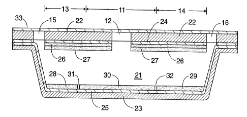
One example of a panel in accordance with this description is shown in FIGS. 1
and 2. The top view of FIG. 1 shows a central circular test area 11 with a
sample
introduction port 12 in the center of the test area. A positive control zone
13 is located
to one side of the test area, and a ~egativ~~ control zone 14 to the other. At
the extreme
outer ends of each of these controzones ire air vents 15, 16. The side cutaway
view of
FIG. 2 shows the chamber 21 formed between two structural sheets of solid
material 22,
23, which are formed of a transparent polymer, conveniently made from a single
sheet
folded over. The inner surfaces of each of the upper and lower walls of the
chamber are
coated with layers of adhesive 24, 25. The upper wall also contains a Layer of
indicator
26 and a layer of reagent 27. These two layers extend the full length and
width of the
CA 02202993 1997-04-17
WO 96/15255 PCT/US95/13790
chamber, surrounding the sample introduction port 12. On the bottom wall of
the
chamber, a reagent for the positive control 28 is in alignment with the
positive control
zone 13, and a reagent for the negative control 29 is in alignment with the
negative
control zone 14. A test reagent 30 as needed to perform the test for the
presence or
5 absence of the hydrolase or its inhibitor occupies the region in alignment
with the test
area 11. The three reagents on the bottom wall are separated by gaps 31, 32 to
prevent
mutual diffusion which might obscure the observed result. Although not shown,
gaps
.can also be present on the top wall. A peel-off protective sheet 33 covers
the top surface
of the panel.
10 2. Liquid-Penetrable Panel
In this construction, the panel itself is a free-standing, self supporting
card or
sheet of porous material, preferably flat and thin, and the reagents are
applied to opposite
sides of the panel. To use the panel, the sample is applied to the reagent on
one side and
permitted to diffuse through the panel to the reagent on the other. Penetrable
panels can
15 be fabricated in a variety of ways. For example, bibulous materials like
particles of
cellulose or other organic or inorganic, natural or synthetic materials can be
suspended in
a liquid medium, forming a thin film of the suspended materials by filtration.
When the
residual liquid is removed by evaporation, the product is a porous film which
can be
separated from the receptable in which it was formed and used as a free-
standing panel.
20 Penetrable panels can also be constructed of woven or non-woven polymer
fibers or
threads, with or without additional binders.
Microporous membranes can also be used as penetrable panels. Microporous
membranes are often fabricated by casting thin films of a polymer, like
nitrocellulose, in
a solvent mixture, and allowing differential evaporation of the solvent
mixture to create
defined pores or channels throughout the film. Pore size, number and density
can be
carefully controlled by proper formulation of the solvent system and, in
addition, by ,
controlling the evaporation temperature.
Penetrable panels can be made from a film-forming polymer by adding porous or
non-porous solids to a solution or suspension of the polymer during the
casting of a film.
Solids which can be used in this manner include, but are not limited to, the
following:
diatomaceous earth, Fuller's earth, Kaolin, microporous glass beads, fumed
silica,
molecular sieves and numerous other materials which allow penetration of a
liquid
sample through the film. In some cases, a penetrable film-forming polymer can
be made
by incorporating specimen-soluble additives including, for example, salts,
sugars,
polymers or, other materials which are soluble in both the sample and the
solvent
CA 02202993 1997-04-17
WO 96!15255 PCT/ITS951~3790
21
mixture used to cast the polymeric film. When the sample is added to the film
containing such additives, the soluble additive dissolves, thereby permitting
passage of
the sample through the film.
In addition, porous polymeric sheets can be fabricated from non-sample-
permeable films, sheets or supports by mechanically or chemically generating
holes or
channels in the otherwise solid films, materials or supports. Lasers or
nuclear particles,
for example, can be used to create precisely defined pores in suitable
polymeric sheets.
Small holes, pores or channels can be created in otherwise solid materials by
etching,
punching, drilling, boring, puncturing, perforating, cutting or abrading. One
can treat
the entire film, sheet or support in this manner or, alternatively, treatment
can be
.restricted to a small defined area where sample is to be applied.
In this type of test device, it may be desirable to prevent the reagents from
migrating in the lateral or radial direction, i. e. , parallel to the flat
surfaces of the device,
during the test. This can be achieved by a radial migration barrier. Such a
barrier can
be generated by depositing a solution or suspension of a sample-impermeable
material on
the upper and lower surfaces of the panel in a defined geometric or iconic
pattern and in
a manner which does not cover specific areas on the upper and lower surfaces
of the
panel. The solution or suspension is permitted to penetrate the thickness of
the panel,
and the solvent is removed by evaporation. To deposit the migration barrier
solution or
suspension in identical aligned patterned regions on both surfaces of the
panel, it is only
necessary that each application penetrate slightly more than half the
thickness of the
panel. In this manner, the solutions or suspensions will penetrate the
complete thickness
of the panel, rendering it impermeable except in the areas specifically left
untreated.
Due to the thinness of the panel, vertical penetration by the migration
barrier suspension
:?5 or solution is rapid, and lateral migration of such solution or suspension
into untreated
test zones is minimized, thereby allowing the geometric length and width of
the untreated
sample application and test interpretation zones to be precisely defined. The
radial
migration barrier leaves a vertical diffusion channel with well-defined
dimensions. This
channel minimizes the quantity of sample needed to produce a test result. The
channel
can also be used to achieve an iconic or geometric readout of the test result.
Radial migration barriers can be prepared from various materials and applied
in
- various ways. For water-based samples, a water-insoluble barner is
preferred.
Examples are organic solvent solutions or suspensions of paraffins, waxes, and
oils,
water-insoluble cellulose derivatives, polyacrylate polyester, and polyamide
derivatives,
~5 water-insoluble adhesives, radiation curable polymeric compositions, and
numerous other
water-insoluble materials. A barrier of paraffin, for example, may be applied
as a
solution in toluene. A barrier of ethylcellulose may be applied as a solution
in alcohol.
Water-insoluble hot melt adhesives can also be used.
CA 02202993 1997-04-17
WO 96115255 PCT/LTS95I13790
22
A solution or suspension of the barrier material can be applied onto each
surface
of the panel by printing, spraying, brush application, or any other kind of
deposition.
With an appropriate selection of solvent systems and control of the amount
applied and
of the temperature and air flow across the panel, the barrier material applied
to both
surfaces of the panel will penetrate the panel. The solvent can then be
evaporated to
result in barrier of defined shape enclosing a porous vertical channel whose
lateral
dimensions reflect the contours of the applied barrier. With the length, width
and shape
of the vertical channel thus defined, detectable changes in the indicator can
be confined
to a defined, geometric pattern.
The solution or suspension of barrier material can further include pigments,
either
dissolved or suspended, which permit visualization of the barrier, as well as
differentiation among the sample application areas) and the test and control
interpretation
zones. .The migration barrier can also be formulated to permit further
printing of
instructions and other indicia on either or both surfaces of the support.
With a visible migration barrier in a geometric pattern, the reagent and
indicator
can be applied on the upper and lower surfaces of the support in complementary
patterns
defined by the barrier pattern. If the reagent is sample-soluble, the sample
when applied
to the reagent side of the support will dissolve the reagent and diffuse
through the
channel to the indicator. The reporter group carried by the sample then causes
a
detectable change in the indicator if the analyze of interest is present in
the sample. The
migration barrier directs essentially all of the sample and reagent toward the
indicator,
and consequently, the test result is readily detectable in a manner permitting
geometric or
iconic readout.
FIGS. 3a, 3b and 4 illustrate an example of a panel of the type described in
this
section. The top or front view as shown in FIG. 3a shows the panel 41 which is
a
porous sheet, with an oval-shaped sample application area 42 indicated by
means of
indicia on its upper surface. The bottom or back view of FIG. 3b shows the
outline of
the lateral sample migration barrier 44 and printed outlines of the test zone
45, the
positive control area 46, and the negative control area 47. The sample
migration barrier
44 extends to all regions except an outer periphery and the test zone and
control areas.
The exploded view of FIG. 4 shows the porous sheet 41 and the shapes of the
upper and lower outlines 43, 44 of the lateral sample migration barner.
Although only
the outlines are shown, the barrier extends through the full thickness of the
porous sheet.
The outlines indicate where the barrier material was applied to each surface
to penetrate
the sheet. Each outline surrounds three untreated zones 48, 49, 50 which form
individual channels in top-to-bottom alignment with the test and control areas
to permit
the sample to diffuse through the sheet only in these areas. On the top or
front surface
of the panel 41, in alignment with the three diffusion channels, are deposits
of a first
CA 02202993 1997-04-17
WO 96!15255 PCTlUS951I3790
23
reagent 51, 52, 53 for the assay. Above the deposit of the first reagent in
the positive
control area 46 is a deposit of a positive control reagent 54. Likewise, above
the deposit
of the first reagent in the negative control area 47 is a deposit of a
negative control
reagent 55. On the bottom or back surface of the panel, in alignment with the
three
diffusion channels, are deposits of the second reagent 55, 56, 57 for the
assay. A
protective cover layer 59 protects the upper surface reagent areas. Printed
outlines and
indicia 60, 61 guide the user in both applying the sample and reading the test
results.
For a test for proline iminopeptidase activity, for example, the first reagent
in
zones 51, 52, 53 may be L-prolyl-beta-naphthylamide, L-prolyl-beta-methoxy-
naphthylamide or hydroxy-L-prolyl-beta-naphthylamide, the second reagent in
zones 55,
56, 57 may be the visual indicator Fast Garnet, the positive control reagent
54 may be
proline iminopeptidase, and the negative control reagent 55 may be a copper
salt.
3. Panel Designed for Use With Swab
The third type of test device construction does not rely on forming a liquid
migration path for the reagents, either by filling a void space with the
liquid sample or
diffusing the sample through a penetrable solid material. Instead, a sample
applicator
which retains a substantial quantity of the sample is used as the transport
medium.
. The reagents, which in the case of the specific examples discussed above are
the
conjugate and the indicator, are deposited in separated regions on a common
surface so
that the swab or other sample-absorptive device, which is wet with the sample,
can be
drawn across the surface to contact first one and then the other region. Two
or more
different indicators in discrete regions can be used rather than just one, to
permit the
simultaneous performance of multiple tests, by drawing the swab across each of
the
indicator regions subsequent to its contact with the conjugate region.
The reagents may be applied by any of the various different methods discussed
above, and may be either immobilized in their respective regions or simply
deposited. In
the immobilization case, the reagents will remain in their respective regions
throughout
contact with the sample-laden applicator, except for fragments cleaved from
the reagents
by one of the assay reactions, such as reporter groups liberated by the action
of a
hydrolase. In the nonimmobilized case, the first-contacted reagent may be
soluble in the
sample, and will travel with the sample to the region of the second-contacted
reagent,
although the presence (or where appropriate, the absence) of the analyte will
still be
required to achieve a detectable change in the indicator.
The regions can be arranged in geometric patterns for a variety of reasons.
The
patterns may for example serve as a guide to the user to indicate the
direction along
CA 02202993 1997-04-17
WO 96/15255 PCT/US95/13790
24
which the applicator is to be drawn. Various other effects to be achieved by
the
geometric pattern will be readily apparent to those skilled in the art.
An illustration of a panel of the type described in this section appears in
FIGS. 5,
6a and 6b. The top view of FIG. S shows the panel 71 with two sample
application
zones -- a test zone 72 and a control zone 73, and indicia indicating how the
zones will
appear for a positive test result 74 and for a negative test result 75. The
control zone 73
includes a reagent which will give the same appearance when the sample is
applied,
regardless of the whether the sample is positive or negative for the analyte,
as long as the
indicator is functioning. The test zone 72 shows a visible plus sign when the
test result
is positive and remains blank when the test result is negative.
The components of the test zone 72 are shown as individual layers in FIG. 6a
in
the order in which they are applied. Using a test for bacterial vaginosis as
an example,
the bottom layer 76 is the visual indicator Fast Garnet; the layer 77 applied
over the
indicator is a wetting layer; the layer 78 above the wetting layer is a mask
layer which
with a hole shaped like a plus-shaped area 79 exposing the underlying
indicator for view;
and the upper layer 80 is one of the three conjugates.
The components of the control zone 73 as shown in FIG. 6b, again for bacterial
vaginosis, are the visual indicator Fast Garnet as the bottom layer 83; the
wetting layer
84 applied above the indicator; the mask layer 85 applied above the wetting
layer, with a
hole 86 in the center; and the control reagent 3-amino naphthoate 87 as the
top layer.
To perform the test, a swab 88 (FIG. 5) saturated with sample is applied to
each
of the two zones. In each zone, the swab is first rubbed over the zone
perimeter,
preferably by a circular motion, and then rubbed over the central area of the
zone. The
appearance of the two zones can then be compared to the indicia to determine
whether
the test result is positive or negative.
4. General Considerations Pertaining to All Test Devices
In each type of test device, the reagents contained in, or deposited on, the
device
are preferably selected such that all that is needed to complete the test is
the addition of
the sample plus a minimal number of additional reagents such as, for example,
a
developer. In particularly preferred embodiments, however, the device will
contain all
reagents needed other than the sample, so that performance of the test
requires nothing
more than addition of the sample.
The indicator is preferably a composition which is substantially insoluble in
the
liquid sample for which the test is designed, so that the indicator remains
stationary
throughout the test. For test samples in either aqueous or water-soluble
media,
CA 02202993 1997-04-17
WO 96/15255 25 PCTlUS95/13790
therefore, the indicator preferably forms a layer (continuous or
discontinuous) which is
substantially insoluble in water, or the indicator is incorporated in a matrix
which is
substantially insoluble in water. In the first of the three devices described
above, the
indicator is retained in a thin concentrated lamina directly underneath a
light-transmitting
wall, and a change in the indicator is detectable through the light-
transmitting wall in a
short period of time, thereby providing both high sensitivity and a fast
result.
To improve access of the free reporter group to a water-insoluble indicator,
the
indicator composition can also include penetration enhancers such as, for
example,
detergents, salts, sugars, sugar alcohols, polyethylene glycols and other
polymers or
compounds which are soluble in both water and organic solvents. These
penetration
enhancers can be incorporated into the indicator lamina in a manner which
facilitates
access of the reporter group to the indicator, while still maintaining the
indicator itself
insoluble. As mentioned above; alcohol-soluble manganese salts are preferred
penetration enhancers in certain embodiments of the invention.
In addition to. the components and structural features described above,
preferred
test devices of this invention include a built-in positive control, a built-in
negative
control, or both. In certain constructions, these controls can be arranged to
be activated
at the same time that the assay itself is performed, all by a single
application of the
specimen. Detectable indications such as, for example, color changes or the
lack
thereof, which represent both the controls and the test are all detectable. In
the chamber
construction, for example, the controls and the test indicator can be arranged
such that
all color-change indications are detectable through a light-transmitting wall.
The controls can occupy positions on the device which are horizontally
adjacent to
the test area. In the case of the chamber device construction, the controls
are preferably
horizontally adjacent to the sample application port. Regardless of the
construction of
the device and the location of the controls, however, the device can contain
appropriate
indicia to identify the control indicator areas and differentiate them from
the test
indicator areas. Alternatively, the differentiation can be made obvious by the
overall
geometric design of the test.
The controls themselves may consist of individual layers or deposits on the
solid
support, the layers or deposits containing reagents or other appropriate
species which will
either induce the detectable change in the indicator by themselves or prevent
the change
from occurnng, and will do so only when the test sample is present and yet
independently of the presence or absence of the suspect hydrolase or hydrolase
inhibitor
in the test sample. The optimal choice of reagents to be used as controls and
the optimal
locations of these controls on or in the device will vary from one test to the
next.
Further preferred embodiments of the test devices of the invention contain
additional features to enhance the performance of the test. A surface-active
agent for
CA 02202993 1997-04-17
WO 96/15255 PCT/LTS95/13790
26
example may be applied along the sample contact surface or surfaces of the
device. The
surface-active agent may be included as a dry solute in a support matrix which
forms the
lamina immediately underneath the surface, or a coating applied over the
surface and
beneath any reagents present on the surface. In some cases, the lamina will
also contain
one or more reagents taking part in the test reactions. In other cases, the
surface-active
agent will be the sole functional component of the lamina.
Surface-active agents will be useful for specimens which are water-based, as
most
biological specimens are. Suitable surface-active agents can be solids, semi-
solid waxes,
or liquids absorbed into solids, and a wide variety of substances which have a
surface-
active effect may be used. The substances will generally be detergents,
wetting agents or
emulsifiers, and will vary widely in chemical structure and electronic
character,
including anionic, cationic, zwitterionic and nonionic substances. Examples
are alkyl .
alkoxy sulfates, alkyl aryl sulfonates, glycerol fatty acid esters, lanolin-
based derivatives,
polyoxyethylene alkyl phenols, polyoxyethylene amines, polyoxyethylene fatty
acids and
esters, polyoxyethylene fatty alcohols and ethers, polyethylene glycol) fatty
acids and
esters, polyoxyethylene fatty esters and oils,
polyoxypropylene/polyoxyethylene
condensates and block polymers, sorbitan fatty acid esters, sulfo derivatives
of
succinates, alkyl glucosides, and cholic acid derivatives. Trade names of some
of the
products falling within these classes are Lubrol, Brij~, Tween~, Tergitol~,
Igepal~,
Triton~, and Teepol.
Test devices may also include additives which enhance stability and shelf life
and
which facilitate dissolution of the catalytically active hydrolase or other
components
which function best when in solution. Examples of such additives are
antioxidants
(BHT, BHA, ascorbate, dithiothreitol), metal binding components and chelators
(EDTA,
EGTA), other components which facilitate the dissolution of the various
components
(mannitol, sorbitol, polyethylene glycol, lactose), and buffers.
In test devices where the solid-phase reagents are applied as laminae, these
laminae may be formed by .applying the lamina material in liquid form followed
by
. drying or other solidification. The liquid form of the reagent may be, for
example, a
solution or suspension of the reagent, or an uncured liquid form of the lamina
matrix
precursor, and the solidification step may thus be an evaporation of the
solvent or
suspending liquid or a curing of the matrix precursor. Additional materials
may be
included in the lamina for a variety of purposes, such as for example:
(1) to facilitate the application of the liquid to the surface by modifying
the
viscosity of the liquid,
(2) to help form a continuous smooth solid layer which remains uniform and
does not disintegrate or granulate over time or upon the application of
additional
layers over it,
CA 02202993 1997-04-17
WO 96115255 PCTYUS95/~3790
27
(3) to modify the solubility of the layer with solvents used in layers to be
applied over it or to make the layer soluble in solvents which do not dissolve
layers applied underneath,
or all of these. Polymeric materials are preferred additives to serve one or
all of these
purposes. Examples are celluloses or polyacrylates and various derivatives
thereof, with
the substitutions appropriately selected to achieve the desired solubility
characteristics.
For those test devices designed for aqueous or other water-based samples, the
indicator
. lamina preferably contains the indicator retained in a matrix of solid
material which is
largely insoluble in water. This prevents the indicator from migrating out of
the lamina
and thereby dropping in concentration. Alternatively, an indicator which is
insoluble in
water and which will form a coherent lamina which will remain intact by itself
can be
used.
For those embodiments of the invention in which a positive control indicator,
a
negative control indicator or both are included in the device, one or more
additional
reagents will be included for each control. These additional reagents will
either be
incorporated within one of the existing laminae in a horizontally defined
portion of that
lamina or applied as a separate, vertically adjacent lamina over a
horizontally defined
portion of the existing lamina. By virtue of their position on or in the
device, these
additional reagents define control areas which are horizontally separated from
each other
and from the test area.
The selection of an appropriate reagent for a positive or negative control
will
depend on a variety of factors, including the analyte which the test is
designed to detect,
the availability of control materials, the type of indicator used to detect
the presence of
the analyte, and whether the reagent is intended to serve as a positive
control or a
negative control. By utilizing known chemistries, the selection of an
appropriate reagent
will in most cases be apparent to those skilled in the art.
A positive control for a hydrolase test, for example, may be a sample of the
hydrolase itself, a hydrolase with similar hydrolytic action, the reporter
group or one of
its analogues, or any other species with a parallel mode of action which
initiates or.
induces the reaction or reaction sequence which culininates in a detectable
change in the
indicator. While the preferred positive control for hydrolase tests on human
specimens is
a hydrolase isolated from human sources, human hydrolase is often not
available. A
similar hydrolase isolated from animal, plant or microbial sources can then be
employed.
When the positive control is a sample of the reporter group or an analog or
derivative of
the reporter group, the control reacts vqith the indicator to generate a
signal. This type
of positive control can be used to assure the performance of the indicator,
which is often
the most critical or the least stable test component of the system.
CA 02202993 1997-04-17
WO 96/15255 PCT/iTS95/13790
28
The negative control for a hydrolase test may be a denaturing, inhibiting or
otherwise hydrolase-inactivating agent which prevents or blocks the reaction
or reaction
sequence, and thereby prevents the detectable change from occurring regardless
of
whether or not the catalytically active hydrolase is present in the sample or
specimen.
Alternatively, the negative control reagent may preferentially react with the
free reporter
group or the indicator, thereby preventing any interaction between them which
would
generate a detectable signal. In test devices designed for hydrolases, the
preferred
negative control is a highly specific inhibitor of the hydrolase whose
presence is being
detected. If such an inhibitor is not available, a less specific hydrolase
inhibitor can be
used, one which inhibits a number of hydrolases, including the hydrolase whose
presence
is being sought. Inhibitors of this type include class-specific protease
inhibitors such as
for example thiol, serine, metallo- and aspartic protease inhibitors,
denaturants such as,
for example, detergents, inactivating metallo-compounds, and acids and bases.
Alternatively, the negative control can incorporate a component which
preferentially
reacts with the reporter group or with the indicator, thereby preventing their
interaction
to generate a signal. The choice of appropriate positive and negative control
elements
for use in the test devices of the present invention are based on factors
known to those of
skill in the art.
Positive and negative controls serve at least three important functions:
(a) First, a properly functioning positive control provides assurance that
all test elements (the conjugate, the indicator and the reaction conditions)
are performing correctly, and can be relied upon to detect a hydrolase in a
specimen, if the hydrolase is indeed present. Similarly, a properly
functioning negative control provides assurance that the test reagents can
be relied upon not to generate a positive result in the absence of the
hydrolase. As such, the positive and negative controls serve as reagent
controls.
(b) Second, a properly functioning positive control provides assurance
that the specimen being tested does not contain interferents which can
inhibit the hydrolase or, by other means, prevent it from generating a
positive test result. Similarly, a properly functioning negative control
provides assurance that the specimen being tested does not contain
interferents which can generate a positive signal in the absence of the
hydrolase. As such, the positive and negative controls serve as specimen
controls.
(c) Third, if a test device and its controls are designed and constructed
such that the control elements of the device are geometrically and
temporally the last portions of the test device to be contacted by the
CA 02202993 1997-04-17
WO 96115255 PCT/LTS95/13790
29
specimen, a properly functioning positive control provides an indication
that the test device has been filled, or properly wetted, with the specimen.
Thus, in this case, the positive control serves as a procedural control.
This consideration is particularly important with clear or colorless
specimens (such as, for example, vaginal fluid or saliva), in devices
designed to contain small volumes and in devices in which specimen flow
paths are partially obstructed from view.
Analogous functions for analytes other than hydrolases are similarly served by
the
inclusion of positive and negative controls.
Preferably, the test device is designed such that both controls are activated
when
the specimen is applied to the test device for the test itself. In some cases,
this is
achieved most effectively by placing the control reagents and the test
reagents on the
same surface of the device. In devices which have a chamber construction,
described
above as the first of the three types of device, the control areas may for
example be
arranged as extensions of the test area or sample application port, all
contained in the
same chamber with unobstructed fluid communication between the various areas.
When
both positive and negative control areas are included, the control areas can
be separated
from each other by the test area or by the sample application port which is
positioned in
between the two. All areas are then filled with sample by a single sample
application.
When the upper wall of the chamber is of a light-transmitting material, the
detectable
changes, or absence thereof, are readily detectable, and the identification of
areas as
positive and negative controls is conveniently achieved by placing appropriate
indicia on
the outer surface of the device. In some cases, effective results can also be
achieved
when the control reagents are placed in laminae on the chamber surface
opposite that
which bears the other reagent(s), such that the control reagent and the
remaining
reagents) are separated by the air gap.
In devices of the invention in general, the control areas of the device will
contain
all components and reagents used in the test area with the addition of the
control
reagents, either incorporated in horizontally delineated sections of one or
more of the
same laminae used in the test area or applied as separate laminae over such
horizontally
delineated sections. To achieve sharp boundaries for the control areas and to
prevent the
control reagents from activating or deactivating the test area, it is often
beneficial to
place discontinuities in the laminae at the boundaries separating the control
areas from
the test area to minimize or eliminate the possibility of lateral diffusion of
the control
reagents out of their respective control areas.
Test devices of the present invention are preferably flat and thin and of a
size
which are easily held by hand. For those test devices which include a chamber,
the
chamber depth is not critical to the invention and may vary. In most cases,
however, a
CA 02202993 1997-04-17
WO 96/15255 PCT/LTS95/13790
chamber ranging from about 3 mil to about 50 mil (0.003-0.050 inch; 0.0076-
0.127 cm)
in depth, preferably from about 5 mil to about 15 mil (0.005-0Ø015 inch;
0.0127-
0.0381 cm), will give the best results. For any given depth, the lateral
dimensions of the
chamber (i. e. , the spacing between its side walls) will define the size of
the sample
5 which the device will accommodate, and are otherwise unimportant except to
define the
size and shape of the visible test area on the outer surface of the device.
The lateral
dimensions should thus provide a test area which is large enough to be seen,
and yet
small enough that the chamber which will be completely filled by a specimen of
reasonable size. The specimen size will vary with the type of specimen and its
source
10 and method of sampling. In typical structures, it is contemplated that the
lateral area of
the chamber will range from about 0.1 cm2 to about 10 cm2, or preferably from
about
0.3 cm2 to about 3 cmz. The internal volume of the chamber in typical
structures will
likewise vary, and for most types of samples, volumes ranging from about 3~,L
to about
300uL will be the most appropriate and convenient. ,
15 ~ In all types of test devices disclosed herein, cover sheets serving as
barriers to
air, moisture, light or all three may be included to provide the device with
desired
rigidity and to enhance long-term storage properties. In devices in which the
support
panel itself is porous, for example, the panel containing the reagent laminae
and indicator
laminae can be laminated or otherwise sealed between two non-porous cover
sheets of
20 plastic, aluminum foil, waterproofed cardboard or other comparatively rigid
materials to
provide structural strength as needed. These rigid cover sheets can be light-
transmitting
or opaque, as desired. If the sheet is opaque, holes can be cut in the
material to permit
either sample addition, test evaluation, or both. For light-transmitting cover
sheets, a
sample entry hole can be made in the sheet for sample application, and test
interpretation
25 can be made directly through the same sheet, preferably through a clear,
transparent zone
on the sheet. Indicia can be printed or labeled onto cover sheets in a manner
which
provides the user with instructions for using the device. Cover sheets can
also be
peel-off laminae of aluminum foil, metallized Mylar~ or other material to
prevent or
minimize exposure of reactive components in the device to light, air, and
moisture.
30 The test device of the present invention is highly versatile and can be
used for a
wide range of assays and chemical reactions, depending on the particular
analyte whose
presence or absence is sought to be determined. The reagents occupying the
laminae in
the test device may thus be enzymes, co-factors, enzyme substrates such as
cleavable
conjugates, proteins and smaller organic molecules, and organic reagents in
general.
Likewise, the reaction which the analyte initiates in, or undergoes with, the
reagent may
be an enzymatic or non-enzymatic reaction such as a hydrolysis or other type
of
cleavage, an oxidation reaction, a reduction reaction, or any of a wide array
of other
types of reactions. The analyte may be any species whose presence, absence or
level is
CA 02202993 1997-04-17
WO 96115255 PCT/i1S95/13790
31
indicative of a condition. Analytes may thus be organic compounds, inorganic
compounds, enzymes, cofactors, proteins of various kinds, viruses,
microorganisms, and
any other species which might be present in a sample.
The following examples are offered by way of illustration only.
CA 02202993 1997-04-17
WO 96/15255 PCT/US95/13790
32
EXAMPLES
Materials used in the experiments were prepared as follows.
A. PREPARATION OF DIAZONIUM DYE INDICATOR LAMINAE
1. MATERIALS
a. Fast Garnet GBC, Fast Red B, Fast Black K, Fast Dark Blue, Fast
Bordeaux, Fast Violet or other diazonium dye.
b. 1.7 M MnCl2 in ethanol.
c. 10% (wt/wt ethylcellulose in ethanol).
d. 10 % (wt/vol) Lubrol in water.
. e. # S, # 10, # 20 or # 30 Meyer Rods.
f. Mylar~ sheets either 5 mil, 10 mil, or 14 mil thick.
g. Mylar~ (5 or 7 mil):polyethylene (2 or 3 mil) laminated sheets.
2. PROCEDURES
The solid dyes were weighed into test tubes (10 - 100 mg) and 100 p,L MnCh
solution and 900 ~cL ethylcellulose solution were added to each tube. The
suspensions
were heated to dissolve the dyes, cooled, and centrifuged to remove insoluble
debris.
Supernatant dye solutions were formed into thin coatings on Mylar~ sheets or
on
Mylar~:polyethylene laminates with Meyer Rods. The coatings were air dried and
stored
at room temperature until used. Coating thickness was controlled by the Meyer
Rod
used (coating thickness increasing with Meyer Rod number).
B. PREPARATION OF CONJUGATE LAMINAE
1. MATERIALS
a. Conjugates:
(1) L-prolyl-beta-naphthylamide (PRO.NAM).
(2) L-glycyl-1.-prolyl-4-methoxynaphthylamide
(GLY-PRO. MNA) .
(3) N-benzyloxycarbonyl-L-prolyl-beta-naphthylamide
(Z-PRO. NAM).
(4) hydroxy-L-prolyl-beta-naphthylamide
(HYDROXYPRO.NAM).
(5) L-arginyl-beta-naphthylamide (ARG.NAM).
(6) N-benzyloxycarbonyl-L-arginyl-L-prolyl-beta-naphthylamide
(Z-ARG.PRO.NAM).
(7) Tert-butyloxycarbonyl-1.-valyl-L-leucyl. L-lysyl-7-amino-4-
methyl coumarin (T-boc.VAL.LEU.LYS. AMC).
b. Other materials:
CA 02202993 1997-04-17
WO 96/15255 PCTlUS951I3790
33
(1) 0.8 M NaOH.
(2) 10% (wt/vol) Lubrol in water.
(3) 7 % (wt/wt) hydroxypropylcellulose in ethanol.
(4) Mylar~ sheets either 5 mil, 10 mil, or 14 mil thick.
(5) Mylar~ (5 or 7 mil):polyethylene (2 or 3 mil) laminated
sheets.
2. PROCEDURES
Solid conjugate was suspended in a mixture of 100 microliters of solvent
(water,
ethanol, or dimethyl formamide), 800 microliters hydroxypropylcellulose
solution, and
, 50 microliters of sodium hydroxide solution. The suspension was heated to
completely
dissolve the conjugate, and the cooled solution was coated onto the solid
sheet as
described in PREPARATION A.
C. PREPARATION OF MANUALLY ASSEMBLED HYDROLASE TEST
DEVICES
1. MATERIALS
a. diazonium dye indicator laminae on 5 mil Mylar~ sheets
(PREPARATION A).
b. conjugate laminae on 5 mil Mylar~ sheets (PREPARATION B).
c. untreated 14 mil Mylar~ sheets.
~ d. double sided adhesive tape.
2. PROCEDURES
Double sided tape was affixed over the entire top and bottom surfaces of a 14
mil
thick solid Mylar~ sheet, and a 0.3125 inch in diameter hole was punched
through the
entire triple laminate (adhesive tape-Mylar~-adhesive tape). The 14 mil Mylar~
sheet
served as a spacer between a top sheet of 5 mil Mylar~ containing a diazonium
dye
indicator lamina, and a bottom 5 mil Mylar~ sheet containing a conjugate
lamina. After
being adhered to the 14 mil Mylar~ spacer sheet by means of the double sided
adhesive,
the top and bottom sheets served as top and bottom of a chamber. The conjugate
lamina
on Mylar~ was adhered to the bottom surface of the double sided, adhesive-
covered 14
mil Mylar~ sheet with the conjugate lamina facing upward. The walls of the
hole
punched in the 14 mil Mylar~ sheet and the bottom mylar sheet formed a small
receptacle, 14 mil deep and 0.3125 inches in diameter. The bottom surface the
receptacle was covered with a lamina containing a conjugate. A smaller
circular hole
(0.125 inches in diameter) was punched in the third Mylar~ sheet containing
the
diazonium dye indicator lamina, and this third Mylar~ sheet was adhered to the
top
surface of the 14 mil Mylar~ sheet covered with double sided tape in such a
fashion that
the diazonium dye indicator lamina formed the inner surface of a top of the
receptacle
CA 02202993 1997-04-17
WO 96!15255 PCT/US95/13790
34
described above, and also such that the small hole in the third sheet was
centered over
the receptacle described above. The resulting laminated panel functioned as a
test
device, containing a receptacle with a top and bottom. The inner surface of
the
receptacle top included a thin, largely insoluble indicator dye lamina
containing a visual
indicator in a water insoluble matrix, and the inner surface of the receptacle
bottom
included a thin lamina of water soluble conjugate. The receptacle was a void
space until
a liquid sample was added. When an aqueous sample was added to the device, the
water
soluble conjugate was solubilized, but the diazonium dye indicator lamina
remained intact
as a thin filin. If a hydrolase capable of hydrolyzing the conjugate was
present in the
sample, the conjugate was hydrolyzed to release free reporter group (4-methoxy
naphthylamine, beta-naphthylamine, or 7-amino-4-methyl coumarin), which
reacted with
the insolubilized diazonium dye indicator Lamina to form a color in a lamina
on the inner
top surface of the device.
D. PREPARATION OF PARADIMETHYLAMINOCINNAMALDEHYDE
(PDMAC) HIGH BUFFER CAPACITY CHROMOGEN SOLUTION
MATERIALS AND PROCEDURES. The following solutions were mixed
sequentially as indicated and the resulting solution stored at 4 degrees C
until needed.
a. 100 ~,L 25 mM para-dimethylaminocinnamaldehyde in ethanol.
b. 6.75 mL 1 M citric acid solution in water.
c. 2.25 mL 2 M disodium phosphate in water.
d. 900 ~,L 10 % (wt/wt) sodium dodecyl sulfate in water.
E. PREPARATION OF PARADIMETHYLAMINOCINNAMALDEHYDE
(PDMAC) CONCENTRATED ACID CHROMOGEN SOLUTION
MATERIALS AND PROCEDURES. The following materials were mixed
sequentially as indicated and the resulting solution stored at 4 degrees C
until needed.
a. 80 mL distilled water.
b. 5.6 mL glacial acetic acid.
c. 435 ~cL 5 M NaOH (to adjust pH to 4.5).
d. 13 mL distilled water.
e. dry sodium dodecyl sulfate to make a final concentration of 1.0
(wt/wt). '
f. 1 mL 50 mM para-dimethylaminocinnamaldehyde.
CA 02202993 1997-04-17
WO 96!15255 PCT/US95/13790
F. PREPARATION OF PARADIMETHYLAMINOCINNAMALDEHYDE
(PDMAC) DILUTE ACID CHROMOGEN SOLUTION
MATERIALS AND PROCEDURES. The following materials were mixed
sequentially as indicated and the resulting solution stored at 4 degrees C
until needed.
5 a. 30 mL distilled water.
b. 2.8 mL glacial acetic acid.
c. approximately 435 ~cL 5 M NaOH (to adjust pH to 3.0).
d. 10 mL (10% wt/wt) sodium dodecyl sulfate solution in water.
e. 1 mL 2S mM para-dimethylaminocinnamaldehyde in ethanol.
10 f. distilled water to make a total volume of 50 ml.
G. PREPARATION OF BACTERIAL CELL SUSPENSIONS
Pure cultures of Mobiluncus curtisii, ATCC strain 35241 and Mobiluncus
mullieris ATCC strain 35239 were grown anaerobically on sterile Brucella Blood
Agar in
petri dishes at 37°C for 4 - 8 days. The cells scraped from the surface
of 10 petri dishes
15 were suspended in 2.5 mLs 150 mM saline and frozen until used.
Pure cultures of Candida albicans, ATCC strain 28366 were grown in liquid
culture medium as indicated by Odds, F.C., J. Gen. Microbiol., 129:431-438
(1983).
The cells were suspended in 2.5 mL 150 mM saline and frozen until used.
EXPERIMENT I
:ZO This experiment tests the comparative ability of Fast Garnet GBC solutions
and
Fast Garnet GBC chromogenic indicator laminae to detect the reporter group, 2-
naphthylamine.
A. MATERIALS
1. 100 mM Tris buffer, pH 7Ø
25 2. 0.15 % (wt/vol) Fast Garnet GBC solution in water.
3. The reporter group 2-naphthylamine dissolved in water at the
concentrations shown.
4. Fast Garnet GBC indicator laminae (# 20 Meyer Rod, PREPARATION
A).
:30 B. PROCEDURES
1. Testing of Fast Garnet GBC indicator solutions
Twenty five ~L Tris buffer and 25 ~.L water were added to duplicate wells of
microtiter plates, followed by 50 IcL of the reporter group, 2-naphthylamine,
solutions at
CA 02202993 1997-04-17
WO 96/15255 PCT/US95/13790
36
the concentrations shown. Finally, 50 ~,L Fast Garnet GBC solution was added
to each
well, and the color formation noted after 5 minutes.
2. Testing of Fast Garnet GBC indicator laminae
Twenty five ~L Tris buffer and 25 ~.L water were added to duplicate wells of
microtiter plates, followed by 50 ~.L of the reporter group, 2-naphthylamine,
solutions at .
the concentrations shown. After mixing, 20 ~,L of the mixture was transferred
to a Fast
Garnet GBC indicator lamina, and the color formation noted after 5 minutes.
TABLE 1
COLOR SCORE: INTERPRETATION
- ' no red color formation
+/- possible red color formation
+ clearly visible red color
formation
+ + extremely intense red color
formation
C. RESULTS
1. Fast Garnet GBC Chromogenic Liquid Reagent
A 2-naphthylamine solution at 1.25 micrograms/ 100 microliters produced a
clearly detectable red color in the solution, but no color formed at lower
concentrations
of 2-naphthylamine.
2. Fast Garnet GBC chromogenic indicator laminae
A 2-naphthylamine solution at 0.3 micrograms/100 microliters produced a
clearly
detectable red color in the Fast Garnet GBC indicator laminae, but no color
formed at
lower concentrations of 2-naphthylamine.
CA 02202993 1997-04-17
WO 96115255 PCT/US95/13790
37
TABLE 2
ADDITIONS MICR OLITER ELLS
W
VOLUME (~.L) 1 2 3 4 5
Tris Buffer 25 25 25 25 25
Water 25 25 25 25 25
Naphthylamine 50 50 50 50 50
(ug Naphthylamine) 2.5 1.25 0.6 0.3 0.15
Fast Garnet 50 50 50 50 50
Fast Garnet Color + + - - -
Liquid Reagent + + - - -
Fast Garnet Color + + + + -
Solid Reagent + + + + -
D. INTERPRETATION
The Fast Garnet GBC chromogenic indicator laminae were at least four times as
1:5 sensitive to the reporter group, 2-naphthylamine, as the Fast Garnet GBC
liquid
chromogenic reagent used in published clinical studies. This increased
sensitivity can be
utilized in either of two ways: (I) it can be utilized to detect lower
concentrations of the
reporter group, 2-naphthylamine, hydrolytically released from a conjugate; or,
(2) it can
be used to decrease the incubation time required to detect hydrolase-catalyzed
release of
the reporter group, 2-naphthylamine, from a conjugate.
EXPERIMENT II
This experiment tests the comparative ability of a liquid Fast Garnet GBC
chromogenic indicator and a Fast Garnet GBC laminae-based chromogenic
indicator to
2o detect proline iminopeptidase activity in Mobiluncus curtisii ATCC strain
35241 cells.
A. MATERIALS
1. Buffers:
a. 100 mM Tris buffer, pH 7Ø
b. 100 mM sodium phosphate buffer, pH 7.0
2. 0.2 % (wt/vol) conjugate, e. g. , L-prolyl-beta-naphthylamide (PRO. NAM)
dissolved in water.
3. 0.15 % (wt/vol) Fast Garnet GBC solution in water.
CA 02202993 1997-04-17
WO 96/15255 PCT/US95/13790
38
4. Fast Garnet GBC chromogenic indicator laminae (# 20 Meyer Rod,
PREPARATION A).
5. Mobiluncus cunisii ATCC strain 35241 suspension (PREPARATION G).
B. PROCEDURES '
1. Testing of solution-based systems
Twenty five microliters of Tris buffer and twenty five ~cL conjugate solution
(i. e. ,
PRO. NAM) in water were added to duplicate wells of a microtiter plate,
followed by 50
~,L Mobiluncus curtisii ATCC strain 35241 cell suspension diluted as shown.
The
suspension was incubated at 25 °C for four hours, and 50 ~,L Fast
Garnet GBC solution
was added to each well. Color formation was noted after 5 minutes.
2. Testing of Fast Garnet GBC chromogenic indicator laminae
Twenty five microliters of phosphate buffer and twenty five ~,L conjugate
solution
(e. g. , PRO. NAM) in water were added to duplicate wells of a microtiter
plate, followed
by 50 uL Mobiluncus curtisii ATCC strain 35241 cell suspension diluted as
shown. The
suspension was incubated at 25 °C for four hours, and a 20 ~cL aliquot
was removed and
added to Fast Garnet GBC chromogenic indicator laminae. Color formation was
noted
after 5 minutes.
TABLE 3
COLOR SCORE: INTERPRETATION
- no red color formation
+/- possible red color formation
+ clearly visible red color
formation
+ + extremely intense red color
formation
ND not done
C. RESULTS
1. Fast Garnet GBC Liquid Reagent
The liquid Fast Garnet GBC chromogenic indicator produced a barely detectable
red color with the undiluted Mobiluncus curtisii ATCC strain 35241 cell
suspension. No
red color was seen when the cell suspension was diluted 1:2 with saline.
2. Fast Garnet GBC chromogenic indicator laminae ,
The Fast Garnet GBC chromogenic indicator laminae produced a clearly
detectable red color even when the Mobiluncus curtisii ATCC strain 35241
suspension
was diluted 1:4 with saline prior to incubation with the proline
iminopeptidase-
CA 02202993 1997-04-17
WO 96/15255 PCTIU995/~3790
39
hydrolyzable conjugate, PRO.NAM. No red color was seen when the cell
suspension
was diluted 1:8 with saline.
TABLE 4
OOMPONFNTS AMOUNT
PFR
W>ZL
Wl~3L NO.: 1 2 3 4 5 6 7 8 9 10
Tiis Bu$'a 25 ?5 25 ?5 25 ND ND ND ND ND
(~iL)
1~ (~cL) ND ND ND ND ND 25 25 25 25 25
Cells (~cL) 50 50 50 50 50 50 50 50 50 50
I
Cell D~ (~L) 0 2 4 8 16 0 2 4 8 16
C.~ju~ (~iL) 25 25 25 25 25 25 25 25 25 25
Fast C~et G)iC50 50 50 50 50 ND ND ND ND ND
(EcL)
h~a~ti~ (haus)4 4 4 4 4 4 4 4 4 4
INDICATOR TEST
RESULTS
(DUPIlCATES~
Fast C~ G)3C +/ - _ _ _ ~ ~ Np
Color
Lid Reaga'~t +~ _ _ _ _ Np ND ND ND ND
Fast Cruet ND ND ND ND ND + + + - -
Coh
- _
D . INTERPRETATION
Mobiluncus curtisii ATCC strain 35239 cells exhibit the ability to hydrolyze
the
proline iminopeptidase-hydrolyzable conjugate, PRO.NAM, releasing the reporter
group,
2-naphthylamine. The reporter group is able to produce a red color upon
interaction
with Fast Garnet GBC, either in solution, or as a dry lamina. The Fast Garnet
GBC
chromogenic indicator laminae are at least four times as sensitive to
Mobiluncus curtisii
ATCC strain 35241 proline iminopeptidase activity as the Fast Garnet GBC
liquid
l5 chromogenic reagent utilized in published clinical studies. This increased
sensitivity can
be utilized in either of two ways: (1) it can be utilized to detect lower
concentrations of
proline iminopeptidase activity; or, (2) it can be used to decrease the
incubation time
required to detect proline iminopeptidase activity.
EXPERIMENT III
This experiment tests the capacity of alternative dried diazonium dye laminae
to
detect the reporter group, beta-naphthylamine and to detect Mobiluncus
curtisii ATCC
strain 35241 cell-catalyzed hydrolysis of the proline iminopeptdase-
hydrolyzable
conjugate, L-proIyl-beta-naphthylamide, the conjugate also present as a dried
lamina.
CA 02202993 1997-04-17
WO 96/15255 PCT/US95/13790
A. MATERIALS
1. Fast Garnet GBC, Fast Red B, Fast Black K, Fast Dark Blue, Fast Violet,
and Fast Bordeaux indicator laminae prepared as described in
PREPARATION A ( # 20 Meyer rod).
5 2. pure Mobiluncus curtisii ATCC Strain 35241 culture grown as described in
PREPARATION G and suspended in 100 mM NaCI solution.
3. proline iminopeptidase-hydrolyzable conjugate, i. e. , the L-prolyl-beta-
naphthylamide, laminae prepared as described in PREPARATION B
(Number 20 Meyer Rod).
10 4. manually assembled prototypes using each of the diazonium dye indicator
laminae (item 1 above) and L-prolyl-beta-naphthylamide laminae (item 3)
as described in PREPARATION C.
5. 50 mM reporter group, beta-naphthylamine, in water
B. PROCEDURES
15 Chromogenic indicator laminae of Fast Garnet GBC, 15 mg; Fast Black K, 57
mg: Fast Dark Blue, Fast Bordeaux, Fast Violet, and Fast Red B, 21 mg prepared
as,
described in PREPARATION A and proline iminopeptidase-hydrolyzable conjugate,
L-
prolyl-beta-naphthylamide, (13 mg/ml) prepared as described in PREPARATION B
were
manually assembled into test devices (PREPARATION C).
20 1. Tests of device performance with the reporter group, beta-naphthylamine.
Approximately 40 microliters of 500 micromolar beta-naphthylamine solution was
added to the manually assembled kits. After 10 minutes, color formation was
noted.
2. Test kit performance with proline iminopeptidase activity in Mobiluncus
curtsii ATCC strain 35241 cells.
25 Approximately 40 microliters of the Mobiluncus curtisii ATCC strain 35241
suspension were added to each test kit. After 10 minutes at room temperature,
the
appearance of color was determined.
C. RESULTS
Strong color formation occurred when either a solution of the reporter group,
30 beta-naphthylamine, or a Mobiluncus curtisii ATCC strain 35241 suspension
was added
to the test devices employing dried lamina containing the proline
iminopeptidase-
hydrolyzable conjugate (PRO.NAM) and chromogenic indicator laminae of Fast
Garnet
GBC, Fast red B, Fast Black K, Fast Dark Blue and Fast Bordeaux. No color
formed
when a solution of the reporter group, beta-naphthylamine, or a suspension of
35 Mobiluncus curtisii ATCC strain 35241 cells were added to test devices
containing Fast
Violet indicator laminae. Water did not produce a color with any of the
devices tested.
CA 02202993 1997-04-17
WAD 96/15255 PCTIUS95/I3790
41
TABLE 5
beta- Mobiluncus
Dyes Water NaphthylamineCurtisii
Fast Garnet no colorred red
GBC
Fast Black no colorblack black
K
Fast Red B no colorpurple purple
Fast Dark no colorblue blue
Blue
Fast Violet no colorno color no color
Fast Bordeauxno colorred red
D . INTERPRETATION
1. Chromogenic indicator laminae containing Fast Garnet GBC, Fast Black
K, Fast Red B, Fast Dark Blue, and Fast Bordeaux can detect beta-naphthylamine
colorimetrically.
2. Mobiluncus curtisii ATCC strain 35241 cells contain proline
iminopeptidase activity which hydrolyzes the conjugate, L-prolyl-beta-
naphthylamide,
present in a dried lamina to release the reporter group, beta-naphthylamine.
3. Dry chromogenic indicator laminae containing appropriate diazonium dyes
detect the released reporter. group, 2-naphthylamine, thereby detecting
proline
iminopeptidase activity.
4. Test devices can be constructed using a dried diazonium dye chromogenic
indicator lamina and a dried L-prolyl-beta-naphthylamide conjugate lamina to
permit
detection of proline iminopeptidase activity upon addition of a liquid
specimen.
5. Not all diazonium indicators can be used as chromogenic indicators for the
reporter group, 2-naphthylamine (e.g., Fast violet). The selection of a
specific
2;5 chromogen will depend on many factors known to those skilled in the art,
including
stability, test color intensity with a reporter group, color of specimen, and
cost.
EXPERIMENT IV
This experiment tests alternative conjugates for hydrolysis by microbial cells
(Mobiluncus cunisii ATCC strain 35241, Mobiluncus mulieris ATCC.strain 35239,
Candida albicans ATCC strain 28366) and enzymes.
CA 02202993 1997-04-17
WO 96/15255 PCT/US95/13790
42
A. MATERIALS
1. Fast Garnet GBC chromogenic indicator laminae prepared as described in
PREPARATION A (# 20 Meyer rod).
2. organisms:
a. pure Mabiluncus curtisii ATCC Strain 35241 culture grown as
described in PREPARATION G and suspended in 100 mM NaCI
solution.
b. pure Mobiluncus mullieris ATCC Strain 35239 culture grown as
described in PREPARATION G and suspended in 100 mM NaCI
solution.
c. pure Candida albicans ATCC Strain 28366 culture grown as
described in PREPARATION G and suspended in 100 mM NaCI
solution.
3. enzymes:
a. proline iminopeptidase from Bacillus coagulans (Sigma).
b. Pronase (2 commercial sources)
4. conjugates:
a. 10 mM L-prolyl-beta-naphthylamide (PRO.NAM).
b. 10 mM L-glycyl.L-prolyl-4-methoxynaphthylamide (GLY.PRO-
MNA).
c. 10 mM N-benzyloxycarbonyl-L-prolyl-beta-naphthylamide (Z-
PRO.NAM).
d. 10 mM hydroxy-L-prolyl-beta-naphthylamide
(HYDROXYPRO. NAM) .
e. 10 mM L-arginyl-beta-naphthylamide (ARG.NAM).
f. 10 mM N-benzyloxycarbonyl-L-arginyl.L-prolyl-beta-naphthylamide
(Z-ARG.PRO. NAM).
g. T-butyloxycarbonyl-L-valyl.z-leucyLL-lysyi-7-amino-4-methyl
coumarin (T-boc.VAL.LEU.LYS.AMC).
5. buffers:
a. 100 mM tris buffer, pH 8Ø
b. 500 mM 2-(N-morpholino) ethanesulfonic acid (MES) buffer, pH
5.0 '
c. 200 mM acetate buffers, pH 3.0, 3.5, 4.0, 5.0, and 5.5.
6. para-dimethylaminocinnamaldehyde indicators (PREPARATIONS D, E,
and F).
CA 02202993 1997-04-17
WO 96!15255 PCT/ITS95/13790
43
B. PROCEDURES '
1. Devices containing Fast Garnet GBC chromogenic indicator laminae
uL aliquots of the indicated conjugate solution (10 mM) was added to 10 ~.L
buffer at the pH shown. 20 ~.L ~of the indicated cell suspension or enzyme
solution
5 (proline iminopeptidase from ~. coagulans or pronase from two separate
vendors) was
added, and the suspension agitated to mix the contents. After a 10 minute
incubation at
room temperature, a 10 ~cL aliquot was added to a Fast Garnet GBC chromogenic
indicator lamina. Red color formation was observed at 5 minutes.
2. Tests with PDMAC chromogen in solution
10 50 1cL distilled water, 25 ~.L buffer, and 25 1cL microbial suspension were
mixed.
5 1cL conjugate was added, and the mixture allowed to incubate for 10 minutes
at room
temperature. 100 ~,L PDMAC was added to each incubation, and the color score
noted.
TABLE 6
COLOR SCORE: INTERPRETATION
i
1S _ no red color formation
0
0.25 Faintest red color detectable
visually
0.5 Distinct red color
1.0 Red color between 1.0 and 2.0
in intensity
2.0 Darkest red color possible in
test system
nd not done
CA 02202993 1997-04-17
WO 96/15255 PCT/L1S95/13790
44
C. RESULTS
TABLE 7
M. MULLIERISM. CURTISII M. CURTISIIC. ALBICAIVS
FAST GARNET FAST GARNET PDMAC FAST GARNET
COLORSCORE COLORSCORE COLOR COLORSCORE
CONJUGATES (pH) (pH) (PREP., (pH)
pH)
PRO.NAM 1 (pH 8.0) 2 (pH 5.0); 2 (D,E, 0.50 (pH
5.0,
i (pH 8.0) pH 4.5-8.0)8.0)
PRO.METHOXY. ND 0 (pH 3.0 0 (E, pH ND
- 4.5) 3.0-
NAM
7.0)
GLY.PRO.MNA 0 (pH 8.0) 2 (pH 8.0) ND 0 (pH 8.0)
Z-PRO.NAM ND 0 (pH 8.0) 0 (E, pH ND
5.0-
8.0)
HYDROXYPRO. ND 2 (pH 5.0) ND 0 (pH 5.0)
1 O NAM
ARG.NAM ND 1 (pH 8.0); 2 (D, pH ND
8)
0 (pH 5)
Z-ARG.PRO.NAM ND 2 2 (D, pH ND
8)
T-boc. VAL. ND ND 2 ND
LEU.LYS.AMC
TABLE 8
PRONASE PRONASE PROLINE
(VENDOR 1) (VENDOR 2) IMINOPEPTIDASE
FAST GARNET FAST GARNET FAST GARNET
COLOR SCORE GBC (pH) COLORCOLOR SCORE
CONJUGATES (pH) SCORE (pH)
PRO.NAM 1.5 (pH 8.0) 2 (pH 8.0) 2 (pH 8.0)
PRO.METHOXY.NAM ND ND 2 (pH 8.0)
2 (pH 5.0)
GLY.PRO.MNA 0 (pH 8.0) 0 (pH 8.0) 0 (pH 8.0)
Z-PRO. NAM ND ND ND
HYDROXYPRO.NAM ND ND 2 (pH 8.0)
2 (pH 5.0)
ARG.NAM ND ND ND
Z.ARG.PRO.NAM ND ND ND
T-boc. ND ND ND
VAL.LEU.LYS.AMC
CA 02202993 1997-04-17
WO 96!15255 PCTlUS95113790
D. INTERPRETATION
The Mobiluncus curtisii ATCC strain 35241 cells tested contained one or more
cell-bound enzymes capable of hydrolyzing the following conjugates: L-prolyl-
beta-
naphthylamide; L-glycyl-L-prolyl-4-methoxy-naphthylamide; L-hydroxyprolyl-beta-
5 naphthylamide; L-arginyl-beta-naphthylamide; Z-L-arginyl.L-prolyl-beta-
naphthylamide;
and, T-boc-L-valyl.L-leucyl.L-lysyl-7-amino-4-methyl coumarin. Reporter groups
were
released upon enzymatic hydrolysis of these conjugates, and were detected
either in a
dried chromogenic indicator lamina containing Fast Garnet GBC or with a liquid
system
containing PDMAC. The Mobiluncus curtisii ATCC strain 35241 cells tested
lacked the
10 enzymes needed to hydrolyze the conjugates L-prolyl-4-methoxynaphthylamide
or Z-L-
prolyl-beta-naphthylamide. With these conjugates, free reporter groups were
not
released, and no color formed either in the chromogenic indicator laminae or
in the
chromogenic liquid reagent solutions.
Like Mobiluncus curtisii ATCC strain 35241, the Mobiluncus mullieris ATCC
15 strain 35239 cells contained an enzyme capable of hydrolyzing the
conjugate, L-prolyl-
beta-naphthylamide, and thereby releasing the reporter group, beta-
naphthylamide, which
was detectable with Fast Garnet GBC indicator laminae. Unlike Mobiluncus
curtisii
ATCC strain 35241, Mobiluncus mullieris ATCC strain 35239 cells lacked the
enzymatic
capacity needed to hydrolyze the conjugate, L-glycyl-L-prolyl-4-
methoxynaphthylamide,
20 at pH 8Ø If desired, this differential hydrolytic capacity could be used
to make tests
which would detect either both species of Mobiluncus (i. e. , PRO. NAM), or to
distinguish between them (i.e., GLY.PRO.NAM).
The Candida albicans ATCC strain 28366 cells tested hydrolyzed the conjugate,
L-prolyl-beta-naphthylamide. Hydrolysis of L-prolyl-beta-naphthylamide by C.
albicans
25 ATCC strain 28366 cells was considerably slower than that by Mobiluncus
curtisii ATCC
strain 35241 and Mobiluncus mullieris ATCC strain 35237 cells. Unlike
Mobiluncus
curtisii ATCC strain 35241 cells, C. albicans ATCC strain 28366 cells lacked
the
capacity to hydrolyze the conjugates, hydroxy-L-prolyl-beta-naphthylamide and
L-glycyl-
L-prolyl-4-medzoxynaphthylamide. This differential enzymatic capacity could be
used to
30 make tests employing selective conjugates (e. g. , L-prolyl-beta-
naphthylamide), which
would either detect both species of Mobiluncus, or selectively detect
Mobiluncus curtisii
ATCC strain 35241 cells in the presence of Candida albicans ATCC strain 28366
and
Mobiluncus mullieris ATCC strain 35239 cells (L-gly.L-pro-4-
methoxynaphthylamide).
Purified pronase preparations from two separate vendors and purified proline
35 iminopeptidase from Bacillus coagulans hydrolyzed the conjugates L-prolyl-
beta-
naphthylamide, thereby releasing the free reporter group, 2-naphthylamine
which reacted
with Fast Garnet GBC indicator laminae to produce a color. The purified B.
coagulans
proline iminopeptidase also released 4-methoxy naphthylamide from L-prolyl-
beta-
CA 02202993 1997-04-17
WO 96/15255 PCT/US95/13790
46
methoxynaphthylamide and beta-naphthylamine from hydroxy-L-prolyl-beta-
naphthyl-
amide, thereby producing a red color. Therefore, all of these enzymes could be
used as
positive enzyme controls for proline iminopeptidase activity tests where L-
prolyl-beta-
naphthylamide is the conjugate, and Fast Garnet GBC is the indicator. However,
only
the B. coagulans enzyme could be used as a positive control in test methods
which
employed 1.-prolyl-4.-methoxynaphthylamide or hydroxy-L-prolyl-4-
methoxynaphthylamide
as conjugates. Also note that purified prolineaminopeptidase from B. coagulans
hydrolyzes PRO.METHOXY.NAM but the M. curtisii cell do not. Hence to the
extent
that this B. coagulans enzyme represents a bacterial proline iminopeptidase,
it differs
from the Mobilincus enzyme or enzymes. This suggests that the purified B.
coagulans
prolineaminopeptidase enzyme may be one which only exhibits
prolineaminopeptidase
activity .
EXPERIMENT V
This experiment tested vaginal fluid specimens from normal, uninfected women,
and women with vulvovaginal candidiasis for proline iminopeptidase activity
using dried
Fast Garnet GBC chromogenic indicator laminae, and L-prolyl-beta-naphthylamide
solution as the conjugate.
A. MATERIALS:
1. Fast Garnet GBC chromogenic indicator laminae prepared as described in
PREPARATION A (# 20 Meyer rod).
2. standard dacron clinical swabs containing undiluted vaginal fluid samples
from women diagnosed as follows:
a. normal (i. e. , not having any form of infectious vaginitis); or,
b. infected with vulvovaginal candidiasis, but not bacterial vaginosis.
The swabs were frozen at -70°C until tested.
3. conjugate solution, 10 mM L-prolyl-beta-naphthylamide (PRO.NAM), in
water.
B. PROCEDURES
The swabs containing vaginal fluid were thawed and the fluid was removed from
each clinical swab by centrifugation. 20 ~.L aliquots of vaginal fluid from
each specimen
was added to test tubes containing 5 ~,L PRO.NAM. The mixture was allowed to
incubate for 10 minutes at room temperature, and a 20 ~,L aliquot was removed
from
each incubation mixture and added to a Fast Garnet GBC indicator lamina. After
10
minutes, color formation was noted on the Fast Garnet GBC indicator laminae.
CA 02202993 1997-04-17
WO 96/15255 PCT/US95113790
47
TABLE 9
( COLOR SCORE: INTERPRETATION
0 no red color formation
0.25 faintest red color detectable
visually
~i 0.5 distinct red color
1.0 red color between 1.0 and 2.0
in intensity
2.0 darkest red color possible in
test system
C. RESULTS
No color formation was generated in the Fast Garnet GBC chromogenic indicator
laminae by any of the vaginal fluid specimens from either patient population.
TABLE 10
SAMPLE 10 mM
SPECIMEN CLINICAL VOLUME PRO.NAM COLOR
NUMBER DIAGNOSIS (~cL) VOLUME (~,L) SCORE
1 normal 20 5 0
2 normal 20 5 0
3 normal 20 5 0
4 candidiasis20 5 0
5 candidiasis20 5 0
6 candidiasis20 5 0
7 candidiasis20 ~ 5 0
8 candidiasis20 5 0
D . INTERPRETATION
Vaginal fluid from normal women lacked sufficient proline iminopeptidase
activity
under the conditions of the assay to hydrolyze the conjugate, PRO.NAM, and
failed to
release the reporter group, beta-naphthylamine. Since no beta-naphthylamine
was
released, no color formation occurred in the Fast Garnet GBC chromogenic
indicator
laminae. The same can be said of vaginal fluid from women with clinically
diagnosed
symptomatic vulvovaginal candidiasis. Based on these results, vaginal fluid
from normal
women, or women with symptomatic vulvovaginal candidiasis alone would not be
CA 02202993 1997-04-17
WO 96/15255 PCT/L1S95/13790
48
expected to produce a positive test result in a positive test for proline
iminopeptidase
activity under the reaction conditions defined above.
EXPERIMENT VI
This experiment tests the ability of proline iminopeptidase activity test
devices
employing dried laminae containing z-prolyl-beta-naphthylamide as the
hydrolyzable
conjugate for proline iminopeptidase activity and Fast Garnet GBC chromogenic
indicator '
laminae to detect proline iminopeptidase activity in vaginal fluid specimens
from
uninfected women, women with Bacterial Vaginosis and women with other forms of
infectious vaginitis.
A. MATERIALS
1. 266 undiluted vaginal fluid samples:
a. 39 samples from women diagnostically uninfected women
b. 32 samples from women with diagnostically proven bacterial
vaginosis, but no other form of infectious vaginitis
c. 28 specimens from women with diagnostically proven bacterial
vaginosis and at least one additional vaginal infection
d. 167 specimens women with diagnostically proven vaginal or
cervical infections, but not including bacterial vaginosis.
The specimens were obtained on dacron clinical swabs and frozen and stored at -
70°C until tested. For testing, the swabs were thawed and centrifuged
to extract the
vaginal fluid. The swabs were discarded, the undiluted vaginal fluid was
immediately
resuspended and used in the devices as outlined below.
2. proline iminopeptidase test devices made as described in PREPARATION
C.
B. PROCEDURES
Vaginal fluid specimens removed from the clinical swabs were vortexed to
resuspend any particulate matter in the undiluted supernatant vaginal fluid,
and was
added to the test devices with a volumetric micropipette. After 5 minutes at
room
temperature, the devices were examined for the formation of a red color.
CA 02202993 1997-04-17
WO 96!15255 PCT/US95/I3790
49
TABLE 11
COLOR SCORE: INTERPRETATION
0 no red color formation
- 0.25 faintest red color detectable
visually
0.5 distinct red color
1.0 red color between 1.0 and 2.0
in intensity
2.0 darkest red color possible in
test system
C. RESULTS
The vast majority (93 % ) of vaginal fluid specimens obtained from women with
diagnostically proven bacterial vaginosis, alone, or with other forms of
vaginal infectious
vaginitis produced a distinct red color in five minutes or less. Almost all
(94%) vaginal
fluid specimens from women without bacterial vaginosis, whether, uninfected,
or
multiply infected with other agents, failed to produce a red color.
TABLE 12
1S TEST
EVALUATION
CRITERIA'
Number of Correct Test Positives
1. SENSITIVITY _ Total Number of Positive Patients
Number of Correct Test Negatives
2. SPECIFICITY _ Total Number of Negative Patients
PREDICTIVE VALUE Number of Correct Test Positives
3' POSITIVE _ Total Number of Positive Patients
PREDICTIVE VALUE Number of Correct Test Positives
4' NEGATIVE _ Total Number of Negative Patients
Number of Correct Positives+Number
of Correct Ne>satives
5. EFFICIENCY _ Total Number of Tests Carried Out
'From:
E.G.
Evans,
et
al.,
Eur.
J.
Obstet.
Gvn.
Revrod.
Biol.,
1986,
22:365-371
CA 02202993 2001-03-07
TABLE 13
SOLID PHASE TEST FOR
PROLINE
. IMINO PEPTIDASE
EVALUATION OF RESLTLTS
ON
CLINICAL SPECIMENS
sensitivity 93.3
specificity 93 .
8
predictive value positive90. 3
predictive value negative95.7
10 efficiency 93.6
%
D . INTERPRETATION
The process employed to detect vaginal fluid proline iminopeptidase activity
in
published procedures ~ required sample extraction from. swabs with saline,
centrifugation.
reconstitution, four hour incubation at elevated temperatures, and multiple
components
15 and steps. The proline iminopeptidase activity test devices permitted
detection of proline
iminopeptidase activity, and thereby bacterial vaginosis with excellent
sensitivity and
specificity in five minutes at room temperature with untreated vaginal fluid
specimens
and minimal manipulation. The sensitivity, specificity, predictive values and
overall
efficiency of the devices in detecting bacterial vaginosis are defined and
presented above.
20 ~ EXPERIMENT VI_I
This experiment tests the ability of four detergents to inhibit Mobiluncus
cunisii
ATCC strain 35241 proline iminopeptidase activity.
A. MATERIALS
1. detergents ( 10 % wt/wt) in distilled water:
25 a. sodium dodecyl sulfate.
b. Sipon~ ESY.
c. Neodol~ 26-3A.
d. sodium octadecyl sulfate.
2. 10 mM conjugate, L-prolyl-beta-naphth.ylamide (PRO.NAM).
30 3. para-dimethylaminocinnamaldehyde liquid chromogenic reagent
(PREPARATION E).
4. distilled water.
CA 02202993 2001-03-07
' S1
5. Mobiluncus cunisii ATCC strain 35241 cell suspension (PREPARATION
G)
B. PROCEDURES
To one set of four test tubes were added 20 ~cL cell suspension, 5 ~L
conjugate,
and 65 ~cL distilled water. After 10 minutes incubation at room temperature,
10 uL
detergent and 90 ~cL chromogen solution was added and the color formation
noted.
To a second set of four test tubes were added 20 ~.L cell suspension, 5 ~,L
conjugate, and 65 ~L detergent solution. After 10 minutes incubation of these
components at room temperature, i0 ~cL water and 9~0 ~.L chromagen solution
was added
and the color formation noted.
C. RESULTS
A red color formed in the first set of tubes containing only cells and
conjugate
upon addition of the chromogen solution. No red color formed in the second set
of tubes
in which the cells and conjugate were incubated with detergent prior to
addition of the
PDMAC chromogenic indicator.
TABLE 14
FIRST ADDITIONS (~L)
CELLS B tV D
PRO.NAM A WATER
(uL) DETERGENT (~,L) (~L)
20 S none b5
20 5 none 65
20 5 none 65
20 5 ne~ne 65
20 5 sodium dodecyl sulfate65
( I O)
20 5 SIPON~ ESY ( 10) 65
20 5 NEODOL 25-3a (10)
20 5 sodium octa~decyl sulfate65
( 10)
CA 02202993 2001-03-07
52
TABLE 15
SECOND ADDITIONS (AFTER.
10 MINUTE
INCUBATION)
DETERGENT CHROryIOGEN COLOR
SDS ( 10) Sb red
sipori esy (10) S~ red
~ neodol~ 26-3a (10) Sb red
sodium octadecyl sulfate5~0 red
( 10)
none 5b yellow
none 90 yellow
none ~ 5b yellow
none 5>0 yellow
D. INTERPRETATION
Red color formed in the first set of tubes because the Mobiluncus curtisii
ATCC
strain 35241 cells contained proline iminopeptidase activity. PRO.NAM was
hydrolyzed,
releasing the reporter group, beta-naphthylamine, which reacted with the PDMAC
chromogenic indicator to change from a yellow color t;o red. Although
detergent was
added to this set of test tubes after the 10 minute incubation, hydrolysis of
the
PRO.NAM had already occurred. and the detergents d.id not interfere with red
color
formation.
Red color did not form in the second set of tubes because the proline
iminopeptidase enzyme activity of the Mobiluncus cuwisii ATCC strain 35241
cells was
inactivated by the added detergents, which were present throughout the entire
incubation
period. PRO.NAM was not hydrolyzed, and therefore: the reporter group, beta-
naphthylamine was not released, and there was no change from a yellow color to
red.
Hence, any of the detergents added might be ernploye<i to inactivate proline
iminopeptidase activity of Mobiluncus curtisii ATCC strain 35241 cells, and
thereby
serve as a negative control for a proline iminopeptidase activity-based test
where
PRO.NAM was employed as the conjugate.
EXPERIMENT VIII
This experiment tests the ability of enzyme inhibitors to inhibit Mobiluncus
cunisii ATCC strain 35241 hydrolysis of L-prolyl-beta-naphthylamide (proline
iminopeptidase activity).
CA 02202993 1997-04-17
WO 96!15255 PCTli1S95/13790
53
A. MATERIALS
I. Enzyme inhibitors dissolved in solvents indicated:
a. 100 mM ethyleneglycol-bis-(beta-aminoethyl ether)-N,N,N',N'-
tetraacetic acid (EGTA) in water.
" 5 b. 20 mM dithiothreitol (DTT) in water.
c. 10 mM I~'-tosyl-L-phenylalanine chloromethyl ketone (TPCK) in
dimethylformamide.
d. 10 mM N-tosyl-L-lysine-chloromethyl ketone (TLCK) in
dimethylformamide.
e. 40 mM N-ethylmaleimide (NEM) in water.
f. 10 mM pepstatin in ethanol.
g. 10 mM para-chloromercuribenzoate (PCMB) in water.
h. 10 mM phenyl methyl sulfonyl fluoride (PMSF) in water.
2. Other materials:
1S a. distilled water.
b. dimethylformamide.
c. ethanol.
d. 100 mM Tris buffer, pH 8Ø
e. Mobiluncus curtisii cell suspension (PREPARATION G).
f. para-dimethylaminocinnamaldehyde indicator (PREPARATION E).
g. 10 mM conjugate, L-prolyl-beta-naphthylamide in water.
B. PROCEDURES
150 ~,L cell suspension, 10 ~L conjugate, (PRO.NAM), 10 ~.L inhibitor, and 30
p.L buffer were added to test tubes, and the suspension incubated for 1.5
hours at room
2.> temperature. 80 ~cL of the suspension was removed from the incubation
tubes and added
to 80 IcL of the chromogen solution in a second set of tubes and the color
formation
noted.
CA 02202993 1997-04-17
WO 96/15255 PCT/US95113790
54
TABLE 16
COLOR SCORE: INTERPRETATION
0 no red color formation
0.25 faintest red color detectable
visually
0.5 distinct red color
1.0 red color between 1.0 and 2.0
in intensity
2.0 darkest red color possible in
test system
C. RESULTS
A red color formed in all tubes containing chromogen.
~ TABLE 17
CONCENTRATION TEST
INHIBITOR (mM) COLOR
water none 2
ethanol 5 % vol/vol 2
DMF 10 % vol/vol 2
~ EGTA 5 2
DTT 1 2
TPCK 0.5 2
NEM 0.5 2
PMSF 2 2
pepstatin 0.5 2
PCMB 0.5 2
D. INTERPRETATION
Known inhibitors of enzymes, including metallohydrolases (e. g. , EGTA),
disulfide-requiring hydrolases (e. g. , DTT), thiol group-requiring hydrolases
(e.g. , NEM,
PCMB), aspartic hydrolases (e. g. , PEPSTATIN), and serine hydrolases (e. g. ,
TCPK,
TLCK, and PMSF) did not inhibit the proline iminopeptidase activity of
Mobiluncus
curtisii ATCC strain 35241 cells. Hence, these classes of hydrolase inhibitors
can be
incorporated into test reagents, test methods or test devices designed to
detect proline
CA 02202993 1997-04-17
WO 96115255 PCTlUS95/I3790
' S5
iminopeptidase activity, and thereby minimize interference from other types of
hydrolases, and increase specificity for proline iminopeptidase.
EXPERIMENT X
This experiment tests the ability of enzyme inhibitors to inhibit Mobiluncus
curtisii ATCC strain 35241 cell hydrolysis of Z-L-arg.L-arg.AFC and t-boc-L-
val.L-leu.L-
lys.7-amino-4-methyl coumarin.
A. MATERIALS
1. Enzyme inhibitors dissolved in solvents indicated:
a. 200 mM ethyleneglycol-bis-aminoethyl ether)-N,N,N',N'-tetraacetic
acid (EGTA) in water.
b. 20 mM dithiothreitol (DTT) in water.
c. 10 mM N-tosyl-L-phenylalanine chloromethyl ketone (TPCK) in
dimethylformamide.
d. 10 mM N-tosyl-L-lysine-chloromethyl ketone (TLCK) in .
dimethylformamide.
e. 40 mM N-ethylmaleimide (NEM) in water.
f. 10 mM pepstatin in ethanol.
g. 10 mM para-chloromercuribenzoate (PCMB) in water.
h. 10 mM phenyl methyl sulfonyl fluoride (PMSF) in water.
2,0 2. Other materials:
a. distilled water.
b. dimethylformamide.
c. ethanol.
d. 100 mM Tris buffer, pH 8Ø
:ZS e. Mobiluncus curtisii ATCC strain 35241 cell suspension
(PREPARATION G).
f. para-dimethylaminocinnamaldehyde indicator (PREPARATION.E).
g. 10 mM conjugate, Z-ARG.ARG.AFC, in dimethylfortnamide
(DMF).
:30 h. 10 mM conjugate, t-boc-VAL.LEU.LYS.AFC, in
dimethylformamide (DMF).
B. PROCEDURES
50 ~.L cell suspension, 5 ~,L conjugate, 10 ~,L inhibitor, and 35 ~,L buffer
were
added to test tubes, and the suspension incubated for 1.5 hours at room
temperature. 90
CA 02202993 1997-04-17
WO 96/15255 PCT/LTS95/13790
56
~.L of the incubated suspension was added to 90 ~,L of the chromogen solution
and the
color formation noted.
TABLE 18
COLOR SCORE: INTERPRETATION
0 no red color formation
0.25 faintest red color detectable
visually
0.5 distinct red color
1.0 red color between 1.0 and 2.0
in intensity
2.0 darkest red color possible in
test system
C. RESULTS (Z-ARG.ARG.AFC as the conjugate)
A red color formed in all the tubes treated with water, ethanol, DMF, EGTA,
DTT. PMSF, pepstatin and NEM. Very little color formed in the tubes containing
TPCK, or TLCK.
TABLE 19
INHIBITOR CONCENTRATION (Mm) TEST COLOR
water none 2
ethanol . 5 % vol/vol 2
DMF 5 % vol/vol 2
EGTA 5 2
DTT 1 2
TPCK 0.5 0.5
TLCK 0.5 0.5
NEM 2 2
PMSF 0.5 2
pepstatin 0.5 2
D. INTERPRETATION
Mobiluncus curtisii ATCC strain 35241 cells have the enzymatic capacity to
hydrolyze the conjugate Z.ARG.ARG.AFC. Small amounts or added water, DMF, or
ethanol did not significantly inhibit the ability of the cells to hydrolyze
Z.ARG.ARG.AFC. Similarly, EGTA, DTT, PMSF, NEM, and pepstatin had no
CA 02202993 1997-04-17
WO 96/15255 PC'T/US95/13790
57
significant inhibitory effect. However, TPCK and TLCK inhibited hydrolysis
almost
completely. When compared to the inhibition pattern seen with PRO.NAM as a
conjugate (EXPERIMENT VIII), it is clear that different enzymes in Mobiluncus
curtisii
ATCC strain 35241 cells are responsible for hydrolyzing PRO. NAM and
Z-ARG.ARG.AFC. These enzymatic activities can be distinguished on the basis of
inhibition pattern.
E. RESULTS (t-boc.VAL.LEU.LYS.7-AMC)
The color forming pattern observed was similar to that seen with Z-
ARG.ARG.AFC as conjugate.
:l0 TABLE 20
INHIBITOR CONCENTRATION TEST COLOR
(Mm)
none - 2
EGTA 5 2
DTT 1 2
1.5 TPCK 0.5 0
TLCK 0.5 0
NEM 2 2
PMSF 0.5 1
pepstatin 0.5 2
2;0 DMF 5 % vol/vol 2
ethanol 5 % vol/vol 2
F. INTERPRETATION
Mobiluncus curtisii ATCC strain 35241 cells have the enzymatic capacity to
hydrolyze the conjugate T-boc-VAL.LEU.LYS.7-AMC. Small amounts of water, DMF,
25 or ethanol added to the incubation mixtures did not significantly inhibit
the ability of the
cells to hydrolyze T-boc-VAL.LEU.LYS.AMC Similarly EGTA, DTT, NEM and
pepstatin had no significant inhibitory effect. PMSF inhibited activity
approximately
50 % . However, TPCK and TLCK essentially inhibited hydrolysis completely.
This
hydrolysis and inhibition pattern was essentially the same as that seen with Z-
30 ARG.ARG.AFC, suggesting that the same enzyme or very similar enzymes are
used to
hydrolyze both conjugates. It would be difficult to distinguish between these
enzymes
using the inhibitors tested.
CA 02202993 1997-04-17
WO 96/15255 PCT/US95/13790
58
However, when compared to the inhibition pattern seen with PRO.NAM as a
conjugate (EXPERIMENT VIII), it is clear that at least two different enzymes
are
responsible for hydrolyzing PRO.NAM and the other two conjugates. These
enzymes
can be distinguished on the basis of inhibition pattern.
EXPERIMENT XI
This experiment tests the ability of metal ions to inhibit Mobiluncus curtisii
.ATCC strain 35241 cell hydrolysis of the conjugate, L-prolyl-beta-
naphthylamide.
A. MATERIALS
1. Metal salts dissolved
in water:
a. 100 mM HgCh.
b. 100 mM CuCh.
c. 100 mM CrCl2.
d. 100 mM ZnClz.
e. 100 mM CaCh.
f. 100 mM MgCl2.
g. 100 mM MnCI~.
h. 100 mM NaCI.
2. Other materials:
a. distilled water.
b. Mobiluncus curtisii ATCC strain 35241 cell suspension
(PREPARATION G).
c. 10 mM conjugate, z-prolyl-beta-naphthylamide (PRO.NAM) in
water.
d. Fast Garnet GBC chromogenic indicator laminae (#20 Meyer Rod)
(PREPARATION A).
B. PROCEDURES
~,L cell suspension, 10 ~cL conjugate, PRO. NAM, and 10 ~,L salt solution or
water were added to test tubes, and the suspension incubated for 2 minutes at
room
temperature. 20 IcL of the suspension was removed from the incubation tubes
and added
30 to Fast Garnet GBC chromogenic indicator laminae. After 3 minutes, the Fast
Garnet
GBC indicator laminae were examined for a red color.
CA 02202993 1997-04-17
WO 96115255 PCTlUS95I33790
59
TABLE 21
COLOR SCORE: INTERPRETATION
i
0 no red color formation
' 0.25 faintest red color detectable
visually
0.5 distinct red color
1
1.0 red color between 1.0 and 2.0
in intensity
2.0 darkest red color possible in
test system
ND not done
C. RESULTS
A dark red spot formed when the suspension of Mobiluncus curtisii ATCC strain
35241 cells. conjugate, PRO.NAM, and metal salt solutions were incubated on
the Fast
Garnet GBC chromogenic indicator laminae, except for those suspensions
containing
mercuric chloride and cupric chloride. When either of these two metal salts
were
present, no red color formed.
a 5 TABLE 22
METAL ION CONCENTRATION TEST COLOR
W)
water none 2
HgCh~ 1 p
CuCh 5 0
CrCI, 20 2
ZnCl2 20 2
CaCl2 20 2
MgCl2 20 2
MnCI, 20 2
I NaCI I 20 I 2
D . INTERPRETATION
The chloride salts of chromium, zinc, calcium, magnesium, manganese, and
sodium did not inhibit the proline iminopeptidase activity of Mobiluncus
curtisii ATCC
strain 35241 cells. Hence, beta-naphthylamine was released from the conjugate,
L-
prolyl-beta-naphthylamide, and a red color formed on the Fast Garnet GBC chro:
v. ~genic
indicator laminae. Therefore, these salts can be incorporated into proline
iminopeptidase
CA 02202993 1997-04-17
WO 96/15255 PCT/LTS95/13790
activity test reagents or devices to minimize interference from other types of
hydroIases,
thereby increasing specificity for proline iminopeptidase activity.
Alternatively, because mercuric and cupric chlorides inhibited the proline
iminopeptidase activity of Mobiluncus curtisii ATCC strain 35241 cells, they
can be
5 incorporated into test systems' or devices as a negative control for this
activity.
EXPERIMENT XII
This experiment tests the ability of metal ions to inhibit Mobiluncus curtisii
ATCC strain 35241 hydrolysis of the conjugate, L-arginyl-beta-naphthylamide
(ARG. NAM).
10 A. MATERIALS
1. Metal salts dissolved in water:
a. 100 mM HgCl2.
b. 50 mM CuCI,.
c. 100 mM CrCl2.
15 d. 50 mM ZnCl2.
e. 50 mM CaCl2.
f. 50 mM MgCl2.
g. 100 mM MnCl2.
h. 200 mM NaCI.
20 i. 50 mM CoClz
2. Other materials:
a. 350 mM Tris buffer, pH 8Ø
b. distilled water.
c. Mobiluncus curtisii ATCC strain 35241 cell suspension
25 (PREPARATION G).
d. PDMAC (high buffer capacity, PREPARATION D)
e. 10 mM conjugate, 1.-arginyl-beta-naphthylamide (ARG.NAM) in
water
B. PROCEDURES
30 20 ~,L cell suspension, 50 ~,L water, 10 p.L salt solution, 10 p.L buffer,
and 5 ZcL
conjugate, ARG.NAM, were added to test tubes, and the suspension incubated for
1 hour
at room temperature. 100 ~cL of the PDMAC chromogen reagent was added, and red
color formation after 5 minutes was noted.
CA 02202993 1997-04-17
WD 96115255 PCT/I1S95/13790
61
TABLE 23
COLOR SCOR E INTERPRETATION
0 no red color formation
0.25 faintest red color detectable
visually
0.5 distinct red color
1.0 red color between 1.0 and 2.0
in intensity
2.0 darkest red color possible in
test system
ND not done
C. RESULTS
A dark red color formed when the suspension of Mobiluncus curtisii ATCC strain
35241 cells, conjugate, ARG.NAM, and water or chloride salts of sodium and
calcium
were exposed to the PDMAC reagent. No color formation was seen in the tube
containing 5 mM cupric chloride, and significantly less color was seen in the
tube
containing 1 mM cupric chloride. Significantly less color was also seen in the
tubes
containing chlorides of zinc, manganese and cobalt.
TABLE 24
METAL ION CONCENTRATION TEST COLOR
y)
water none 2
HgCI, ND ND
CuCh 5 0
CuCl2 1 1
ZnCl2 5 0.5
ZnCl2 1 1
CaClz 5 2
CoCl2 2.5 1
MnCh 5 1
NaCI 10 2
D . INTERPRETATION
Of the salts tested, chlorides of calcium and sodium did not inhibit the
ability of
Mobiluncus curtisii ATCC strain 35241 cells to hydrolyze the conjugate, z-
arginyl-beta-
CA 02202993 1997-04-17
WO 96/15255 PCT/US95/13790
62
naphthylamide. Hence, the reporter group, beta-naphthylamine was released, and
a red
color formed after addition of the PDMAC reagent.
Alternatively, 5 mM cupric chloride inhibited the ability of Mobiluncus
curtisii
ATCC strain 35241 cells to catalyze hydrolysis of the conjugate, z-arginyl-
beta-
naphthylamide, thereby preventing release of the reporter group, beta-
naphthylamine, and
hence subsequent color formation. Copper salts can be incorporated into test
systems or
devices as a negative control for this enzyme, under the conditions studied.
The
inhibition of hydrolysis by the other salts suggest that hydrolysis of the
conjugate, L-
arginyl-beta-naphthylamide, is catalyzed by different enzymes from the enzyme
which
catalyzes hydrolysis of the conjugate, L-prolyl-beta-naphthylamide (EXPERIMENT
XI).
The effect of Zinc and Manganese salts is examined further in EXPERIMENT XIII.
EXPERIMENT XIII
This experiment tests the ability of manganese and zinc chlorides to inhibit
Mobiluncus curtisii ATCC strain 35241 hydrolysis of the conjugate, L-prolyl-
beta
naphthylamide, and the conjugate, L-arginyl-beta-naphthylamide at high and low
pH.
A. MATERIALS
1. Metal salts dissolved in water:
a. 100 mM ZnCl2.
b. 100 ~mM MnCh.
c. 350 mM Tris buffer, pH 8Ø
d. 200 mM acetate buffer, pH 5.5
2. Other materials:
a. distilled water.
b. Mobiluncus curtisii ATCC strain 35241 cell suspension
(PREPARATION G).
c. Fast Garnet GBC chromogenic indicator laminae (# 20 Meyer Rod,
Procedure A).
d. 10 mM solutions of L-prolyl-beta-naphthylamide (PRO.NAM) and
1.-arginyl-beta-naphthylamide (ARG.NAM).
B. PROCEDURES
20 ~.L cell suspension, 40 ~.L water, 10 ~,L salt solution, and 10 ~L
conjugate
solutions (PRO.NAM or ARG.NAM) were added to test tubes, and mixed thoroughly.
Immediately thereafter, 20 p,L aliquots were removed from each tube, and added
directly
to Fast Garnet GBC chromogenic indicator laminae. After S minutes at room
CA 02202993 1997-04-17
WO 96115255 PCT/ITS95/13790
63
temperature, the Fast Garnet GBC sheets were examined for the formation of a
red
color.
TABLE 25
COLOR SCORE: INTERPRETATION
0 no red color formation
0.25 faintest red color detectable
visually
0.5 distinct red color
_ 1.0 red color between 1.0 and 2.0
In intensity
2.0 darkest red color possible in
test system
a 0 ND not done
C. RESULTS
A red color formed on Fast Garnet 3GBC chromogenic indicator laminae when
the suspension of Mobiluncus curtisii ATCC strain 35241 cells were incubated
with
either of the two conjugates at pH 8.0, in the absence of added metal salts.
T.5 The most intense red color was seen with PRO. NAM as the conjugate, and at
pH
5. With PRO.NAM as the conjugate, less color was seen at pH 8Ø Neither zinc
chloride nor manganese chloride affected color formation with the PRO.NAM
conjugate
at either pH.
With ARG.NAM, as the conjugate, a red color was seen at only at pH 8.0, and
20 then only in the absence of added metal salts. At pH 5.0, no red color
formed with
ARG.NAM as the conjugate.
CA 02202993 1997-04-17
WO 96/15255 PCT/US95/13790
64
TABLE 2b
VOLUMES
ADDED
(~.L)
COLOR
PRO.NAM ARG.NAM MnCI_, Z.nCI=
M. CfIRTTSd2.5 mM 2.5 mM 100 100 BUFFER WATER 5 MINUTES
mM mM
20 10 0 0 0 20 ~cL,50 0.5 .
pH 8
20 10 0 to 0 20 ~t, 40 0.5
pH 8
20 10 0 0 10 20 EcL 40 0.5
pH 8
20 10 ~0 0 0 10 /cL.50 I
pH 5
20 10 0 10 0 10 ~d. 40 I
pH 5
20 . 10 0 0 10 10 /ct,40 1
pH 5
20 0 10 0 0 20 N.L 50 0.5
pH 8
0 10 10 0 20 /cI,40 p
pH 8
20 0 10 0 10 20 ~.L,40 0
pH 8
20 0 10 0 0 10 ~L. 50 0
pH 5
20 0 10 10 0 10 fcL 40 p
pH 5
15 20 0 10 0 10 10 fcL 40 0
pH 5
D . INTERPRETATI ON
1. Mobiluncus curtisii ATCC strain 35241 cells hydrolyzed the conjugate,
PRO.NAM thereby releasing the reporter group, beta-naphthylamine. The
reporter, beta
naphthylamine, group reacted with the Fast Garnet GBC chromogenic indicator
laminae
20 to form a red color. The Mobiluncus cunisii ATCC strain 35241 enzyme
catalyzing this
hydrolysis is more efficient at pH 5.0 than at pH 8.0, and a more intense
color formed at
pH 5Ø
CA 02202993 1997-04-17
WO 96115255 PCTlLTS95/I379~
2. Neither zinc chloride nor manganese chloride inhibited the Mobiluncus
curtisii ATCC strain 35241 enzyme which hydrolyzed the conjugate for proline
iminopeptidase, PRO.NAM.
3. At pH 8.0, but not at pH 5.0, Mobiluncus curtisii ATCC strain 35241
5 cells hydrolyzed the conjugate, ARG. NAM, thereby releasing the reporter
group, beta-
naphthylamine. The reporter group, beta-naphthylamine, reacted with the Fast
Garnet
GBC chromogenic indicator laminae to form a red color. The Mobiluncus curtisii
ATCC
strain 35241 enzyme catalyzing this hydrolysis functioned over the time
interval studied,
at pH 8.0, but not at pH 5Ø
10 4. Both zinc chloride and manganese chloride inhibited the Mobiluncus
curtisii ATCC strain 35241 enzyme which hydrolyzed the conjugate, ARG.NAM at
pH
8Ø
5. The demonstrated differences in pH profile, and the differing responses to
zinc chloride and manganese chloride indicate that the conjugates, PRO.NAM and
15 ARG.NAM are hydrolyzed by different enzymes which are distinguishable on
the basis
of pH profile and inhibition by metal salts.
EXPERIMENT XIV
This experiment was designed to identify the capacity of naphthalene
derivatives
to react with Fast Garnet GBC chromogenic indicator laminae, and thereby serve
as
20 positive control elements capable of testing the performance of the Fast
Garnet GBC
chromogenic indicator laminae.
A. MATERIALS
1. Fast Garnet GBC chromogenic indicator laminae (PROCEDURE A).
2. 2-naphthylamine.
2S 3. 1-naphthylamine.
4. 3-amino-2-naphthoic acid.
5. 3-amino-2-naphthoyl-L-aspartic acid.
6. 4-methoxy-2-naphthylamine.
7. 6-amino naphthol-3-sulfonic acid.
30 8. 8-amino-2,7-naphthalene disulfr.nic
acid.
9. 2-amino-1-naphthalene sulfonic :~~.id.
10. 2-amino-3,6,-naphthalene disulfonic
acid.
11. 6-amino-naphthalene sulfonic acid.
12. 7-amino-1,3-naphthalene disulfonic
acid.
3:5 13. 3-amino-2,7-naphthalene disulfonic
acid.
14. 10 % (wt/wt) ethylcellulose solution
in methanol.
CA 02202993 1997-04-17
WO 96/15255 PCT/LTS95/13790
66
15 . 10 % (wt/wt) ethylcellulose solution in ethanol.
16. 1.7 M manganese chloride in water.
17. 10 % (wt/wt) hydroxypropylcellulose in ethanol.
19. 10 % (wt/vol) octyl glucoside in water.
20. 1.6 M Tris buffer, pH 8.5.
B. PROCEDURES
1. Preparation of Fast Garnet GBC chromogenic indicator laminae:
3.5 mg Fast Garnet GBC was added to 180 ~.L ethylcellulose solution in
methanol
and 720 ~.L ethylcellulose in ethanol. The mixture was stirred, and 50 ~,L
MnCI2
solution were added. After thorough mixing, the solution was drawn into thin
chromogenic indicator laminae on the polyethylene surface of a 7 mil Mylar~:3
mil
polyethylene laminate with a # 10 Meyer Rod. The indicator laminae were dried
with a
stream of warm air, and stored sealed in plastic bag until used.
2. Preparation of Naphthalene-derivative laminae:
Sufficient solid naphthalene derivative to prepare a 1 mL 5 mM solution was
added to 50 ~,L water and 50 ~,L Tris buffer, and mixed until the solid was
dissolved.
900 ~.L hydroxypropylcellulose solution in ethanol was added, and the mixture
stirred
until homogenous. After thorough mixing, the solution was drawn into thin
laminae on
the polyethylene surface of a 7 mil Mylar~:3 mil polyethylene laminate with a
# 10
Meyer Rod. The laminae was dried with a stream of warm air, and stored sealed
in
plastic bag until used.
3. Preparation of Manually Assembled Test Devices:
Devices were assembled as described in PREPARATION C, with Fast Garnet
GBC chromogenic indicator laminae on the inner surface of the top, and the
naphthalene
derivative laminae on the inner surface of the bottom of the device.
Sufficient water was
added to the devices to fill the reaction chamber, and the formation of color
on the top
sheet was noted.
CA 02202993 1997-04-17
WO 96/15255 PCTlUS95/I3790
67
TABLE 27
COLOR SCORE INTERPRETATION
0 no red color formation
0.25 faintest red color detectable
visually
0.5 distinct red color
1.0 red color between 1.0 and 2.0
In intensity
2.0 darkest red color possible in
test system
ND not done
C. RESULTS
Devices containing the following compounds produced a color on the Fast Garnet
GBC chromogenic laminae of the device: 2-naphthylamine, 1-naphthylamine, 3-
amino-2-
naphthoic acid, 3-amino-2-naphthoyl-L-aspartic acid, 4-methoxy-2-
naphthylamine, 6-
amino naphthol-3-sulfonic acid, 8-amino-2,7-naphthalene disulfonic acid, and 2-
amino-1-
naphthalene sulfonic acid.
1S No color formation was seen in the Fast Garnet GBC chromogenic laminae of
devices containing the following derivatives: 2-amino-3,6-naphthalene
disulfonic acid, 6-
amino-naphthalene sulfonic acid, 7-amino-1,3-naphthalene disulfonic acid, and
3-amino-
2,7-naphthalene disulfonic acid.
CA 02202993 1997-04-17
WO 96/15255 PCT/US95/13790
68
TABLE 28
COMPOUND COLOR SCORE
2-naphthylamine 2
1-naphthylamine 2
3-amino-2-naphthoic acid 2
3-amino-naphthoyl-i-aspartic 2
acid
4-methoxy-2-naphthylamine 2
6-amino-1-naphthol-3-sulfonic 1.5
acid
8-amino-2,7-naphthalene disulfonic0.5
acid
2-amino-1-naphthalene disulfonic0.25
acid
2-amino-3,6,naphthalene disulfonic0
acid
6-amino-naphthalene sulfonic 0
acid
7-amino-1, 3-naphthalene disulfonic0
acid
3-amino-2,7-naphthalene disulfonic0
acid
D. INTERPRETATION
The compounds which produced a red color on the inner surface of the devices
(2-naphthylamine, 1-naphthylamine, 3-amino-2-naphthoic acid, 3-amino-2-
naphthoyl-L-
aspartic acid, 4-methoxy-2-naphthylamine, 6-amino naphthol-3-sulfonic acid, 8-
amino-
2,7-naphthalene disulfonic acid, and 2-amino-1-naphthalene sulfonic acid)
reacted with
the Fast Garnet GBC chromogenic laminae, and could be utilized as a positive
control to
detect appropriate performance of the Fast Garnet GBC indicator lamina
component of
the device. Selection of the final derivative would be contingent on factors
like
commercial availability, cost, stability, rate of solubilization, toxicity,
and other
considerations known to those skilled in the art.
Not all naphthylamine derivatives, however, would be suitable as positive
control
elements. Those compounds which failed to produce a red color (2-amino-3,6-
naphthalene disulfonic acid, 6-amino-naphthalene sulfonic acid, 7-amino-1,3-
naphthalene
disulfonic acid, and 3-amino-2,7-naphthalene disulfonic acid) would not
function in this
capacity.
The foregoing is offered for purposes of illustration. It will be readily
apparent
to those skilled in the art that the operating conditions, materials,
procedural steps and
other parameters of the methods and test devices described herein may be
further
CA 02202993 1997-04-17
WO 96/15255 PCTlUS95/13794
69
modified or substituted in ways without departing from the spirit and scope of
the
invention.