Note: Descriptions are shown in the official language in which they were submitted.
CA 02204161 1997-04-30
- 1 -
Ultrasonic Processes and Circuits for
Carrying Out Those Processes
Sumanary of the Invention
The invention relates to ultrasonic processes for
imaging and, optionally, for evaluation of a Doppler
spectrum of objects having limited resistance to sound
intensity, and to circuits for carrying out those
processes.
In ultrasonics, ultrasonic waves are radiated into an
inspection zone for selective imaging and/or evaluation
of the Doppler spectrum. In processes and apparatus for
material testing and for examining biological tissue,
combined transmitter/receiver sound heads are normally
used. By means of the crystals of the oscillators and
the apparatus electronics, a sound frequency (fo) is
fixed, which is the same for transmitting and receiving.
A typical 5 MHz sound head has a frequency range of
approximately from 3 to 7 MHz with a maximum at fo =
5 MHz. In the case of the pulse echo technique, the
reflected and/or backscattered signal is received in the
same frequency range. Such apparatus and processes are
also employed in the examination of biological tissue
using ultrasonic contrast agents. Signal components
outside the given frequency range, such as, for example,
vibrations that are in a harmonic relationship with
respect to the transmission frequency, are not used for
imaging the object under inspection and other analyses,
such as, for example, Doppler measurements. Furthermore,
in order to cover a larger frequency range, the processes
and apparatus-systems hitherto known use several sound
heads, which are changed during the inspection.
CA 02204161 1997-04-30
- 2 -
EP-A2-0 147 955 discloses an ultrasonic process in which
the object to be inspected is exposed to a measuring
pulse of high frequency and a pump pulse of low frequency
but high sound pressure.
In the evaluation, use is made of the pressure
dependence of the sound velocity. The pressure in the
object to be inspected is varied by the pump pulse.
The measuring pulse, which is superimposed on the pump
pulse, undergoes a phase change, which is ultimately used
for the evaluation. The important factor in this known
process is therefore the phase relation. There is no
evaluation of the harmonic, subharmonic or ultraharmonic
frequencies.
In addition, in the known process it is necessary to
evaluate a reference signal without a pump pulse in order
to be able to measure the phase shift.
A process is known from EP-A3-0 072 330 in which the
pressure in the object to be inspected is measured. For
that purpose, bubbles are produced in the object under
inspection solely by exposure to ultrasonic waves. A
low-frequency ultrasonic source in the range below
approximately 100 MHz down to, typically, about 20 MHz
produces in the object to be inspected, in the low-
pressure phase, vapor bubbles in gas-free fluids or, if
dissolved gases are present, gas bubbles.
The ultrasonic power is increased until cavitation
bubbles form in the body to be inspected. Such bubbles
may be very large (easily visible with the naked eye),
remain caught in the sound field, and pose a risk of
embolism. If they are produced in the tissue, accompanying
reactions such as those encountered with decompression
sickness are to be expected. Owing to the unavoidable stray
low-frequency ultrasonic waves there is a risk especially of
lung damage.
EP-A2-0 068 399 describes a process for determining the
CA 02204161 1997-04-30
- 3 -
ultrasonic attenuation or absorption coefficient in tissue.
To that end, the variation in the average frequency of the
backscatter spectrum in time, or the spatial variation
therein in the direction of propagation, is determined.
Because of the approximately frequency-proportional
attenuation, the average frequency slowly shifts towards
lower frequencies as the distance of travel of the pulse of
ultrasonic energy increases. The shift from fT to fc and fR
is relatively slight.
In the process according to US-A-3 640 271, blood
pressure and flow velocity are measured. For that purpose,
individual bubbles of a controlled size within a range of
from 10 to 100 m diameter are injected and their resonant
frequency is determined before and after injection. This is
effected either with a damped transducer and a frequency
sweep, or with a shock excitation from a weakly damped
transducer. According to the size of the bubbles,
frequencies in the range of from 60 to 600 KHz, that is to
say wavelengths of from 2.5 to 25 mm, must be used. The
bubbles used are large, so that they are unable to pass
through the capillaries. The velocity of the bubbles is
measured by means of the Doppler effect or from the time
taken to pass between two points.
It is known from literature reference L. Germain,
J.O.N. Cheeke, J. Acoust. Soc. Am. 83 (1988) 942, to improve
image quality in ultrasonic microscopy using harmonic
Inultiples of the excitation frequency. For that purpose,
however, ultrasonic waves of very high amplitude must be
radiated in order to produce non-linear vibrations on
the way into the inspection zone, energy from the vibrations
having the fundamental frequency being converted into higher
harmonic vibrations as a result of that non-linearity.
That literature reference, like the literature
reference Journal of the Acoustical Society of America, Vol.
69, No. 4, April 1981, pp. 1212, W.K. Law et al.,
CA 02204161 2005-08-23
' ~ - 4 -
relates to the non-linear propagation of ultrasound,
which occurs in water and tissue only at high
intensities.
In the case of non-linear propagation, no subharmoriic
vibrations occur, and harmonic vibrations occur only
after a minimum distance of travel of several centimete4s in
the medium.
However, those processes cannot be used in the
ultrasonic inspection, using frequencies, for example, in
the range of from 1 to 10 MHz, of objects that are not
resistant to high sound intensities, such as,. especially,
biological tissue.
The problem underlying the invention is to extend the
field of application of ultrasonic.processes for objects
having limited resistance to sound intensity, especially
biological tissue, to selective imaging and evaluation of
the Doppler spectrum, and to provide circuits for
carrying out those processes.
By introducing materials or media that produce a non-
linearity into the inspection zone to be exposed to low
sound intensities, which are not harmful, to obtain,
in addition to the excitation frequency, fo, intensive and
greatly frequency-shifted scatter and/or transmission-
signals. 'These scatter and/or transmission signals are
intensive especially at the harmonics (2 fo, 3 fo . ..),
,the subharmonics (1/2 fo, 1/3 fo, 3/4 fo) and the ultra-
harmonics (3/2 f,, 5/4 fo ...) of the excitation
frequency. With this process; low frequencies can be
radiated-in, so that a greater depth. of penetration is
obtained, and received signals of higher frequencies can
be evaluated.
In advantageous manner, selective evaluation of the
signal components affected by the materials or media
which have been introduced and selective representation
CA 02204161 2005-08-23
, ` .
- 5 -
of the regions filled with those agents are possible
without, as was hitherto necessary, finding the difference
between two or more conditions recorded before and after
application of the materials or media. In particular, the
Doppler effect that has been produced can be evaluated free
of artifacts. ,
Advantageously, non-linear scattering bodies are
introduced into the inspection zone, but a non-linear ultra-
sonic contrast agent in the form of a solution or
suspension and, especially, microbubbles or agents that
produce microbubbles may also be introduced into the
inspection zone.
Suitable nonlinear ultrasonic contrast media are, for
example, the media, disclosed in EP 0 365 457, based on galactose particles
containing fatty acid.
But under certain conditions - - explained in more detail below - -
contrast media as they are described in DE 38 03 972, WO 93/25242 and
WO 94/07539, are also suitable. These media contain
microparticles consisting of a gas core and a polymeric
shell and show an ambivalent behavior. At low sonic
pressures, they show a linear backscatter behavior, at
higher sonic pressures (whose intensity is still in the .
diagnostic range), a nonlinear backscatter-behavior. They
can therefore be used in the nonlinear range.
The introduction of a microbubble-suspension having a
concentration of from 10'3 % by weight to 30 % by weight
dry substance in the suspension medium produces good
results. Processes according to some embodiments of the invention
and circuits according to some embodiments of the invention surprisingly
achieve the low bottom limit of 10-3 % by weight.
Under certain conditions, especially when using the
media described in DE 38 03 972, WO 93/25242 or WO 94/07539,
CA 02204161 1997-04-30
- 6 -
a further increase of sensitivity is possible. These media
surprisingly show a superproportional level boosting of the
transient backscatter signals with increase in the amplitude
of the irradiated signal above a certain threshold value.
This superproportional level boosting can be observed not
only at the frequency of irradiated signal (fa), but
especially also at 1/2 fa, 3/2 fo, 2 fo, 5/2 fo, 3 fo, 7/2 fo
and 4 fo. Since the backscatter signal at 2 fo at
supraliminal excitation reaches almost the intensity of fo,
this signal is preferably detected. By supraliminal
excitation in the diagnostic range, a detection of
individual particles or gas bubbles is possible. The dose
necessary for a space-filling contrasting can be lowered in
the area examined up to a particle (gas bubble)
concentration of 10 ppb. Taking into consideration the
relative density to 1 ppb, this concentration corresponds to
about 1000 particles, preferably 100 to 1000, per cm3 of the
body region examined. Also, concentrations of 1000 to
100,000 particles per cm3 may be used.
The reduction of the contrast media concentration
results in a decrease of the acoustic damping caused by the
contrast medium, by which the penetration depth of the
irradiated ultrasonic signal in the tissue is increased.
Thus, the sonographic examination of the lower-lying body
regions is also possible.
This effect is additionally enhanced in that a
d.estruction of the particles (or bursting of the gas
bubbles) is caused by the irradiation of ultrasound with an
energy above the above-mentioned threshold value, so that
the particle (bubble) concentration in the tissue constantly
decreases in the course of the examinations. In this case,
first the particles (bubbles) are destroyed, which exhibit
the smallest distance to the source of sound. With a
progressive examination period, the ultrasonic signal also
penetrates subjacent layers, by which a uniform contrasting
CA 02204161 2005-08-23
, k .
- 7 -
through all tissue (organ) layers is possible. Since these
processes occur especially in the smallest contrast medium
concentrations in very short time intervals, a recording of
the detected signals by modern data acquisition memory
techniques is particularly preferred.
The energy necessary for the destruction of the
particles (bubbles) varies as a function of the contrast
medium selected. In the case of the contrast media
disclosed in EP 0 365 467, the energy must lie above a
threshold value of 0.03 MPa, in the case of the contrast
media disclosed in WO 93/25242 and WO 94/07539, above a
threshold value of 0.1 MPa. The energy necessary for other
contrast media can be determined easily by one skilled in
the art and generally lies in the range of 0.01 to 1 MPa,
and the threshold value increases with increasing stability
of the bubbles.
The reduction of the contrast medium concentration,
possible by the process according to certain embodiments of the
invention, further allows the imaging of body regions that are deficient in
particles, e.g., those that do not belong to the RES. Thus,
the tissue perfusion can be represented, i.a., by the
detection of the contrast medium in very fine blood vessels,
which by their small cross section are able to take up only
small amounts of contrast media (e.g., in the myocardium,
liver, kidney, muscles, skin, ocular fundus, lymph vessels,
lymph nodes, urinary tracts, tubes, small and large body
,cavities).
The advantages of the processes according to described embodiments
of the invention become especially clear if site-, structure- or tissue-
specific
contrast media are to be detected: Such specific contrast media are
disclosed, e.g., in WO 94/07539.
Since in specific contrast media, generally only a small
part of the administered dose attaches to the desired target
tissue (organ), a detection by usual ultrasonic methods is
problematical. The detection of these small amounts of
CA 02204161 2005-08-23
8 -
contrast medium is possible, however, problem-free, by using
the processes and circuits described herein, especially if the
contrast media are irradiated with an energy above its threshold value.
Based on the drastic increase of sensitivity of the
process according to the invention in combination with the
above-mentioned contrast media, the imaging of all body
regions with the exception of lungs, cartilage areas and
bones is thus possible.
To use this special sensitivity of the process
according to the invention in combination with one of the
contrast media mentioned in patent applications EP 0 36.5
457, WO 93/25242, DE 38 03 972 or WO 94/07539, excitation
frequencies in the range of 1- 22 MHz, preferably 2-5 MHz,
are used. The necessary sonic pressure amplitudes-lie in
the range of 0.01-5 MPa, preferably 0.03-0.2 MPa. The HF
bursts in this case have 1-50 pulses, preferably 2-8 pulses.
Preferably, the sound transducer is advantageously excited by means
of a function generator by which HF bursts having an adjustable amplitude
and an adjustable average frequency (fT) in the range of from 0.3 MHz to 22
MHz, preferably from 1 MHz to 11 MHz, and with from 0.5 to 20 cycles,
preferably from 1 to 5 cycles, are generated. It has been found especially
advantageous to evaluate frequencies that are lower than the average
frequency fT of the sound transducer (transmitter).
In the evaluation it is advantageous to select at least
one time interval by means of a computer-controlled gate
circuit and to determine the associated frequency
spectrum in analog or digital manner. In so doing, the
length of the time window and the number of cycles per
burst are adjusted between optimum frequency resolution
and optimum spatial resolution.
Using the process according to embodiments of the invention it is
CA 02204161 2005-08-23
- 9 -
possible, advantageously, to evaluate Doppler effects in
the case of harmonics of the excitation frequency and in
the case of the mixing products, such as the upper
sideband in the case of 2-frequency excitations. This
permits the representation of relatively slow flows
without interference from vessel wall movements.
Moreover, in the evaluation of harmonic signal
components or of signals in the upper sideband, an improved
depth of penetration and/or spatial resolution is obtained,
which is very advantageous in imaging and in Doppler
measurements.
The circuit according to certain embodiments of the invention for carrying out
the process described above comprises a function-generator,
the output of which is connected by way of a T/R
(transmitter/receiver) switch, which.is synchronized by
the function generator and downstream of which there-is
connected a signal processing system, to the oscillator
of an acoustically highly damped, electrically matched,
wide-band transducer element.
In another embodiment of the circuit, the function
generator is connected to the input of a transducer, the
output of which is connected to a signal processing
system.
In the first-mentioned case, when the T/R switch is-
f' 25 switched to "transmit", the burst generated by the
function generator is applied to the oscillator of the
.transducer, and the signal received by the transducer,
when the T/R switch is set to "receive", is passed on to
the evaluation system. In the second case, the input and
the output of the transducer are separate, so that a T/R
switch is not required.
It is especially advantageous to use a transducer
element, the average frequency fT of which is greater
than the upper limit of the working range. The transducer
element is so constructed that the sound intensity it emits,
CA 02204161 2005-08-23
- 10 -
as a function of the frequency, has, in the frequency range
below the excitation or average frequency fT, a positive
first derivative according to the frequency, which
derivative, especially in the working range, is
approximately constant, or that the sound intensity itself
has a constant value in the working range. Owing to this
approximately linear frequency response in the working
range, a similar frequency response, especially damping, in
the inspection zone exposed to ultrasonic waves can be
largely compensated for. As a result of this circuit and
the transfer that is used, it is possible to change the
frequency used for the inspection without changing the
sound head. Moreover, in the evaluation of spectra for
material characterization, especially in tissue
characterization, the optimum ratio of spatial resolution
and frequency resolution can be selected.
The process according to embodiments of the invention can
advantageously.be carried out by means of a circuit which
has a multi-element transducer with transducer elements that
receive signals in phase-delayed manner, in order to carry.
out a phase-array or a dynamically focused process. In this
circuit, the output of a function generator is connected,
by way of an n-way signal divider, n computer-controlled
time-delay circuits and n T/R switches which are controlled
:.. 25 by the function generator or by a computer; to the inputs of
n acoustically highly damped, electrically matched,
wide-band transducer elements, the outputs of which are
connected, by way of n T/R's, each to an m-way signal
divider. These m-way signal dividers are each connected, by
way of m time-delay circuits and m fixed or variable
circuits for frequency band selection, and also by way of a
circuit for phase-correct summation and, if appropriate,
signal division, to a system for the selective further
processing of m frequency bands.
In a further solution to the problem underlying the
CA 02204161 2005-08-23
, ` -
- 11 -
invention, there is introduced into the inspection zone
to be exposed to ultrasonic waves a material by means of
which non-linear vibrations are produced in that zone by
ultrasonic waves which are radiated in, a wide-band,
acoustically highly damped, electrically matched ultrasonic
transducer having one or more transducer elements,
controllable individually or in groups, is excited by
means of two HF bursts, the excitation frequencies of
which are different and are less than half the upper
frequency limit of the working range, and signal
combinations of the two excitation frequencies, especially
their sum or difference frequency, are evaluated from the
ultrasonic signal received by the ultrasonic transducer,
reflected from the inspection zone or scattered back from
that zone. For achieving the above-mentioned threshold
level, it is preferred that at least one of the two
frequencies is provided above the threshold level..
In the above process, the radiating in of two separate
signals produces a stronger received.signal, the frequency
of which is a combination of the frequencies of the signals
radiated in, especially the sum or the difference frequency.
The sum frequency is of particular interest on account of
the higher spatial resolution that can be obtained. In this
process; one transducer element can be excited by means of
~ 25 two HF bursts, but it is also possible to excite two
separate transducer elements with one HF burst each, the
average frequencies of those HF bursts being different and
being less than half the upper limit of the frequency of the
workingrange.
On account of the non-linearity produced in accordance
with embodiments of the invention, the use of, for example, two low-
frequency signals, e.g. f,-. fp m 2 MHz, results in a
stronger received signal at fo + fp, i.e., at approximately
4 MHz, than that obtained when, with the same total power
Io, IP1 only one transmission signal having the frequency f.
CA 02204161 1997-04-30
- 12 -
+ fP is used. This phenomenon permits a greater depth of
penetration at high observation frequencies.
As materials or media that produce the non-linearity
there may be used the same materials and media as are
used in the process for evaluating the harmonic frequencies
of the excitation frequency. It is possible to use
substantially the same circuit elements, with the addition
of a second HF generator.
In the case of the circuit having a multi-element
transducer, in order to reduce the average power radiated
into the inspection zone, the second signal is always
emitted in the direction of the first signal and begins
approximately 1 to 2 cycles earlier and lasts until the
end of the first burst signal. In order to achieve this,
the second signal from the second generator is so
influenced by suitable time-delay circuits that, after
passing through the T/R switch, it passes to the same
transducer elements in the sound head and is emitted in
the same direction as the first transmission signal. The
circuit matrix then receives signals at the sum
frequency. The T/R switch is controlled by the second
transmission signal, which is of longer duration.
Embodiments of the invention will be explained in the
following description, with reference to the Figures
shown in the drawings, in which:
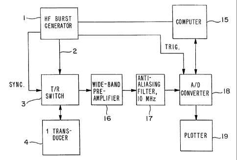
.Figure 1 shows a block circuit diagram,
Figure 2 shows a schematic sectional view of a
sample vessel,
Figure 3 shows a representation of the sound power
curve of the transducer as a function of
frequency,
Figures 4-9 show graphic representations of the
back-scatter signals,
CA 02204161 2005-08-23
- 13 -
Figure 10 shows a further block circuit diagram,
Figures 11-13 Graphic representations of the backscatter
signals (time resolution) and their spectta
(frequency resolution) at various sound
intensities when using a contrast medium as
disclosed in WO 93/25242.
Figure 14 Graphic representation of the scatter signals
at 2 fo as a function of the sound intensity
when using a contrast medium as disclosed in
WO 93/25242.
To produce the signals shown in Figures 4-9, which are
ready for further processing, the circuit shown in Figure 1
is used together with the sample vessel shown in Figure 2,
the wide-band sound head having the power characteristic
shown in Figure 3.
Periodically repeated electrical transmission pulses -
HF bursts - of variable frequency f, in the working range
fo min to fo max (fo min = 0.3 MHz < fo < fo max=22 MHz) and
of variable bandwidth, given by the number n of sine cycles
per burst: 0.5 < n < 20 with adjustable amplitude, are
generated by a function generator 1, which is controlled by
the central computer 15. The central-computer 15 controls
both the 6ourse of the.measurement and its evaluation. The
output 2 of the generator 1 leads to a transmitter/receiver
switch 3 which, as shown schematically, is synchronized by
.the generator 1. The T/R switch 3 may also be controlled
directly by the computer is. The output of the T/R. switch
3 is connected to a wide-band, matched and focused
transducer element 4. The particular features of the
transducer element.4 are shown schematically in Figure 3,.
The transducer is a very wide-band transducer without
interfering resonances in the working range; furthermore, it
has good electrical and acoustic impedance matching and a
transmitter average frequency fT > fo max. In the
CA 02204161 1997-04-30
- 14 -
Example described, ft = 17 MHz. The transducer may also
have spatially and electrically separate transmitter and
receiver transducer elements. In that case the.T/R
switch 3 is unnecessary. Advantageously, there may also
be provided a further transducer element for emitting a
second, independent high-frequency signal.
The signal received by the transducer element 4 is fed
via the switched-over T/R switch to a wide-band preamplifier
16 downstream of which, in the case of digital frequency
analysis, there is connected an anti-aliasing filter 17.
The wide-band pre-amplifier 16 has a bandwidth > fo max.
The filter 17 has, for example, a cut-off frequency of 10
MHz. Downstream of the filter 17 there is connected a
high-speed A/D converter in which the signal is digitized,
for example with a Nyquist frequency of 12.5 MHz. Further
processing of the signals is carried out in a digital
storage oscilloscope and in the central computer.
Downstream of the A/D converter 18 there is connected a
plotter 19.
Figure 1 shows that the A/D converter is triggered by
the function generator 1.
The digitized signal is stored and processed further in
a manner known, per se. It is available especially for
necessary corrections. It is also possible for a signal_
to be branched off before the A/D conversion and
digitized only after analog further processing.
. Figure 2 shows schematically the geometry of vessel 20
with which the measurement results given below were
obtained.
As shown in Figure 2, the sound head 4 is arranged in
the sample vessel 20. It is a 17 MHz sound head, which is
wide-band, matched and focused. The sample vessel 20
contains water. Two films 21 bound a sample region in
which 10 mg of ultrasonic contrast agent are dissolved in
3 ml of H20.
CA 02204161 1997-04-30
- 15 -
The reflected and/or backscattered signals in the
measurement region between the films 21 contain certain
components which were obtained by interaction of the
transmission pulse (at fo) and the non-linear contrast
agent introduced into the object of measurement.
Figure 3 shows schematically the frequency band of the
transducer element in the sound head. It will be seen
that, in the working range, the frequency response of
the oscillator in the sound head is quasi-linear. The
frequency response in the working range can be used to
compensate for a similar frequency response in the
sample under examination, but the frequency response in
the sample under examination may also be corrected
subsequently by weighting.
For measurement, an interesting time interval in the
time range is selected by means of a computer-controlled
gate circuit (not shown). It is also possible to select
several time intervals. The associated spectrum is
calculated by means of an FFT circuit (Fast Fourier
Transformation), and examples of such spectra are shown
in Figures 4 to 9. By selecting a suitable time window
length, it is possible to choose between optimum
frequency resolution and optimum spatial resolution.
Figures 4 to 8 each show the spectrum over the time
0 25 window. In order to show the spectral components clearly
in these Figures, a long time window, that is to say poor
.spatial resolution, was chosen. Figure 4 illustrates the
variation in time of the transmission pulse after
reflection at the coupling window without contrast
agents. fo = 4.0 MHz, + 15 dBm at the sound head. A
clear signal can be seen at 4 MHz. The signal shown in
the upper part of Figure 4 is an averaged power spectrum,
which was obtained behind the low-pass filter with a
Nyquist frequency of 50 MHz.
Figure 5 shows the backscatter signal from the sample
CA 02204161 1997-04-30
- 16 -
chamber without ultrasonic contrast agents. Figure 6
shows the backscatter signal seven minutes after the
addition of 10 mg of contrast agent in 3 ml of H20. A
clear peak can be seen at 2 x fo.
Figure 7 shows a measurement after 21 minutes under the
conditions given in Figure 5. A frequency fo = 3 MHz was
used. The spectrum recorded clearly shows the first and
second harmonics at 6.0 and 9.0 MHz. Figure 8 shows the
backscatter signal 15 minutes after the addition of an
ultrasonic contrast agent in a small concentration. A
frequency fo of 4 MHz + 20 dBm at the sound head was
used. The spectrum shown in the upper part of Figure 8
shows with relatively high frequency resolution the sub-
harmonic at 1/2 fo, the ultraharmonic at 3/2 fo and the
first harmonic at 2 fo.
Figure 9 shows a backscatter signal from linear
ultrasonic contrast agent fo = 4 MHz + 15 dBm at the sound
head. The spectrum shows backscattering only at the
excitation frequency.
It will be seen that the spectra illustrated have clear
amplitudes in frequency ranges that do not occur in the
transmitted spectrum when interaction with a non-linear
contrast agent has taken place. It is possible to
evaluate spectral changes dependent on a Doppler effect._
In order to use the circuit employed in the embodiments
described for imaging ultrasonic processes, additional
zomponents are provided in case a phased-array-type sound
head or a dynamically focused sound head is used. Such a
circuit diagram is shown in Figure 10.
The transmission signal from the function generator 1
(frequency fo) is fed from the output 2 to the n-way
signal divider 5. The signal is divided to one branch
per transducer element. In the embodiment shown, n
transducer elements 4 are provided. The transducer
elements 4.1...4.n receive the signal by way of the time-
CA 02204161 1997-04-30
- 17 -
delay circuits 7.1...7.n and the T/R switches 3.1...3.n
which are controlled by the generator or the computer.
The computer sets the time delay for each transducer
element in such a manner that, at the selected transmission
frequency, the desired directional characteristic is
produced at the sound head. The same directional
characteristic is set by the computer in the receiver part
by corresponding time delays. The signal received by the
sound heads 4.1...4.n is fed by way of the T/R switches
3.1...3.n to wide-band pre-amplifiers 6.1. . . 6.n. Each
pre-amplifier 6.1...6.n supplies a signal to an m-way signal
divider 10, downstream of which are connected suitably
controlled or adjusted time-delay circuits 11 which feed
circuits 12 for frequency band selection. Connected
downstream are circuits for the phase-correct summation of
the frequency bands and, if appropriate, for signal
division. This is followed by
selective further processing of the individual frequency
bands by means of processes known per se.
In particular, evaluation of the frequencies that are
not identical with fo, for example 1/2 fo, 2 fo, is carried
out.
The time-delay circuits may be variable or fixed. The
distribution of the received signals to m-way signal
dividers produces the desired number of frequency bands,
the position and width of which are adjusted by means of
band filters. Alternatively, the division may be
effected in such a manner that the received signal is
mixed with an auxiliary signal, which is derived from the
initial signal-and is different depending on the
frequency band, in such a manner that the individual
bands can work with uniform components in the subsequent
stages.
The frequency band around fo gives the usual results,
while the other bands contain greatly frequency-shifted
. . { . . _. . _. ..... . .
CA 02204161 1997-04-30
- 18 -
and non-linear signal components from interaction of the
transmission signal with the non-linear ultrasonic
contrast agents.
The further processing steps and signal analyses may be
carried out in any desired frequency channel or in
several parallel frequency channels in accordance with
known processes.
In-order to use two transmission frequencies fo and fP,
the second generator, shown on the right-hand side in
Figure 10, is provided, which generator is connected by
way of signal dividers and time-delay lines 15 to the
T/R switches 3.1...3.n. The second generator 1 allows
the exposure to ultrasonic waves of at least that spatial
region in the object under examination which is determined
by the directional characteristic at the time and the
receiver gate. The construction may be such that, in
addition to the wide-band transducer elements described,
the sound head contains at least one further, likewise
wide-band, transmission transducer, which is preferably
electrically separate from the others and is fed by the
second, independent transmission generator 1. However,
the two transmission signals may also be superimposed
electrically in such a manner that the same transducer
elements can be used.
Fig. 11 (upper half of the figure) shows the
backscatter signal, caused by a contrast medium as disclosed
zn WO 93/55242, with weak excitation with a 5 MHz burst of
an amplitude of 0.1 MPa, in the time range.
In the lower half of the figure, the power spectrum of
the same signal is reproduced. The signal in excitation
frequency fo (5 HMz) can be seen clearly; harmonic, sub- and
ultraharmonic signals are drowned out in static.
Fig. 12 shows the backscatter signal at excitation with
an amplitude of 0.34 MPa under otherwise identical test
conditions to those for Fig 11. In this case, the greater
CA 02204161 2005-08-23
, ~ .
- 19 -
backscatter portion of the contrast medium in the time range
can be seen clearly. In the frequency resolution, the
signals can be detected clearly at 2 fo and 3 fo.
Fig.-13 shows the backscatter signal at excitation with
an amplitude of 1 MPa. The backscatter portion of the
contrast medium is, in the time range (upper half of the
figure), clearly greater than the reflexes of the
transmitting pulse, and it is to be noted that 1 scale mark
corresponds to the ordinate here of 50 mV. In the power
spectrum (lower half of the figure), the signals can be seen
clearly at 1/2 fõ fa, 3/2 fa, 2 fo, 5/2 fo, 3 fo, 7/2 fo and
4 f,. Surprisingly, the signal at 2 fo.is of an intensity
similar. to the echo of irradiated frequency (fo).
Fig. 14 shows the intensity of the backscatter.signal
at 2 fa as a function of the irradiated sonic pressure at
-various excitation frequencies (fa) of 2, 3, and.4 MHz.
Also in this case,- a contrast medium.as disclosed in WO
93/25242 were used. Surprisingly, the intensity of the
backscatter detected signal grows above a threshold value of
about 40 dB superproportional to the excitation intensity.
This behavior is observed in an analogous way also for other
contrast medium preparations, e.g., microparticles based on
fatty acid-containing galactose particles or microparticles,
consisting of a gas core and a biodegradable polymeric shell
on which optionally a molecule with site-, structure- and/or
tissue-specific properties is bound.
From the foregoing description, one skilled in the art
can easily ascertain the essential characteristics of this
invention and, without departing from the spirit and scope
thereof, can make various changes and modifications of the
invention to adapt it to various usages and conditions.