Note: Descriptions are shown in the official language in which they were submitted.
CA 02205057 1997-OS-09
WO 97/10056 PCT/US96/14681
DEVICE AND METHOD FOR DNA AMPLIFICATION AND ASSAY
Field of the Invention
The present invention relates generally to devices and methods for carrying
out biological processes on liquid biological samples, and is particularly
concerned with a unitary DNA amplification and homogenous DNA probe assay
device which is suited for fluorescence polarization assays and is capable of
accommodating a plurality of liquid biological samples in discrete, sealed
sample
cells.
Bac and of the Invention
The processes of nucleic acid (DNA) amplificafion and subsequent nucleic
acid probe assay are well known and have been implemented in a variety of
formats. While these formats are highly effective, they are somewhat diffcnlt
to
perform in the clinical laboratory. Generally, DNA amplification and assay
reactions are performed sequentially on the sample to be assayed; that is, the
DNA amplification reaction is first carried out to completion, and the DNA
probe
assay is then performed on the fully amplified sample. This is referred to as
an
end point assay.
One problem with end point assays is that the amplified DNA (ampiicons)
from the DNA amplification reaction must be physically transferred to the
subsequent DNA probe assay. Because of the transfer, the potential exists for
contaminating the laboratory environment with the DNA amplicons. In addition,
the general risk of misidentifying a given sample or confusing it with other
samples increases each time that a physical transfer of the sample takes
place.
There have been previous proposals for self-contained test units that are
capable of carrying out an integrated nucleic acid amplification and nucleic
acid
assay on a liquid biological sample while the sample remains confined within
the
test unit. For example, U.S. Patent No. 5,229,297, to Paul N. Schnipelsky et
al,
CA 02205057 2000-04-27
WO 97/10056 PCT/US96/14681 -
describes a cuvette for DNA amplification and detection which comprises a
plurality of flexible compartments for containing a sample, amplifying
reagents
and detection reagents, together with passageways co~ecti.ng the sample and
reetgent compartments with a detection site and waste compartment A roller is
used to squeeze or compress the sample and reagent compartiments in a desired
sequence, thereby forcing the sample and detection reagents through the
passageways to the detection site and waste compardment,. Temporary seals are
used to isolate the sample and reagent com~eats from the passageways until
snffident pressure is generated by the roller. Although this arrangement is
advantageous in that the sample irmains.within the cnvette daring
amplification
and detection, the need for a roller to break the temporary seals and cease
the
various fluids to flow betwren compartments introduces undesirable complerity
and makes it difficult to aatomate the amplification and assay procedure.
In US Patent No. 5,639,428,
an improved test unit for carrying out integrated
nndeic add amplifications and nucleic acid assays is disclosed. In the
improved
test unit, the flow of sample and reagent liquids is controlled by centrifugal
force
applied by a triatively simple rotating apparatus, thereby avoiding the need
for
rollers and other complex mechanisms. While this represents a substantial
imprnvement over the arrangement disclosed in U.S. Patent No. 5,229,297, the
need to provide for controlled fluid movement within the test unit still
exists and
renders the test unit somewhat more complex than might be desired.
In addition to the end point assays discussed previously, homogenous
methods of nucleic acid assay also exist. Homogeneous methods do not require
the physical transfer of the amplified material to a separate assay site, but
rather
function simultaneously with the amplification reaction. E~Camples of imown
homogenous assay methods include fluorGSCence polarization, fluorescence
energy
transfer and light absorbance. V~hile fluorescence polarization, in
particalar,
functions very well in a research laboratory, it has a significant drawback in
that
-2-
CA 02205057 1997-OS-09
WO 97/10056 PCT/US96/14f81 -
it requires glass sample tubes or cells. This is a result of the fact that
most
plastic processing methods, such as injection molding or thermoforming, create
stresses in the material of the finished part. These stresses have random
polarization effects, and interfere with the transmission of polarized light
that is
required for a fluorescence polarization assay.
As is well lmown, DNA amplification reactions must occur within a certain
temperature range in order to produce the desired number of amplicons. If the
sample and the DNA amplification reagents are allowed to react before the
sample reaches the required temperature, a phenomenon Imown as "mis-priming"
can occur. This can affect the validity of the assay results, both in the case
of an
end point assay and a homogeneous assay.
In view of the foregoing, a need exists for a device or a test unit which is
capable of carrying out an integrated DNA amplification and DNA probe assay
with minimal complexity, and preferably without requiring fluid movements to
occur within the test unit itself. There is also a need for a test unit which
can be
used to carry out a homogenous DNA probe assay using fluorescence polarization
methods, but which does not require the use of glass to properly transmit
polarized light Finally, there exists a need for a test unit which can be used
to
carry out an integrated DNA amplification and DNA probe assay in a simple and
effective manner, while preventing inadvertent mis-priming of the
amplification
reaction. The present invention is directed to fulfilling these objectives.
It is an object of the present invention to provide a DNA amplification and
homogenous DNA probe assay device in a "card" format that can be conveniently
handled by clinical laboratory personnel, and accommodated in a suitable test
apparatus.
It is another object of the invention to provide a unitary DNA
amplification and DNA probe assay device which includes a multiplicity of
sample cells, with each sample cell comprising the elements and reagents
needed
for a DNA amplification reaction and a homogeneous DNA probe assay:
-3-
CA 02205057 1997-OS-09
WO 97/10056 PCT/LTS96/14681 -
It is a further object of the invention to provide a unitary DNA
amplification and DNA probe assay device in which all reagents needed for both
DNA amplification and DNA prnbe assay are contained, in dried form, within the
device, so that the addition of a liquid biological sample is all that is
needed to ,
carry out the amplification and assay procedure.
It is a further object of the invention to provide a test unit and method
that performs a "hot start" of the DNA amplification reaction, thereby
avoiding
an invalid assay result due to mis-priming of the amplification reaction.
It is a further object of the invention to provide a test unit which has the
optical properties necessary for a fluorescence polarization assay, but which
can
be made of inexpensive plastic materials rather than glass.
It is a further object of the invention to provide a test unit and method
that yields instantaneous DNA probe assay data by means of a Idnetic or
dynamic
measurement of DNA amplicons, rather than a conventional end point
measurement
It is a further object of the invention to provide a fluorescence polarization
DNA probe assay device which includes an integral polarizer, allowing for the
use
of a confocal polarization method.
It is a further object of the invention to provide an integral DNA
amplification and homogenous DNA probe assay device that can be permanently
sealed after the introduction of a liquid biological sample, thereby
preventing
amplicon contamination of the laboratory environment.
It is a still further object of the invention to provide an integrated DNA
amplification and DNA probe assay device which can accommodate a plurality
of liquid biological samples in discrete sample cells, and which can provide
DNA
probe assay data in a matter of minutes.
-4-
CA 02205057 1997-OS-09
WO 97/10056 PCT/US96/14G81 -
Summary of the Invention
In accordance with a preferred embodiment of the present invention, the
disadvantages and limitations of the prior art are substantially avoided by
providing a DNA amplification and homogeneous DNA probe assay device which
includes a multiplicity of discrete sample cells in a flat "card" format, with
each
sample cell containing the reagents necessary for both DNA amplification and
homogeneous DNA probe assay. The device is particularly suitable for
fluorescence polarization DNA probe assays, and is preferably provided with an
integral polarizer to avoid the need for polarizing elements in the related
measuring apparatus. The size and geometry of the sample cells allows for a
~hot
start" of the DNA amplification reaction and thereby avoids inadvertent mis-
priming of the amplification reaction.
In one aspect, the present invention is directed to an apparatus for
carrying out a nucleic acid amplification and a homogeneous nucleic acid assay
on a liquid biological sample. The apparatas includes a sample cell for
receiving
the fiquid biological sample. The sample cell includes a sample chamber and a
sample port for admitting the liquid biological sample into the sample
chamber.
A dried nucleic acid amplification reagent and a dried homogeneous nucleic
acid
assay reagent are adhered to the interior of the sample chamber for reacting
with
the liquid biological sample. A sealing member may be attached to the sample
cell for sealing the sample port after the liquid biological sample has been
admitted to the sample chamber. The homogeneous nucleic acid assay reagent
may comprise a fluorescence polarization assay reagent, and in that event a
portion of the sample cell may be made transparent to permit external
detection
of the fluorescence polarization reaction in the liquid biological sample. The
sealing member may be attachable over the transparent portion of the sample
chamber and may be made of a transparent, light-polarizing material to avoid
the
need for polarizing elements in the related measuring apparatus.
-5-
CA 02205057 1997-OS-09
WO 97/10056 PCT/CTS96/14681 '
In another aspect, the present invention is directed to an apparatus for
carrying out a biological process on a liquid biological sample. The apparatus
comprises a substantially flat, card-like member having at least one sample
cell
therein for receiving the liquid biological sample. The sample cell includes a
sample chamber, a sample port for admitting the liquid biological sample into
the sample chamber, an air vent for allowing sir to be displaced from the
sample
chamber during admission of the liquid biological sample into the sample
chamber, and a dried reagent adhered to an internal surface of the sample
chamber for reacting with the liquid biological sample. A sealing member,
preferably in the form of a layer of flexible material carrying a pressure-
sensitive
adhesive, is attachable to the card-like member for sealing the sample port
and
the air vent after a fiquid biological sample has been admitted to the sample
chamber.
In another aspect, the present invention is directed to an apparatus for
carrying out a nucleic acid fluorescence polarization assay on a Liquid
biological
sample. The apparatus includes a sample cell for receiving a liquid biological
sample. The sample cell has a sample chamber and a sample port for admitting
the liquid biological sample into the sample chamber. The apparatus also
comprises a dried nucleic acid fluorescence polarization reagent that is
adhered
to an internal surface of the sample chamber for reacting with the liquid
biological sample. At least a portion of the sample cell is made of a light-
transmissive, light-polarizing material to facilitate external detection of
the
fluorescence polarization reaction in the liquid biological sample without the
need
for separate polarization elements in the related measuring apparatus.
In a further aspect, the present invention is directed to a method for
carrying out an integrated nucleic acid amplification and homogeneous nucleic
acid fluorescence polarization assay on a fiquid biological sample. The method
comprises the steps of introducing a liquid biological sample into a sample
well
having a light-transmissive portion; bringing the liquid biological sample
into
-6-
CA 02205057 1997-OS-09
WO 97/10056 PCT/US96/14681 -
contact with a dried nucleic acid ampliscation reagent and a dried homogeneous
nucleic acid fluorescence polarization assay reagent within the sample cell;
4
sealing the sample cell; incubating the sample cell to allow the liquid
biological
sample to react with the nucleic acid amplification reagent and with the
homogeneous nucleic acid fluorescence polarization assay reagent; and
detecting
fluorescence polarization in the liquid biological sample through the light-
transmissive portion of the sample cell. The detection step may comprise
directing polarized light through the light-transmissive portion of the sample
cell
or, if the light-transmissive portion of the sample cell is made of a light-
polarizing material, directing unpolarized light through the light-
transmissive
portion of the sample cell.
In a still further aspect, the present invention is directed to a method for
carrying out a nucleic acid amplification reaction on a liquid biological
sample.
The method comprises the steps of preheating a sample cell containing an dried
nucleic acid amplification reagent to a temperature suitable for nucleic acid
amplification; introducing a liquid biological sample into the preheated
sample
cell to bring the liquid biological sample into contact with the dried nucleic
acid
amplification reagent; equilibrating the temperature of the liquid biological
sample to the temperature of the preheated sample cell; and, after the
equilibration is substantially complete, commencing the nucleic acid
amplification reaction in the sample cell. Preferably, the step of
equilibrating the
temperature of the liquid biological sample to the temperature of the
preheated
sample cell comprises forming a thin layer of the fiquid biological sample in
the
sample cell to enhance heat transfer between the sample cell and the liquid
biological sample.
-
CA 02205057 1997-OS-09
WO 97/10056 PCT/CTS96/14681 -
Brief Description of the Drawing
The various objects, advantages and novel features of the invention will be
more readily appreciated from the following detailed description when read in
coqjunction with the appended drawing figures, in which:
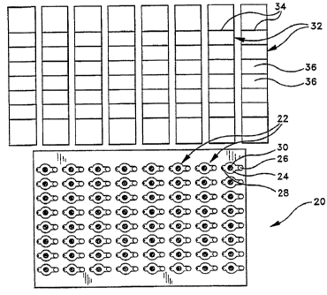
Fig. 1 is a top view of a DNA amplification and homogeneous DNA probe
assay card constructed in accordance with a preferred embodiment of the
present
invention, illustrating separate sealing strips which are used to seal the
individual
sample cells of the card atlter liquid biological samples have been introduced
into
the sample cells;
Fig. Z is a side view of the DNA amplification and DNA probe assay card
of Fig. 1, illustrating its relatively small thickness;
Fig. 3 is a top view of the DNA amplification and DNA probe assay card
and sealing strips of Fig. l, with some of the sample cells showy filled with
liquid
biological samples and sealed using the sealing strips;
Fig. 4 is an enlarged cross-sectional view of a po~on of the DNA
amplification and DNA probe assay card of Figs. 1- 3, with solme of the
discrete
sample cells shown filled and others left empty to illustrate the locations of
the
dried amplification and assay reagents;
Fig. S is a plan view of the top layer of the DNA amp~cation and DNA
probe assay card of Figs. l - 4, illustrating the manner in which sample ports
and
air vents are provided for the discrete sample cells;
Fig. 6 is a plan view of the middle layer of the DNA amplification and
_y
DNA probe assay card of Figs. 1 - 4, illustrating the keyhole-shaped apertures
which define the side walls of the discrete sample cells;
Fig. 7 is a plan view of the bottom layer of the DNA amplification and
DNA probe assay card of Figs. 1 - 4;
Fig. 8 is an exploded perspective view illustrating the relationship of the
top, middle and bottom layers of the DNA amplification and DNA probe assay
card of Figs. 1 - 4; '
_g_
CA 02205057 1997-OS-09
WO 97/10056 PCT/US96/14681 -
Figs. 9A and 9B are top view of two different embodiments of the sealing
strips that are used to seal the sample ports and air vents of the DNA
h
amplification and DNA probe assay card of Figs. 1 - 4;
Fig. 10 is an enlarged sectional view through one of the discrete sample
cells in the DNA amplification and DNA probe assay cx~rd of Figs 1 - 4,
illustrating the manner in which the use of a light-polarizing material for
the
sealing strips allows a confocal detection method to be used;
Figs. ll and 12 are graphs depiMing the change in fluorescence intensity
with time during a DNA amplification and homogeneous DNA fluorescence
polarization assay; and
Figs. 13 - 15 illustrate an exemplary measuring apparatus which can be
used to measure fluorescence intensity in the sample cells of the DNA
amplification and DNA probe assay card of Figs. 1 - 4.
Throughout the drawings, like reference numerals will be understood to
refer to Like parts and components.
Detailed Description of the Preferred Embodiments
A DNA amplification and homogeneous DNA probe assay device 20
(hereinafter referred to as a "DNA card") constructed in accordance with a
preferred embodiment of the present invention is illustrated in Fig. I.
Although
the specific dimensions and geometry of the DNA card may be varied in
accordance with the requirements of particular applications, the card 20 of
the
preferred embodiment is rectangular with a length of approximately 5.025
inches
and a width of approximately 3362 inches. The card contains a rectangular
array
of discrete sample cells 22, spaced evenly across the length and width of the
card.
" Each sample cell 22 includes a closed sample chamber 24 (the top wall of
which
is transparent) for receiving a liquid biological sample, an open sample port
26
which communicates with the sample chamber 24, and an air vent 28 which also
-9-
CA 02205057 1997-OS-09
WO 97/10056 PCT/US96/14681 -
communicates with the sample chamber 24. Dried DNA amplification and assay
reagents are adhered to the upper interior wall of each sample cell 22 in the
form
of a single, discrete spot 30. The sample ports 26 provides the means by which
liquid biological samples (not shown in Fig. 1) can be introduced into each of
the ,
sample chambers 24 (preferably by pipetting), and the air vents 28 allows air
to
be displaced firm the sample chambers 24 as the liquid biological samples are
being introduced. As will be described hereinafter, the liquid biological
sample
that is introduced into the sample chamber 24 of each sample cell 22 makes
contact with, and dissolves, the dried reagent spot 30 in the sample cell,
thereby
initiating the desired DNA amplification and assay reactions. Measurement of
the assay results takes place while the liquid biological samples remain
sealed
within the sample cells 22, also in a manner to be described hereinatlter. In
the
illustrated embodiment, the DNA card 20 contains sixty-four (64) identical
sample cells 22, arranged in a rectangular array of eight rows by eight
columns,
on vertical centers of approximately 0354 inches and horizontal centers of
approximately 0.628 inches.
With continued reference to Fig. 1, sealing strips 32 (one for each column
of sample cells 22 in the card 20) are provided for sealing the sample ports
26
and air vents 28 of the sample chambers 24 after liquid biological samples
have
been introduced into the sample cells 22. Preferably, the strips 32 are
segmented
along score lines 34 to define segments which can be used individually, if
desired,
to seal some of the sample cells 22 and not others. In the illustrated
embodiment, the sealing strips 32 are approximately 0.628 inches in width and
approximately 3362 inches in length. Each sealing strip 32 seals a column of
eight sample cells 22, with eight strips 32 being used to seal all sixty-four
sample
cells 22. The individual segments or seals 36 of each sealing strip 32 are on
the
same vertical centers (i.e., approximately 0354 inches apart) as the sample
cells .,
22 themselves. The sealing strips 32 are preferably about 0.015 inches thick
and
are provided with a layer of pressure sensitive adhesive on their lower
surfaces '
-10-
CA 02205057 1997-OS-09
WO 97/10056 PCT/US96/14681
(not visible in Fig. I). In practice, the sealing strigs 32 are applied in a
manner
similar to adhesive tape, and serve to permanently seal the sample cells 22 by
covering the sample ports 26 and air vents 28. The sealing strips 32 are
generally
used whole for convenience, and are only subdivided as necessary along the
score
lines 34.
The sealing of the sample cells 22 by means of the sesIing strips 32
provides several advantages. First, the sealing strips 32 prevent evaporation
of
the liquid biological samples firm the sample cells 22 during the DNA
amplification and homogeneous DNA probe assay. Given that the volume of the
liquid biological sample wiU typically be very small (about 20 ~L) and that
the
amplification and assay reactions will usually take place at an elevated
temperature (up to about 75° C), such evaporation may otherwise result
in
significant lass of the liquid biological sample. The second advantage of the
sealing strips 32 is that they prevent the release of DNA amplicons from the
sample cells 22, thereby preventing contamination of the laboratory
environment.
Finally, in accordance with a particularly preferred embodiment of the present
invention, the sealing strips 32 may be made of a transparent, light-
polarizing
material so as to serve as polarization elements during the detection or
measurement step. This avoids the need to provide separate polarization
elements in the related measuring apparatus.
In the description which follows, it will be assumed that all of the sample
cells 22 and seals 36 are identical, and that the description of any one
sample cell
22 or seal 36 will apply to all. Although this is true in the preferred
embodiment,
the invention should not be regarded as being limited to this arrangement. It
is
within the scope of the invention to provide sample cells 22 andJor seals 36
that
are different from one to the next, including (but not limited to) different
reagents, different dimensions, different volumes and different optical
properties.
The reagents may differ for either the DNA amplification or the DNA probe
r assay, or both, with exemplary homogeneous DNA probe assay methods including
-11-
CA 02205057 1997-OS-09
WO 97/10056 PCT/L1S96/14681
fluorescence polarization reactions, fluorescence energy transfer reactions
and
light absorbance reactions. Any or all of the foregoing differences may exist
within a single card 20 and/or from one card 20 to the next. Thus, for
example,
different types of DNA cards 20 could be provided for carrying out different
types
of assays, with such cards retaining only a generic or functional similarity
(e.g.,
to the extent necessary to fit into the same type of measuring instrument).
Fig. 2 is a side or edge-on view of the DNA card 20, which in the preferred
embodiment has a generally flat or planar configuration. As will be apparent
from Fig. 2, the DNA card 20 can be made extremely thin if desired. In the
preferred embodiment, the thiclmess of the DNA card 20 is approximately 0.047
inch.
Fig 3. illustrates the DNA card 20 as it might appear during actual use,
with fourteen of the sample cells 22 filled with liquid biological samples 38.
In
these filled sample cells 22, the liquid biological samples 38 have dissolved
the
dried reagent spots 30 of Fig. 1. The filled sample cells 22 are covered by
the
respective segments or seals 36 of the sealing strips 32. One complete sealing
strip 32 has been applied to the left-hand column of sample cells 22, and a
second
sealing strip 32 has been subdivided along one of the score lines 34 into a
first
portion 32A which has been applied to the upper six sample cells 22 of the
second
column, and a second portion 33B which has been retained for future use. The
unused portion 32B can be used to seal the two lowermost sample cells 22 of
the
second column during a subsequent use of the DNA card 20.
Typically, the various liquid biological samples 38 shown in Fig. 3 will
consist of blood samples or other body fluid samples from different patients,
all
of which are being tested for the same pathogen by identical amplification and
assay reagents 30. However, it will be understood that embodiments are
possible
in which more than one of the liquid biological samples 38 are drawn from the
.,
same patient, and in which the reagents 30 differ fi-om one sample cell 22 to
the
next.
-12-
CA 02205057 1997-OS-09
WO 97/10056 PCT/US96/14681 -
Fig. 4 is a partial cross-sectional view of the DNA card 20, taken along the
Line 4-4 in Fig. 3. In this view, the laminated construction of the DNA card
20
in the preferred embodiment can be readily appreciated. The DNA card 20 is
made up of a bottom layer 40, a middle layer 42 and a top Layer 44. The seals
36 of the sealing strip 32 are adhered to the upper surface 46 of the top
Layer 44.
Each of the layers 40, 42 and 44 is preferably made of a plastic film having a
thickness of approximately 0.015 inches, and the seals 36 are of a similar
material and thickness. The layers 40, 42 and 44 (together with the seals 36)
are
held together by a pressure-sensitive adhesive (not shown) that is typically
about
O.OOI inch thick. Referring for convenience to the empty sample cell 22A in
Fig.
4, the sample port 26 is preferably about 0.125 inch in diameter and
communicates with a narrow section 48 of the sample chamber 24 that is
preferably about 0.125 inch wide. The narrow section 48, in turn, communicates
with a larger, substantially circular portion of the sample chamber 24 which
is
approximately 0.250 inch in diameter. The dried reagent spot 30 is adhered to
the upper wall of the circular portion of the sample cell 24 (corresponding to
the
lower surface of the top layer 44 of the DNA card) and is situated
approximately
at the center of this circular portion. The circular portion of the sample
chamber
24 rnmmunicates with another narrow section 52 of the sample chamber which
is approximately 0.125 inch in diameter. The section 52, in turn, communicates
with the air vent 28 located at the opposite end of the sample cell 22A from
the
sample port 26. The air vent 28 is preferably about 0.040 inch in diameter.
The
height of the interior of the sample chamber 24 is defined by the thickness of
the
middle layer 42 of the DNA card 20, and by the thickness of the adhesive on
either side of this layer. This results in an overall height of about 0.017
inch for
the interior of the sample chamber 24.
With continued reference to Fig. 4, the sample cell 22B is shown as it
would appear during use. Thus, the sample cell 22B is filled with a Liquid
biological sample 38 and sealed with a seal 36 which covers the sample- port
26
-I3-
CA 02205057 1997-OS-09
WO 97/10056 PCT/US96/14681 -
and air vent 28. The dried reagent spot 30 of Fig. 1 has been dissolved by the
liquid biological sample 38.
In order to use the DNA card 20, a suitable measuring instrument is
required. Depending upon whether the DNA card 20 contains assay reagents for
fluorescence polarization reactions, fluorescence energy transfer reactions or
light
absorbance reactions, and whether (in the case of a fluorescence polarization
assay) the DNA curd 20 has integral polarizing elements, the instrument may be
either a conventional instrument, such as a microplate fluorometer or a
microplate reader, or a specialized instrument of the type to ~e described
shortly
in connection Figs. 13 - 15. In either case, suitable temperature controls
must
be provided, together with means for optically addressing the ~adividual
sample
cells 22.
In order to perform an integrated DNA amplification and homogeneous
DNA fluorescence polarization assay, the DNA card 20 is planed oa the heated
carrier of the instrument. The cmrier is a heated tray whir can be extended
outside the instrument to receive the cx~rd, and which then '~ thdraws into
the
instrument in order to perform the desired readings of fluorescence
polarization,
fluorescence intensity or light absorbance. Typically, the instrument is
provided
with means for moving the card in both the x and y directions so that each
sample cell 22 can be read individually. During the entire operation, the
heated
carrier maintains the DNA card 20 at as optimum temperature, typically between
25° C and ?5° C.
Initially, an empty DNA card 20 is placed on the extended, heated carrier
of the instrument and is allowed to equilibrate to the catTier temperature.
This
equilibration may take approximately one minute. Once the DNA card 20 is
equilibrated to the carrier temperature, liquid biological samples 38 are
pipetted
into the sample ports 26 of one or more of the sample cells 22. The liquid
biological samples 38 instantly fill the sample chambers 24 due to a
combination
-14-
CA 02205057 1997-OS-09
WO 97/10056 PCT/US96/14681 -
of hydrostatic and capillary force. In the preferred embodiment, the pipetted
volume of each liquid biological sample is approximately 20 pL.
As soon as liquid biological samples 38 have been pipetted into all of the
~ sample chambers 24, the sample cells 22 may be sealed using the seals 36,
and
the measuring instrument may be started. The carrier is then drawn into the
instrument for fluorescence polarization, fluorescence intensity or light
absorbance reading. Due to the extreme thinness of the sample chambers 24, and
the large surface area of the sample chambers 24 with which the liquid
biologicxtl
samples 38 come into contact, the liquid biological samples 38 heat up within
seconds of being pipetted into the sample cells 22 to the optimum temperature
desired for DNA amplification. Thas, by the time the dried reagent spots 30
dissolve and diffuse throughout the liquid biological samples 38 to begin
"priming" of the DNA amplification, the reagents are already up to the optimum
temperature. It is in this way that the DNA card 20 effects a "hot start" of
the
DNA amplification reaction.
Figs. 5 - 7 depict the individual layers of plastic film that the DNA card
20 is composed of. In the preferred embodiment, each layer is approximately
0.015 inch thick. The top layer 44, shown in Fig. 5, contains the holes that
form
the sample ports 26 and air vents 28. The lower surface of the top layer 44
forms
the upper walls of the sample chambers 24. The middle layer 42, shown in Fig.
6, contains keyhole-shaped apertures 54 that form the side walls of the sample
chambers 24. The middle layer 42 is coated on both sides with a pressnre-
sensitive adhesive (not shown). The bottom layer 40, shown in Fig. 7, is a
solid
rectangular sheet that forms the bottom of the DNA card 20. The upper surface
of the bottom layer 40 forms the lower wall of each of the sample chambers 24.
The exploded view of Fig. 8 depicts the relative alignment of the top layer
44, middle layer 42 and bottom layer 40 during assembly of the DNA card 20.
In practice, the top layer 44 and middle layer 42 are laminated together with
a
suitable adhesive, and the partial assembly is then inverted so that the
-15-
CA 02205057 2000-04-27
WO 97110056 PCT/US96/14681 -
amplification and assay reagents can be pipetied and dried onto the underside
of
the top layer 44 to create the dried reagent spots 30. With reference to Fig.
4, the
sectioned edge of the DNA card 20 can be seen to include a dried reagent spot
30
that is adhered to the underside of the top Layer 44 of the card 20. After the
dried reagent spot 30 is formed, the bottom layer 40 is Laminated to the
middle
layer 42, as shown in Fig. 8, to complete the assembly 20.
The dried reagent spot 30 contains both DNA ampLificadon and
homogeneous DNA assay reagents, the latter' preferably consisting of
fluorescence
polarization assay reagents. E~camples of suitable DNA amplification and DNA
flnocescence polarization assay reagents are disclosed in
US Patent No. 5,593,867
and entitled 'Fluorescence Poiarizatioa Detection of Nucleic Acid
Amplification",
The chemical reagents in the dried spot 30 are carried in a readaly soluble
matrix,
such a trehalose or another carbohydrate.. These reagents will i pontaneonsly
re-
suspend when exposed to as aqueous sample introdnaed into t~e sample chamber
24. It will be understood that more than one dried reagent spot 30 may be
provided in each sample cell 22 if desired, as for example by providing the
amplification reagents in one spot and the assay reagents in ~ different spot.
In
the case of a DNA amplification and homogeneous DNA assay, however, the
reagent spots (if separated) should be positioned in such a way that they are
dissolved by the liquid biological sample 38 at essentially tli ~ same time.
If the homogeneous DNA assay that is to be used ia~the DNA card 20 is
a fluorescence polarization assay, the top layer 44 of the card 20 must be
made
of a material that does not interfere with the transmission of polarized
light. Two
eacamples of materials that satisfy this requirement are cellulose acetate
butyrate
(CAB) and triacetate cellulose (TAC).
Two alternative embodiments of the sealing strip 32 are illustrated in Figs.
9A and 9B, respectively. Ia Fig. 9A, the sealing strip 32 is made either of a
-16-
CA 02205057 1997-OS-09
WO 97/10056 PCT/US96/14681 -
transparent CAB having a thickness of about O.OIS inch, with an optically
clear
pressure-sensitive adhesive (such as Adhesives Research type 8154) applied to
its
back surface, or of a transparent light-polarizing film with an optically
clear
pressure-sensitive adhesive applied to its back surface. A suitable adhesive-
backed polarizing film is available from Nitto Denko as product number 1220
DU. As previously mentioned, score lines 34 are provided to allow the sealing
strip 32 to be separated into individual seals or segments 36 and shown in
Fig.
3. In Fig 9B, a modified sealing strip 32' is shown in which each of the
segments
or seals 36 has a central hole or aperture 56. The holes 56 align with the
central
circular areas of the sample chambers 24 when the seals 36 are applied to the
sample cells 22 as illustrated in Figs. 3 and 4. The sealing strip 32' of Fig.
9B is
similar to the sealing strip 32 of Fig. 9A in that it carries a layer of
pressure-
sensifive adhesive on its back surface, but the sealing strip 32' of Fig. 9B
may be
made of an opaque material (such as black PVC or CAB) since the holes 56 allow
for light transmission to and from the sample chambers 24 through the top
Layer
44 of the DNA card 20. The sealing strip 32' of Fig. 9B is advantageous in
that
the light emitted by the liquid biological samples 38 during the fluorescence
polarization assay is required to travel only through the top layer 44 of the
DNA
card 20, rather than through the top layer 44 and the sealing strip 32 as in
the
embodiment of Fig. 9A.
In fluorescence polarization assays, a polarized excitation beam of a given
wavelength of light is used to excite the fluorescent DNA probes. The
intensity,
at a given wavelength, of fluorescent emission from these excited probes is
measured in the plane polarized parallel to the excitation polarization, and
also
in the plane polarized perpendicular to the excitation polarization. When a
fluorescent DNA probe hybridizes to a DNA amplicon, the intensity of
fluorescent
emission in the plane parallel to the excitation plane increases. Typically,
both
parallel and perpendicular intensities are measured. The changes in total
intensity are then compensated for by applying the formula:
-17-
CA 02205057 1997-OS-09
WO 97/10056 PCT/US96/14681
p - ~IPARA ' IPER) ~ ~IPARA + IPER)~
where:
Ip~ = Fluorescent intensity in the plane polarized in the plane
polarized parallel to the plane of excitation polarization; and
Ip~ = Fluorescent intensity in the plane polarized in the
plane polarized perpendicular to the excitation
polarization.
This formula yields the dimensionless quantity referred to as the polarization
ratio (P).
Since it is the polarization intensity in the plane parallel to the excitation
polarization which increases with increased hybridization, measuring the
intensity
of the polarization in the plane parallel to the excitation polarization over
time
will show the increase in hybridization over time. This is a lanetic or
dynamic
approach to the measurement of fluorescence polarization, which is also
suitable
for use with fluorescence energy transfer and light absorbance assays. By
using
such a kinetic or dynamic approach, compensation for absolute intensity
becomes
somewhat less important because each sample is measured against itself and is
thus a relative measurement. In the case of a fluorescence polarization assay,
therefore, it becomes necessary to measure fluorescence intensity only in the
plane polarized parallek to the plane of the excitation polarization.
The Idnetic or dynamic approach described above allows for the use of a
confocal polarization method, where the polarizer far the excitation beam is
also
used as the polarizer for the fluorescence emitted by the sample, thereby
reducing
the number of required polarizing elements to one. This differs from the
rnnventional approach, in which separate polarization elements are needed in
the
measuring instrument for both the excitation beam and the sensor used to
detect
the fluorescent emissions from the samples. With only a single polarizing
element being required in the confocal method, this element can be provided in
-18-
CA 02205057 1997-OS-09
WO 97/10056 PCT/CTS96/14681 -
the DNA card 20 itself (i.e., in the form of a polarized sealing strip 32 or
top
layer 44) and need not be provided in the measuring instrument as in the prior
art. Thus, standard microplate fluorometers containing no polarization
elements
can be used in the fluorescence polarization assay of the p~sent invention. In
the case of fluorescence energy transfer assays, standard microplate
fluorometers
can also be used, and in the case of light absorption assays, standard
n>icroplate
readers can be used.
The enlarged cross-sectional view of Fig.10 illustrates the sample chamber
24 of one sample cell 22 filled with a liquid biological sample 38 during a
fluorescence polarization assay. In this example, the seal 36 ~s made of a
plastic
polarizing film, and serves as the confocal polarizer during ~he assay. The
top
layer 44 of the DNA card 20 is made of transparent, non-polarizing CAB. In
operation, an uapolarized light beam 58 is directed toward the sample cell 22
containing the liquid biological sample 38. When the unpolarized light beam
passes through the polarizing seal 36, the transmitted light ''Beam 60 is of
single
polarization. The fluorescent DNA probes in the Liquid biohba'cal sample 38
are
excited by the polarized beam 60 and emit light (indicated by the arrow 62) of
various polarizations. However, the same polarizing seal 36 polarizes these
emissions in a plane parallel to that of the excitation beam. 60, resulting in
a
polarized beam 64 being detected by the fluorometer. In this way, a confocal
polarization method is implemented without requiring any polarization elements
in the fluorometer itself.
_.
Fig. 11 is a graph of the typical relationship that homogeneous DNA
amplii"lcation and assay reactions exhibit with respect to time. This
relationship
is similar whether assay reagents for fluorescence polarization, fluorescence
energy transfer (fluorescence intensity) or light absorbance are used. The
graph
shows that the reactions exhibit an initial exponential portion 66, and a
final
linear portion 68. Fig. 12 depicts a graph similar to that of Fig. 11, but
includes
curves for three different concentrations of genomes. The concentrations used
are
-19-
CA 02205057 1997-OS-09
WO 97/10056 PCT/US96/14681 -
genomes, 100 genomes and 1000 genomes per unit volume. Typical DNA
probe assays are performed on fully amplified samples, in which case they are
end point reactions. DNA amplification reactions typically produce a maximum
number of amplicons that is independent of the starting number of genomes. In
,
Fig. 12, it can be seen that the 1000-genome curve exhibits its exponential
phase
70 at a time before the 100-genome curve exhibits its exponential phase 72.
Similarly, the 100-genome curve exhibits its exponential phase 72 at a time
before
the lOtgenome curve exhibits its exponential phase 74. However, by the time
labelled tend point, the magnitudes of all three curves are very similar and
there
is only a small difference between the 1000-genome, 100.genome and 10-genome
curves, as shown by the points a, b and c on the vertical axis. By
accumulating
fluorescence intensity data during the entire time interval represented by any
given one of the curves in Fig. 12 (i.e., between to and t rather than
end point)
simply taking the final reading at tend poina ~°~ahon about the
fluorescence
polarization assay is available earlier and with better resolution. In
addition,
various different protocols may be used. For instance, by measuring the time
to
a given amplitude (point d in Fig. 12), it can be seen that the 1000.genome
curve
at time tl will be detected first and that its resolution from the 100-genome
curve
(at time ts) is increased, as is the resolution of the 100-genome curve from
that
of the 10-genome curve at time t3. Alternatively, examining the amplitudes of
the
three curves over time indicates that there are many places better than tend
point
to make measurements to resolve the differences in the three curves. If time
tl
is taken, for example, there is much better resolution between the 1000-genome
curve and the 100-genome curve than at tend pointy and a similar increase in
resolution exists between the 100-genome curve and the 10-genome curve at time
t2.
As noted previously, the nature of the measuring instrument with which y
the DNA card 20 is used will vary depending upon the construction of the DNA
card 20 itself. For embodiments of the DNA card 20 containing polarizing
- 20 -
CA 02205057 1997-OS-09
WO 97/10056 PCT/US96/14681 -
elements, a typical microplate fluorometer with suitable thermal control can
be
used. For embodiments of the DNA card 20 that do not contain polarizing
elements, a measuring instrument containing such elements in required. An
example of such an instrument is shown in Figs. 13 - 15.
Referring first to Fig. 13, the measuring instrument 80 is shown in a
perspective view with a portion cut away to illustrate the manner in which the
DNA card 20 is received in the instrument. The DNA card 20 is initially placed
on a heated carrier 81 which extends out over the front portion 82 of the
instrument, and is then drawn automatically into the interior of the
instrument
at the posifion shown. In this posifion, shown in more detail in the enlarged
view
of Fig. 14, the DNA card 20 is located below three polarized laser diode
sources
84 of different wavelengths such as 630 am, 660 nm and 690 nm. A motor 86
selectively rotates a six-position polarized filter wheel 88 to position
polarized
wavelength filters 90, 92, 94 and 96 (and two additional filters which are not
visible) above the sample cell 22 of interest. These filters match the
emissions
of the various fluorescent DNA probes and allow for the detection and
measurement of these wavelengths by a photomultiplier tube (PM'I~ detector 98
in planes both parallel and perpendicular to the polarization plane of the
input
or source beam. The heated carrier 81 is indexed in the x and y directions (by
means not shown) to address different ones of the sample cells 22.
Fig. 15 is a front or edge-on view of the DNA card 20 of Figs. 13 and 14,
showing a single source 84 which provides a specular input beam 100 directed
to
a particular sample cell 22 of the DNA card 20. This beam is monochromatic
and polarized as a consequence of having been generated by a laser diode
source.
If other types of sources are used, a polarizes and wavelength filter are
required.
When excited by the input beam 100, the fluorescent DNA probes in the sample
- cell 22 of the DNA card 20 emit light 102. The light 102 passes through the
polarizes and wavelength filter of the filter wheel 88, to the PMT
photodetector
98 for detection and measurement.
-2I-
CA 02205057 1997-OS-09
WO 97/10056 PCT/US96/14681 -
Examples of several different ways in which the DNA card 20 may be
constructed are provided below. It should be understood that these examples
are ,
merely illustrative and are not intended to Iimit the srnpe of the present
invention.
Example 1
The DNA card 20 comprises a top layer 44 made of transparent, non-
polarizing CAB, a middle layer 42 made of black PVC, and a bottom layer 40
made of transparent, non-polarizing CAB. The sealing strips 32 have the
configuration shown in Fig. 9A, and are made of a plastic polarizing film
coated
on one side with an optically clear pressure-sensitive adhesive. The resulting
DNA card 20 can be used in a standard microplate fluorometer having no
polarizing elements.
Example 2
The DNA card 20 comprises a top layer 44 is made of a plastic polarizing
film, a middle layer 42 made of black PVC, and a bottom layer 40 made of
transparent, non-polarizing CAB. The sealing strips 32' are made of black PVC
and have the configuration illustrated in Fig. 9B. The resulting DNA card 20
can
be used in a standard microplate fluorometer having no polarizing elements.
Example 3
The top layer 44, middle layer 42 and bottom Iayer 40 of the DNA card 20
are all made of transparent, non-polarizing CAB. The sealing strips 32 have
the
configuration shown in Fig. 9A, and are made of a transparent, non-polarizing
CAB with an applied pressure-sensitive adhesive. When constructed with
fluorescence polarization assay reagents, the resulting DNA card 20 is used in
a -
measuring instrument containing palarizing elements as illustrated, for
example,
in Figs. 13 - 15. Alternatively, this embodiment, when constructed with
-22-
CA 02205057 1997-OS-09
WO 97/10056 PCT/IJS96/14681 -
fluorescence energy transfer assay reagents, results in a DNA card 20 that is
measured on a typical microplate fluorometer. This embodiment is also suitable
for rnnstruction with light absorbance assay reagents, which can be measured
on
a typical microplate reader.
Example 4
The DNA card 20 comprises a top layer 44 made of transparent, non-
polarizing CAB, and a middle layer 42 and bottom layer 40 both made of black
CAB. The sealing strips 32 have the configuration shown in Fig. 9A, and are
made of a plastic polarizing film with an applied pressure-sensitive adhesive.
The
resulting DNA card 20 may be used with a conventional microplate fluorometer.
bramnle 5
The top, middle and bottom layers 44, 42 and 40 of the DNA card 20 are
as set forth in Example 4. However, the sealing strips 32' have the
configuration
shown in Fig. 9B, and are made of transparent, non-polarizing CAB with an
applied pressure-sensitive adhesive. The resulting DNA card 20 is used in a
measuring instrument of the type illustrated in Figs. 13 - 15.
Example 6
The top layer 44, middle Iayer 42 and bottom layer 40 of the DNA card 20
are as set forth in Example 3, and the sealing strips 32 have the
configuration
shown in Fig. 9a. However, the sealing strips 32 are made of a plastic
polarizing
film with an applied pressure-sensitive adhesive. The resulting DNA card 20
may
be used with a conventional microplate fluorometer.
The foregoing is illustrative of the present invention, and is not to be
construed as limiting thereof, as. numerous alternatives to the devices and
methods described which incorporate the present invention will be apparent to
_ 23 _
CA 02205057 1997-OS-09
WO 97/10056 PCT/US96/146~1
those skilled in the art. The invention is accordingly defined by the
following
claims with equivalence of the claims to be included therein.
-24-