Note: Descriptions are shown in the official language in which they were submitted.
CA 0220~176 1997-0~-12
WO 97/03613 PCT/US96/11118
WOUND CLOSUR~ APPARATUS AND METHOD
BACKGROUND OF THE INVENTION
Field of the Invention
This invention relates generally to apparatus and
methods for closing a wound or hole in a body wall, and
more specifically to wound closure devices using suture
material.
Discussion of the Prior Art
A puncture-type wound in a body wall can be created
either unintentionally, or intentionally as part of a
surgical procedure. In either case, the wound typically
has the configuration of a hole extending through the body
wall where access to the ~ar side o~ the wall is available
only through the wound.
The wound in the body wall may be accidentally created
but it is more likely that it will be intentionally created
in a surgical procedure associated with interventional
cardiology, for example.
Surgical procedures involving interventional
cardiology c~monly produce a wound in the femoral or iliac
artery of the patient. In this case, the body wall in
which the wound is formed is the wall of the artery which
is disposed some distance beneath the skin of the patient.
When these arteries are punctured, wounds or holes are left
in the tissue wall forming the arteries. Currently, large
caliber access sheaths and dilators are used which me~ely
CA 0220~176 1997-0~-12
WOg7/03613 PCT~Sg6/1l1l8
accentuate the size of the wound and ~em~n~ an appropriate
suturing apparatus and technique for closing the wound.
Wounds of this type have been addressed with direct hand
pressure or specialized weights, bandages and dressings.
These implements, requiring placement for extended
supervised periods of time, also result in considerable
discomfort to the patient. Other types of closure devices
include bioresorbable plugs which in some cases provide a
matrix which facilitates clotting. More recently, devices
have been used which include needles and attached sutures
which have been inserted into the vessel. The needles have
been driven outwardly through the wall of the vessel where
the suture ends have been captured and retrieved for knot
tying.
Similar devices are represented by U. S. Patent No.
5,417,699 which disclose a pair of proximally facing
needles insertable through the wound and radially
expandable so that upon retraction of the device the
needles extend through holes in the surrounding tissue.
The prethreaded holes are then drawn outwardly through the
tissue wall along with the ends of attached sutures. This
construction requires a special needle capture mech~n;.~m to
pull the needles proximally through the body wall.
SU ~ RY OF THE INVENTION
The wound closure device of the present invention
includes a distal c~nnllla which is insertable through the
wound and functions to provide a suture on the far side of
the body wall. A handle of the device rem~;n~ on the near
side of the body wall. A finger tab operable on the handle
manipulates the suture on the far side of the body wall
between radially spaced outer and inner positions. A
needle deployment me~h~n;~m, also operable on the handle
deploys needles from a proximal position to a di~stal
CA 0220~176 1997-0~-12
WO97/03613 PCT~S96/11118
position. In the distal position, the needles extend
through needle holes in the surrounding tissue of the body
wall, and into proximity with the suture on the far side of
the body wall. In this position, the needles are disposed
between the first and second positions of the suture so
that manipulation of the suture between the first and
second positions results in engaging the needle. The
needles can then be withdrawn to capture the ends of the
suture and to bring those ends through the needle holes to
the near side of the body wall. At this point, the entire
wound closure device can be retracted leaving the suture
ends extending through tissue on opposite sides of the
wound. Tying a knot between these ends tightens the suture
across the wound to accomplish closure.
The apparatus for threading the needle on the far side
of the body wall can include a Mallicot structure legs
which is radially expandable and contractible to move the
suture into threading engagement with the needles. This
mechanism is operable from the handle of the apparatus on
the near side of the body wall.
Apparatus for deploying the needles between the
proximal and distal positions is also operable from the
handle. This apparatus not only moves the needle but also
trips the threading mechanism when the needles are
appropriately positioned for threading. Further operation
of the deployment mechanism frees the suture loop from a
tensioning mechanism after the needles have been threaded,
thereby permitting the apparatus to be withdrawn from the
wound with the suture appropriately positioned to
facilitate closure of the wound.
In one aspect of the invention, a wound closure
apparatus is adapted for suturing a wound in a tissue wall.
The apparatus includes a cannula having an axis extending
between a proximal end and a distal end. A handle is
disposed at the proximal end of the c~nn~ . At least one
CA 0220~176 1997-0~-12
WO 97/03613 PCT/US96/11118
pair of needles is disposed in proximity to the distal end
of the ~nnllla, the needle being movable between a proximal
position and a distal position wherein the needle extends
through the tissue wall. A suture manipulator carried by
the c~nnllla is insertable through the wound and operable to
manipulate the suture into an engaging relationship with
the needle when the needle is in the distal position.
An associated method results in suturing a wound
formed in tissue of a body wall having a near side and a
far side. Steps of the method include providing a suture
having a first end, a second end, and a suture loop
extending therebetween. The first and second ends of the
suture are inserted through the wound leaving the suture
loop on the near side of the body wall. Inserting an
unthreaded first needle and an unthreaded second needle
through the tissue places the distal ends of the needles on
the far side of the body wall. The method also includes
the steps of threading the first and second needles on the
far side of the wall with respective ends of the suture,
and withdrawing the threaded needles together with the
associated first and second ends of the sutures to the near
side of the body wall. Then the suture ends can be
tensioned to draw the suture loop to the far side of the
body wall, and tied to draw the tissue into proximity and
thereby close the wound in the body wall.
DESCRIPTION OF THE DRAWINGS
Fig. 1 is a side elevation view of a patient and
illustrating _one embodiment of a wound closure apparatus
of the present invention being used to close a wound in the
femoral artery of the patient;
Fig. 2 is a front elevation view of one embodiment of
the wound closure apparatus illustrated in Fig. l;
CA 0220~176 1997-0~-12
WO97/03613 PCT~S96/l1118
Fig. 3 is an axial cross section view of the apparatus
taken along lines 3-3 of Fig. 2;
Fig. 3A is a radial cross section view taken along
lines 3A-3A of Fig. 3;
Fig. 3B is a radial cross section view taken along
lines 3B-3B of Fig. 3;
Fig. 3C is a cross-section view of a suture tensioning
mechanism taken along lines 3C-3C of Fig. 3;
Fig. 3D is a cross-section view of the suture
tensioning me~h~n;~m illustrated in Fig. 3C, the mechanism
being deactivated to release the suture;
Fig. 4 is an axial cross section view similar to Fig.
3 and showing one embodiment of a suture deployment
me~h~n;~m associated with the present invention;
Fig. 5 is an axial cross section view similar to Fig.
4 and showing one embodiment of a needle deployment
mech~n;.~m associated with the present invention, the needle
mechanism being illustrated in a distal position suitable
for threading the needles;
Fig. 5A iS a radial cross-section view taken along
lines 5A-5A of Fig. 5, and showing the suture deployment
mechanism in an expanded state prior to threading;
Fig. 5B is a radial cross-section view similar to Fig.
5A and showing the suture deployment mechanism in a
radially contracted state with the suture tightly held
against the needles for threading;
Fig. 6 is an axial cross section view similar to Fig.
5 and showing the needles in a retracted position after
being threaded;
Fig. 6A iS an enlarged view of the threaded needles in
the retracted position illustrated in Fig. 6;
Fig. 7 is a side elevation view illustrating the wound
closure apparatus fully removed from the wound with the
suture ends extending through the surrounding body wall;
CA 0220~176 1997-0~-12
WO97tO3613 PCT~S96/11118
Fig. 8 is a side elevation view of the suture ends
tied to close the wound in the body wall;
Fig. 9 is an end view similar to Fig. 5A and showing
an alternate orientation of the suture to facilitate the
threading of needles in an embodiment suitable for forming
more than one pair of needle holes;
Fig. 10 is an elevation view of a further embodiment
o~ the apparatus including two pairs of needles;
Fig. 11 is an elevation view of the wound closure
apparatus being rotated so that a single pair of needles
creates more than one pair of needle holes; and
Fig. 12 is a side elevation view of a further
embodiment of a wound closure apparatus including a pair of
suture capture needles.
DESCRIPTION OF PREFERRED EMBODIMENTS
AND BEST MODE OF THE INVENTION
A wound closure apparatus is illustrated in Figure 1
and designated generally by the reference numeral 10. The
apparatus 10 is illustrated in use for closing a wound 12
in a femoral artery 14 of a patient 16. The wound 12 would
typically have been intentionally created in order to
provide access through the femoral artery 14 to the
cardiovascular system of the patient 16. In this case, the
wound 12 is formed in the tissue of a body wall which forms
the femoral artery 14. Thus, portions o~ the femoral
artery define the hole or wound 12 in the body wall. The
apparatus 10 is typically introduced into the artery 14
over a guidewire 18 which extends through an incision 21 in
the skin 23 of the patient 16.
It will be understood that although Figure
illustrates the suturing of a wound 12 interiorly of the
skin 23, the apparatus 10 is equally adapted for usé in
CA 0220~176 1997-0~-12
W097/03613 PCT~S96/11118
suturing a wound in any body wall including the skin 23.
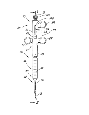
A preferred embo~;m~nt of the wound closure apparatus
lO is illustrated in Figure 2 and consists generally of a
cannula 30 having a distal section 32, a proximal section
5 34, and an intermediate section 36. In this particular
embodiment,the proximal section 34 comprises a handle 37,
the distal section 34 comprises a flexible catheter 45, and
the intermediate section 36 comprises a generally rigid
tube. The cannula 30 is otherwise defined by an axis 38
which extends between a proximal end 41 and a distal end 43
of the apparatus lO. The cannula 30 is generally hollow
in order to facilitate insertion of the apparatus lO over
the guidewire 18 which is shown at both the proximal and
distal ends 41 and 43 respectively. Graduations 47 can be
15 provided on the exterior surface of the intermediate
section 3 6 in order to measure the depth of the wound 12
relative to the skin 23. Radiopaque marker rings can also
be provided on the catheter 45 to facilitate fluoroscopic
guidance and X-ray contrast.
The handle 37 includes a pair of stationary finger
rings 52, and an actuator in the form of an axially movable
finger ring 54 which is discussed in greater detail below.
A finger tab 56 is also movable along the handle 37. This
finger tab 56 rides within an elongate groove 58 which
25 includes a slot 61 having a proximally facing shoulder 63.
The embodiment of Figure 2 is also provided with a Touhy-
Borst valve 65 which can be tightened to seal against the
guidewire 18. Backbleeding through the catheter 45 can be
evidenced through the Touhy-Borst valve 65 to provide a
30 visual indication that the catheter 45 is located within
the artery 14.
The interior regions of the c~nnl~la 30 are illustrated
in Figure 3. From this view it can be seen that the
catheter 45 in this particular embodiment extends from the
35 proximal end 41 and exits the cannula 30 through the distal
CA 0220~l76 l997-0~-l2
WO 97/03613 PCT~96/11118
end 43. Thus the catheter 45 forms a hollow tube within
the hollow tube of the ~nm~la 30. An inner channel 70
associated with the catheter 45 iS adapted to receive not
only the guidewire 18, but also a suture 72 which is
described in greater detail below. An inner ~h~nnel 74
associated with the c~nnllla 30 iS provided with a plug 76
at its distal end 43. This plug 76 has a distally facing
outer surface 78, a proximally facing shoulder 79, and an
annular recess 81 terminating within the channel 74 at a
proximally facing surface 83. Radially outwardly of the
recess 81, a pair of longitll~;n~l needle bores 85, 87 are
provided which extend generally parallel to the axis 38
from the cannula channel 74 to the outer surface 78. A
pair of needles 90, 92 are mounted for reciprocal movement
within the respective bores 85 and 87. These needles 90,
92 are carried by a common radial flange 94 which is
disposed within the ~h~nnel 74 of the c~nnl~la 30 but is
suitably apertured to receive the catheter 45 along the
axis 38. On the distal side of the flange 94, a needle
20 return spring 96 iS disposed to extend from the surface 83
of the recess 81 to the flange 94. On the proximal side of
the flange 94, a travel slack spring 98 extends from the
flange 94 to a distally facing surface 101 of an elongate
cylinder 103. This cylinder 103, which is disposed coaxial
25 with and interiorly of the c~nnlll a 30, and is moveable by
operation of the finger ring 54 within the channel 74 of
the c~nnllla 30. The catheter 45 extends through the
cylinder 103 so that the cylinder 103 occupies a generally
cylindrical space between the catheter 45 and the cannula
30 30.
In this embodiment of the wound closure apparatus 10,
the finger ring 54 and attached cylinder 103 together with
the springs 96, 98, the needles 90, 92 and associated
flange 94 function as a deployment mechanism to initially
35 deploy and ultimately retract the needles 90, 92. As the
CA 0220~l76 l997-0~-l2
WO97/036l3 PCT~S96/11118
finger ring 54 is moved distally, the surface 101 of the
cylinder 103 presses against the spring 98 which in turn
forces the flange 94 and attached needles 90, 92 distally
against the bias of the spring 96. Thus, distal pressure
on of the finger ring 54 moves the needles 90, 92 from a
proximal position best illustrated in Figure 3 to a distal
position best illustrated in Figure 5.
When the finger ring 54 is moved backwardly or
proximally, the cylinder 103 withdraws from the spring 98
and the force of the compressed spring on the flange 94
causes the needles 90, 92 to return from the distal
position illustrated in Figure 5 toward the proximal
position illustrated in Figure 3. The combination of the
cylinder 103 and finger ring 54 is also provided with a
trip mechanism in the form of a tang 105 which rides within
the groove 58 to engage a fl~nge lû7 on the finger tab 56.
This flange 107 has an inclined surface lû9 and a distal
facing shoulder 111, discussed in greater detail below.
A suture deployment mechanism 114 can be formed as
part of the catheter 45 in general proximity to the distal
surface 78 associated with the c~nnllla 30.
In a preferred embodiment, this suture manipulating
me~h~n;sm 114 is formed by a plurality of slits 116 (best
shown in the radial cross section view of Figure 3B) which
are spaced around the circumference of the catheter 45.
These slits 116 define a plurality of fingers 118 perhaps
best shown in Figure SA. Each of the fingers 118 can be
provided with an intermediated living hinge 121 which
facilitates radial expansion of the fingers 118 when the
catheter 45 is axially compressed. This radial expansion
occurs between a first position illustrated in Figure 3,
where the fingers 118 have a relatively low profile, to a
second position illustrated in Figure 4, where the fingers
118 have a relatively high radial profile. In combination,
the expandable fingers 118 form a device comm~nly referred
CA 0220~l76 l997-0~-l2
WO 97/03613 PCT~S96/11118
to as a Mallicot structure. Between the first, low profile
position and the second, high profile position of the
fingers 118, there exists a natural position described
below with reference to Figure 6.
The suture manipulating mechanism 114 also includes
the finger tab 56 and a projection 123 which extends from
the tab 56 through the groove 58 and into the channel 74 of
the c~nnllla 30. An elongate element 125 iS fixed at its
proximal end to the a projection 123 and at its distal end
10 to a flange 127 at the distal tip of the apparatus 10.
This flange 127, which is apertured to receive the
guidewire 18, iS at least as large as the catheter 45 at
the distal end 43 of the apparatus 10. It is the purpose
of the finger tab 56, the element 125 and the flange 127 to
15 move the fingers 118 between the low profile position and
the high profile position. As the finger tab 56 is moved
prox;m~lly, from the solid line position to the dotted line
position in Figure 3, the element 125 iS tensioned thereby
drawing the distal flange 127 proximally. This creates an
20 axial compression force on the catheter 45 which causes
each of the fingers 118 to buckle at its ends and at the
associated living hinge 21 thereby resulting in expansion
of the finger 118 forming the Mallicot structure.
These fingers 118 can be maintained in the second,
25 expanded position automatically by ensuring that the flange
107 associated with the finger tab 56 is approprial_ely
lodged within the slot 61 associated with the groove 58.
Thus, the finger tab 56 can be locked in the proximal,
dotted position when the flange 107 falls into the slot 61
30 and the distally facing surface 111 of the flange 107
engages the proximally facing surface 63 of the slot 61.
The resulting lock, which holds the tab 56 in its proximal
position and holds the fingers 118 in their second expanded
state, is released in a preferred embodiment when the tab
105 associated with the finger ring 54 and cylinder 103
CA 0220~176 1997-0~-12
WO97/03613 PCT~S96/1l1l8
engages the flange 107 forcing it out of the slot 61. When
the lock is released, it is the natural tendency of the
fingers 118 to move back toward the low profile state.
This forces the distal flange 127 distally tensioning the
element 125 and drawing the finger tab 56 distally, from
the dotted position to the solid line position in Figure 3.
Distal finger pressure on the tab 56 can also facilitate
movement of the fingers 118 from the high profile state
toward the low profile state if the elongate element 125
can accommodate a compressive load.
This operation of the suture manipulating mechanism
114 and the expandable fingers 118 make this structure
particularly useful in deploying the suture 72. As best
illustrated in Figures 3 and 4, the suture 72 includes free
ends 130 and 132 which can be embedded in or near the
living hinge 121 of adjacent fingers 118. Between the ends
130 and 132, the suture forms a suture loop 134 which is
relatively long and extends from the fingers 118 into the
channel 70 of the catheter 45 and along substantially the
entire length of the ~nn~lla 30 where it exits the catheter
45 through a pathway or hole 136. Exteriorly of the
catheter 45, the suture loop 134 engages a suture
tensioning device 138 in the form of a tab 141 formed in
the wall of the cylinder 103. This tab 141 includes a post
143 which extends radially outwardly to receive the suture
loop 134. The tab 141 is compressible radially inwardly by
a projection 145 on the inner surface of the handle 37. As
the cylinder 103 moves axially, by operation of the finger
ring 54, the projection 145 engages the tab 141 and moves
it radially inwardly. This causes the suture 72 to clear
the post 143 effectively dislodging the suture loop 134
from the tensioning device 138. This feature is
particularly advantageous during operation of the apparatus
10 for reasons discussed in greater detail below.
CA 0220~176 1997-0~-12
WO 97/03613 PCT~S96/11118
A preferred method for attaching the suture 72 to the
fingers 118 can be best understood with reference to Figure
5A. In this embodiment, the fingers 118 are further
designated by the reference numerals 152, 154, 156, and
158. In the view of Figure 5A, the fingers 152-158 are
fully extended in the second position. The associated
living hinges 121 are disposed at the bends of the fingers
152-158 where the fingers reach their maximum radial
distance from the axis 38.
With such an embodiment, the suture end 130 can be
loosely embedded in the finger 154, the suture can then be
led from the finger 154 and through a slit in the living
hinge 121 associated with the finger 156. From this point,
the suture loop 134 can be fed into the catheter 45 and
15 along the length of the cannula 30. In a similar manner,
the suture end 13 2 can be loosely embedded in the finger
152 and led through a slit in the living hinge 121
associated with the leg 158. With this orientation, each
of the suture ends 130, 132 forms a short section of suture
20 which extends between adjacent legs. Thus, the suture end
130 extends between adjacent legs 154, 156 to form a suture
portion 159, while the suture end 132 extends between
opposing adjacent legs 152, 158 to form a suture portion
160.
Loading of the wound closure apparatus 10 can be
accomplished during the final stage of manufacture. The
finger tab 56 on the handle 37 is initially set to the
distal position illustrated by the solid lines in Figure 3.
This ensures that the fingers 118 forming the Mallicot
structure are in the low profile state. The suture 72 can
then be mounted on the fingers 118 and the suture loop 134
and threaded through the channel 70 of the catheter 45.
After exiting the hole 136 in the catheter 45, the suture
loop 134 is disposed around the post 143 in the suture
35 tensioner 138. Finally, the movable finger ring 54 is
CA 0220~l76 l997-0~-l2
WO97/036l3 PCT~S96/111l8
placed in its proximal position so that the needles 90, 92
are retracted into the associated bores 85, 87. The Touhy-
Borst valve 65 can also be loosened to allow insertion over
the guidewire 18.
Operation of the wound closure apparatus is begun by
inserting the catheter 45 into the wound 12. If the wound
12 is in the skin 23 of the patient 16, the catheter 45 is
introduced directly into the wound 12. If however, as
illustrated in Figure 1, the wound occurs in a body wall,
such as the wall of the femoral artery 14, the small
incision 21 can be made in the skin 23 in order to gain
access to the artery 14. The guidewire 18 will typically
have been placed through the incision 21 and wound 12 as
part of the surgical procedure which developed the wound
12.
With the guidewire 18 in place, the apparatus 10 can
be positioned over the guidewire 18 so that the distal tip
of the catheter 45 follows the guidewire through the
incision 21 and the wound 12 into the artery 14. At this
point, the graduations 47 on the c~nnllla 30 will provide an
indication of the depth of the artery 14 relative to the
skin 23. Radiopaque markers on the catheter 45 can be
useful to facilitate fluoroscopic guidance to an operative
position. The Touhy-Borst valve 65 also provides a visual
indication of backbleeding which would evidence proper
placement within a blood vessel. The valve 65 can also be
provided with a T-fitting to facilitate injections of X-ray
contrast media through the catheter 45.
With reference to Figure 3, it will be noted that the
body wall, in this case a body wall 161 of the artery 14,
has a near side 163 and a far side 165. Since access to
the far side 165 of the body wall 161 is severely limited,
it is of particular advantage to the present invention that
wound closure can be achieved without direct access to the
far side 165 except through the wound 12.
CA 0220~l76 l997-0~-l2
WO97/036l3 PCT~S96/111l8
14
In the pre~erred method, the catheter 45 is inserted
through the wound 12 a distance sufficient that the fingers
118 forming a Mallicot structure are positioned on the far
side 165 of the body wall 161. The cannula 30 including
the needles 90 and 92, remain on the near side 163 of the
body wall 161. With this orientation, it will be noted
that the suture ends 130, 132 illustrated in Figure 4 are
positioned on the far side 165 while the suture loop 134 is
positioned on the near side 163 of the body wall 161.
This preferred operative position for the apparatus 10
is achieved by operation of the handle 37 and associated
stationary rings 52. With the apparatus 10 thus disposed,
the fingers can be expanded from their first low profile
state illustrated in Figure 3 to their second exp~n~
state illustrated in Figure 4. Note that this expansion of
the fingers 118 results in the radial outward movemenl_ of
the suture portion 159, 160.
With the suture portions 159, 160 appropriately
positioned, the needles 90, 92 can now be deployed. These
needles 90, 92 are initially disposed in their associated
bores 85, 87 on the near side 163 of the body wall 161.
Deployment of the needles 90, 92 is accomplished by moving
the finger ring 54 and associated cylinder 103 distally in
the direction of an arrow 167 in Figure 5. The distally
facing surface 101 of the cylinder 103 compresses the
spring 98 which in turn exerts a force on the needle flange
94. This force, which is increased with further distal
movement of the finger ring 54 against the bias of the
spring 96, moves the needles 90, 92 beyond the distal
surface 78 and through needle holes 170 and 172
respectively, in the body wall 161. Distal movement o~ the
needles 90, 92 ceases when the flange 94 bottoms out on the
proximal facing surface 79 of the plug 76.
The needles 90, 92 each have a needle eye 176, 178
respectively, which in a preferred embodiment is in the
CA 0220~l76 l997-0~-l2
WO97/03613 PCT~S96tlll18
form of a French eye. When the needles 90, 92 are fully
extended, these eyes 176, 178 are preferably disposed on
the far side 165 of the body wall 161 and beyond the suture
portions 159, 160. With the suture portions 159, 160 in
5 their radially extended position, the needles 90, 92 in
their distal-most position, and the needle eyes 176, 178
facing the suture portions 159, 160, the suture 72 can now
be manipulated to thread the needles 90, 92.
In a preferred embodiment, this manipulation of the
suture 72 is accomplished by further distal movement of the
finger ring 54 to a second position where the tang 105,
riding within the groove 58, engages the inclined surface
109 of the flange 107. This engagement of the flange 107
by the tang 105 releases the finger tab 56 from its locked
15 proximal position. This relieves tension on the element
125 and enables the fingers 118 to radially compress
naturally as the catheter 45 elongates. The radial
compression of the fingers 118 moves the suture portions
159, 160 against the associated needles 90, 92 at a
position proximal to the eyes 176, 178.
Of course with the radial compression of the fingers
118, the distance between the adjacent living hinges 121 is
reduced. This would normally cause the suture portions
159, 160 to become slack were it not for the suture
25 tensioning mechanism 138 previously discussed. It will be
noted that as the finger ring 54 is moved distally, the
suture tensioning mechanism also moves distally. Since the
hole 136 in the catheter 45 is disposed proximally of the
tensioning mechanism 138, the distal movement of the
30 mech~n;.~m 138 creates proximal movement of the suture loop
through the catheter ch~nn~1 70. This in turn tensions the
suture portions 159 and 160 even as the fingers 118 are
radially contracting.
To this point, distal movement of the finger ring 54
35 has operated to deploy the needles 90, 92 in a first
CA 0220~176 1997-0~-12
W097/03613 PCT~S96/11118
16
position, and to manipulate the fingers 118 in order to
thread the needles in a second position of the finger ring
54. This manipulation of the fingers 118 in the second
position is accomplished while tensioning the sutures 72
5 with the mech~n;~m 138.
Further distal movement of the finger ring 54 to a
third position results in removal of the suture loop 134
from the post 143 of the tensioner 138. This has the
effect of releasing the suture loop 134 from the apparatus
10 so that it is free to be drawn into the wound 12. This
freeing of the suture loop 134 iS accomplished as
illustrated in Figure 3B when distal movement of the
cylinder 103 brings the tab 141 into proximity with the
projection 145 on the handle 3 7. This interfering
15 relationship causes the tab 141 to compress radially
inwardly thus forcing the suture loop 134 from the post 143
as illustrated in Figure 3D.
It will be noted at this point that movement of the
finger ring 54 in a common distal direction operates to
20 perform three sequential steps in a method for operating
the apparatus 10. In a first step associated with first
distal position of the ring 54, the needles 90, 92 are
deployed to a position where they can be threaded. In a
second step associated with a second position of the ring
25 54, the finger tab 56 is released thereby permitting
contraction of the fingers 118 to facilitate threading the
needles 90, 92. In a third step associated with a third
position of the finger ring 54, the suture loop 134 iS
released from the tensioning mechanism 138 to facilitate
30 withdrawal of the apparatus 10.
With the suture portions 159, 160 tensioned radially
against the needles 90, 92 proximally of the eyes 176, 178,
the needles 90, 92 can now be retracted. Initially this
retraction of the needles 90, 92 causes the tensioned
suture portions 159, 160 to enter the eyes 176, I78.
CA 0220~176 1997-0~-12
WO 97/03613 PCT/US96/11118
17
Further retraction of the needles 90, 92 withdraws the
captured suture portions 159, 160 through the associated
needle holes 170, 172 in the skin 23. At this point, both
of the suture ends 130, 132, as well as the suture loop
134, are disposed on the near side 163 of the wall 161.
The apparatus 10 can now be withdrawn from the wound 12.
The needles 90, 92 are preferably retracted until the
eyes 176, 178 enlarged by the presence of the suture
portions 152, 154 are biased against the distal surface 78
at the distal end of the cannula 30. In this position of
the needles 90, 92, illustrated in the enlarged view of
Figure 6A, the suture ends 130, 132 are trapped between the
respective needles 90, 92 and the c~nnllla 30 due to return
spr1ng force.
With the suture loop 134 released from the post 143 of
the tensioner 138, removal of the apparatus 10 will draw
the suture portions 159, 160 further away from the body
wall 161 as illustrated in Figure 7. This causes the
suture loop 134 to be drawn distally through the catheter
45 until it exits from between adjacent legs 156, 158 and
is drawn through the wound 12 to the far side 165 of the
body wall 161.
Once the wound closure apparatus 10 has been fully
removed from the wound 12, the suture ends 130, 132 can be
removed or cut from the fingers 118 and tied with a
surgeon's knot in a conventional manner. Such a knot can
be pushed through the incision 21 and into proximity with
the wound 12 as illustrated in Figure 8.
An additional embodiment of the invention is
illustrated in Figure 10 and includes two needle pairs.
The needles 90 and 92 previously discussed form one of the
needle pairs while an additional needle pair comprises
needles 201 and 203. In this case, the four needles 90,
92, 201 and 203 are displaced 90 from each other around
the distal surface 78 of the ~nnllla 30. The neédle
CA 0220~l76 l997-0~-l2
WO 97/03613 PCT~S96/11118
18
deployment (not shown in Figure 10) can be similar to that
previously discussed where the four needles 90, 92, 201 and
203 are fixed to the common flange 94 (best illustrated in
Figure 4) .
This embodiment of the invention functions to
simultaneously apply two sutures to the wound 12. The
first suture can be that previously referenced with the
numeral 72. This suture can be laced to the suture
manipulating mechanism illustrated in Figure 9 in the same
m~nner as discussed with reference to Figure 5. A second
color-coded suture 205 can also be laced on to the legs
152-158 of the Mallicot structure in the manner illustrated
in Figure 9.
This suture 205 having a first end 207 and a second
end 209 iS also characterized by a suture loop portion 210.
In a preferred method, the end 207 of the suture 205 iS
loosely held in the leg 154 of the Mallicot structure. The
suture is then laced through the living hinge 121 and the
leg 152 with the suture loop 210 extending into the ch~nnel
20 associated with the catheter 45. AS the suture loop exits
this channel, it can be passed through the living hinge 121
associated with the leg 158. Finally, the end 209 of the
second suture 205 can be loosely embedded in the leg 156.
This configuration for the second suture 205 produces
a suture portion 212 between the legs 152 and 154 and a
second suture portion 214 between the legs 156 and 158. In
this manner, the two sutures 72 and 205 can form the four
suture portions 159, 160, 212 and 214 for the respective
30 needles 90, 92, 201 and 203. The resulting suture
deployment mechanism can be operated in the manner
previously discussed to thread each of the four needles
associated with the embodiment of Figure 10.
The dual suture embodiment of the suture deployment
35 mechanism illustrated in Figure 9, can also be used with
CA 0220~176 1997-0~-12
WO97/03613 PCT~S96/11118
19
the two needle embodiment of the apparatus 10 previously
discussed. In a preferred method of operation, the two
needles 90-92 can be operated in the manner previously
discussed to capture the suture portions 159 and 160. Then
the cannula 30 and needles 90, 92 can be rotated 90 to
function with respect to the suture portions 212 and 214.
Of course in this embodiment, means must be provided for
rotating the c~nnl31a 30, and needles 90, 92 relative to the
legs 152-158 forming the Mallicot structure.
Figure 12 illustrates a further embodiment of the
wound closure apparatus 10. This embodiment includes the
cannula 30 and catheter 45, as well as the fingers 118 and
associated suture 72. The illustrated embodiment also
includes the needles 90, 92, but in this case, the needle
eyes (previously designated by the reference numerals 176,
178) are not formed as French eyes, but rather as mere
slots 221, 223. As compared to the French eyes previously
discussed, the slots 221, 223 can be formed with a
significantly lesser depth. As a result, the needles 90,
92 can provided with a much smaller diameter.
The embodiment of Figure 12 provides for effective
suture capture with the provision of suture capture needles
225 and 227. These needles 225 and 227 can be provided
with a tubular configuration and a telescoping relationship
with the associated needles 90, 92. The distal ends of the
suture capture needles 225, 227 can be sharpened to
facilitate their deployment through the body wall 17.
Operation of the Figure 12 embodiment proceeds in the
manner previously discussed with the needles 90, 92 being
deployed through the body wall 17. With the more narrow
configuration of the needles 90, 92, this step of deploying
the needles should be more easily accomplished than in the
Figure 5 embodiment. As the suture 72 is manipulated by
the fingers 118 in the manner previously discussed, the
suture 72 is drawn into the slots 221, 223 of the
CA 0220~l76 l997-0~-l2
W097/03613 PCT~S96tllll8
' 20
respective needles 90, 92. At this point, the suture
capture needles 225, 227 can be deployed by moving them
distally along the associated needles 90, 92. As the
distal end of the capture needles 225, 227 approach the
slots 221, 223, the suture 72 iS pinched between the
associated needles 90, 225 and 92, 227.
With the suture 72 appropriately captured in the slots
221, 223, the needles 90, 92 and associated capture needles
10 225, 227 can be withdrawn toward the cannula 30.
Preferably after the needles 90, 92 and 225, 227 have
cleared the body wall 17, the entire apparatus 10 can be
withdrawn from the wound 12 as the method of closure
continues in the manner previously described.
Given the wide variations in the concept embodied in
the foregoing apparatus and methods, one is cautioned not
to restrict the invention to the embodiments which have
been specifically disclosed and illustrated, but rather is
encouraged to determine the scope of the invention only
20 with reference to the following claims.