Note: Descriptions are shown in the official language in which they were submitted.
CA 022061~6 1997-0~-14
Inventor: Robert Lee Thompson
Title: SURGICAL/DIAGNOSTIC IMAGING DEVICE
TECHNICAL FIELD OF THE lNV~I.LlON
This invention is related to an imaging device for use in
interabdominal, interthoracic, and other surgical and
diagnostic procedures on the human body.
BAC~GROUND
Endoscopic surgery and diagnosis are considerably less
invasive than the conventional procedures. This results in a
lower mortality rate and minimizes both the patient's hospital
stay and recovery time.
Conventional endoscopes include a rigid elongated member,
a lens assembly, and an imaging device mounted either on or
within the endoscope. Examples of such endoscopes are
described in U.S. Patents Nos. 4,697,210 (Toyota et al.),
4,791,479 (Ogiu et al.), and 4,989,586 (Furukawa).
Although a conventional endoscope can be constructed to
have a wide field of view, the picture quality suffers. As a
practical matter, the field of view of conventional endoscopes
must be relatively narrow. As a result, a conventional
endo~cope mu~t be po~itioned carefully at the beginning of a
procedure, then held in position throughout the procedure,
which generally requires the full-time attention of one member
of the operating team. U.S. Patent No. 5,351,678 (Clayton et
al.) addresses the initial positioning problem by providing an
endoscope having a distal end which is offset from the
--1--
CA 022061~6 1997-0~-14
endoscope's longitudinal axis. With the Clayton et al.
endoscope, the surgeon can easily change the area viewed by
rotating the endoscope about its longitudinal axis. However,
the Clayton et al. endoscope must still be held in place
throughout the procedure by a member of the operating team.
SUMNARY OF THE lNv~ION
A surgical/diagnostic imaging device embodying the
invention includes a charge-coupled device ("CCD") and an
associated lens mounted within a camera bore in a camera
housing. The camera housing is pivotally mounted at the distal
end of an elongated camera support. High intensity lights are
also mounted within bores in the camera housing that are
coaxial with the camera bore and thus with the axis of the CCD.
Prior to use, the camera housing and camera support tube
are inserted into a disposable sterile sheath. The distal
portion of the sheath is then inserted into the patient through
an incision in the patient. Electric stepper motors and
associated components are provided to move the camera housing
(and thus the CCD) in elevation and azimuth.
The imaging device is electrically connected to a control
console. The control console is in turn electrically connected
to a display device and a control assembly. The display device
displays the image received by the CCD and the control assembly
allows the surgeon to control the elevation and azimuth of the
camera housing.
The surgical/diagnostic imaging device is easily aimed at
the area of interest within the patient by the surgeon. In
addition, surgical/diagnostic imaging device need not be held
in position in the patient by a member of the operating team.
--2--
CA 022061~6 1997-0~-14
BRIEF DESCRIPTION OF THE DRAWINGS
An embodiment of the invention will be described, by way
of example only, with reference to the accompanying drawings,
in which:
s Fig. 1 is a front view of a surgical/diagnostic imaging
device in accordance with the invention;
Fig. 2 is a partially cutaway side view of the imaging
device of Fig. 1;
Fig. 3 is a cutaway top view of the sheath cap taken
through plane 3-3 in Fig. 1;
Fig. 4 is an enlarged cutaway side view of the upper
housing and the lower portion of the imaging device;
Fig. 5 is a cutaway top view of the upper housing taken
through plane 5-5 in Fig. 4;
Fig. 6 is an cutaway top view of the lower portion of the
imaging device taken through plane 6-6 in Fig. 4;
Fig. 7 is a block diagram of a system for controlling the
imaging device of Fig. 1 and for displaying the images
transmitted by the imaging device;
Fig. 8 is a block diagram of a second control and display
system for the imaging device of Fig. 1;
Fig. 9 is a cutaway side view of a second imaging device
in accordance with the invention;
- Fig. 10 is a front view of the camera housing shown in
25 Fig. 10; and
Fig. 11 is a cutaway side view of the camera housing taken
through plane 11-11 in Fig. 10.
DETAILED DESCRIP~ION
CA 022061~6 1997-0~-14
Figs. 1-3 show a surgical/diagnostic imaging device 1 for
use in interabdominal, interthoracic, and other surgical and
diagnostic procedures. The device 1 comprises an upper housing
3, a camera housing 5, and left and right camera housing
supports 7, 9. Before use, the device 1 is inserted into a
sterile sheath 11. The device 1 and sheath 11 (collectively,
the "camera") are then inserted through an incision (not shown)
into the patient's body (not shown). The camera is inserted
so as place the camera housing 5 in a position from which it
can be pointed at the surgical site or the area to be
diagnosed. The incision is sealed around the camera with a
purse string stitch, thereby preventing leakage of the CO2 gas
which is used to distend the patient's abdomen or chest during
surgery or diagnosis.
In this embodiment, the sheath 11 is constructed of
medical-grade plastic and is intended to be disposed of after
use. Alternately, the sheath 11 can be constructed of heat-
resistant materials in order to allow it to be sterilized using
an autoclave, then reused.
The camera housing S contains a CCD (not shown) and a zoom
lens assembly (not shown). A plurality of high intensity
lights 13 are mounted within a light housing 15 which extends
about the outer circumference of the camera housing 5. The
lights 13 are aligned with the focal axis 17 of the CCD, and
2S they provide illumination of the area at which the camera
housing 5 and hence the CCD are pointed.
When the device 1 is inserted in the sheath 11, the left
and right camera housing supports 7, 9 engage complimentary
locking keys 19, 21 within a sheath cap 23. As a result, the
~0 camera housing 5 is locked into a position in which the CCD's
--4--
CA 022061~6 1997-0~-14
focal axis 17 is aligned perpendicular to an optically-clear
window 25. In addition, as will be described below in
connection with Figs. 4-6, the locking keys 19, 21 cause the
sheath cap 13 to rotate about the longitudinal axis 27 of the
camera when the camera housing supports 7, 9 are rotated about
that axis.
A camera cable 29 extends between the camera housing 5 and
the upper housing 3. The camera cable 29 contains conductors
which carry the CCD's signals to the upper housing 3 and which
supply electrical power to the CCD and lights 13. An imaging
device cable 31 is provided to carry control signals and supply
electrical power to the device 1 and to carry the CCD's signals
to external processing and display devices (not shown).
The length of the camera housing supports 7, 9 and the
length of the sheath 11 are varied to accommodate variation in
the thickness of the abdominal walls of patients and to allow
the camera to be used in thoracic surgery/diagnosis. Three
lengths are provided: 3, 6, and 11 inches below the upper
housing 3.
Referring now to Figs. 4-6, an elevation motor 51 drives
an elevation shaft 53 by means of gears 55, 57. The elevation
shaft 53 extends downwardly through the hollow left camera
support 7. A ring and pinon gear arrangement 59 at the lower
end of the elevation shaft 53 transfe~s the rotary motion of
25 the ele~ation shaft 53 to the camera housing 15, thereby
causing the camera housing 15 to elevate or depress, depending
on the direction of rotation of the elevation motor 51. In
this embodiment of the invention, the camera housing 15 can be
elevated 70 degrees above horizontal and depressed 90 degrees
below horizontal.
--5--
CA 02206l~6 l997-0~-l4
The elevation motor 51 is mounted on a plate 63. The
plate 63 is rotably mounted within the upper housing 3 on a
bearing 65. An azimuth motor 67 is also mounted on the plate
63. The azimuth motor 67 drives an azimuth gear 69. The
5 azimuth gear 69 engages a housing gear 71 which is attached to
the inner surface of the upper housing 3. When the azimuth
motor 67 rotates, the plate 63 rotates within the upper housing
3. In this embodiment, the plate 63 rotates plus or minus 180
degrees in order to minimize the amount the camera cable 21 is
twisted. 360 degree rotation can easily be achieved by using
conventional slip rings.
A zoom/focus motor 72 drives gears 73, 75, which rotate
a zoom/focus shaft 77. The zoom/focus shaft extends downwardly
through the right camera support 9. At the bottom of the focus
15 shaft 77, a ring and pinon arrangement 79 transfers the rotary
motion of the focus shaft 77 to a zoom lens mechanism (not
shown) within the camera housing 5.
Referring now to Fig. 7, the imaging device 1 is connected
to a control console 101 by means of the imaging device cable
20 31. Signals from the CCD of the imaging device 1 are amplified
by circuits in the control console 101 and directed to a
display device 103. In this embodiment of the invention, the
display device 103 is a conventional television set.
A foot pedal control assembly 105 allows the surgeon (not
shown) to control the imaging device 1. The foot pedal control
assembly 105 includes four controls (not shown): (1) camera
housing left and right; (2) camera housing up and down; (3)
zoom in and out; and (4) light intensity up and down. Signals
from the foot pedal control assembly 105 are routed to the
30 control console 101. Circuits (not shown) in the control
--6--
CA 02206156 1997-0~-14
console 103 convert the control assembly signals into signals
which are suitable to control the imaging device 1, then route
the converted signals to the imaging device 1.
In the embodiment of the invention shown in Fig. 8, a
computer 107 is interposed between the control console lol and
the display device 103. A plurality of computer programs
contained in the computer 107 allow operating team personnel
to manipulate and/or store the signals from the imaging device
1.
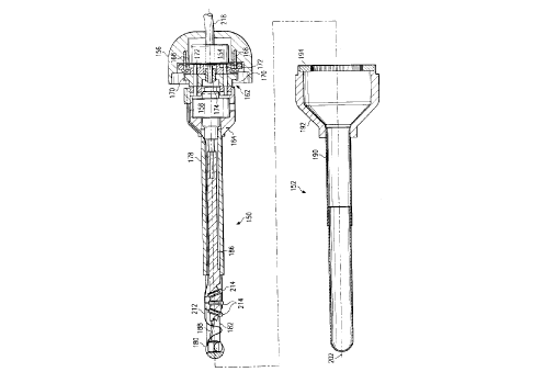
Figs. 9-11 illustrate a second surgical/diagnostic imaging
device in accordance with the invention. Referring first to
Fig. 10, the imaging device comprises two major assemblies: a
camera assembly 150 and a disposable sheath assembly 152.
In the camera assembly 150, a rotary stepper motor 154 is
rigidly mounted in an upper housing 156. A linear stepper
motor 158 and the distal end of a planetary gear assembly 162
are press fitted in a linear stepper motor housing 164. The
proximal end of the planetary gear assembly 162 is attached to
the upper housing 156 by screws 168.
Three planetary gears 170 (only two of which are shown in
Fig. 9) are rotably mounted on pins 172 within the planetary
gear assembly 162. The rotary stepper motor 154 drives the
planetary gears 170 through a sun gear 174.
The proximal end of a camera support tube 178 is press
fitted in the linear stepper housing 164. A camera housing 180
is pivotally mounted between pair of arms 182 (only one of
which is shown in Fig. 9) that are integral with and extend
from the distal end of the camera support tube 178. The linear
stepper motor 158 acts through a pushrod 186 and a fork 188 to
control the elevation of the camera housing 180.
--7--
CA 022061~6 1997-0~-14
The disposable sheath assembly 152 comprises a sheath 190,
a sheath housing 192, and a ring gear 194. The distal portion
of the sheath 190 is optically clear. The proximal end of the
sheath 190 is adhesively attached within the distal end of the
sheath housing 192. The ring gear 194 is adhesively attached
within the proximal end of the sheath housing 192.
Prior to use, the camera assembly 150 is inserted into the
sheath assembly 152, and the planet gears 170 engage the ring
gear. As a result, when the rotary stepper motor 154 is
actuated, the camera assembly 150 rotates in relation to the
longitudinal axis 202 of the sheath assembly .
As is best shown in Figs. 10 and 11, a CCD assembly 204
and a lens 206 are mounted within a camera bore 208 in the
camera housing 180. A pair of high intensity lights 210 are
mounted in bores that are coaxial with the camera bore 208.
A multi-conductor flexcable 212 provides the necessary
connections for the CCD assembly 204, for the camera housing
lights 210, and for three high intensity lights 214 that are
disposed in bores in the pushrod 186. The flexcable 212 extends
from the camera housing 180 to the upper housing 156. In the
upper housing 156, the flexcable 212 is combined with power and
control wires (not shown) for the rotary stepper motor 154 and
the linear stepper motor 158 to form the camera assembly cable
218. The camera assembly cable 218 passes through an orifice
2S 220 in the upper housing 152. As with the embodiment of the
invention shown in Figs. 1-8, the camera assembly cable 218
connects the camera assembly 150 to external display and
control devices (not shown).