Note: Descriptions are shown in the official language in which they were submitted.
CA 02207871 1997-05-30
WO 96/220S2 PCTJIJS9SJ16703
- 1 -
BREATHABLE LIOUID ELIMINATION ANALYSIS
Field of the Invention
T_is invention relates to methods and processes for determining
S and controlling the amount of interaction in a "~;I"""~ n lung between a
breathable liquid contained therein and respiratory gas in the lung. In addition,
this invention relates to ~ "lilication of perfluorocarbon (PFC) volume loss
from a system. This invention also relates to methods and processes for
~l~tecting breathable liquid vapors and employing measured values to detect and
10 control breathable liquid recovery ~aldLus and other ~ n body
functions. T_is invention also relates to methods and processes for (1etecting
breathable liquid vapors and employing measured values to monitor the
efficiency of an oxygen/carbon dioxide e7~ch~nger system.
15Back~round of the Invention
~r~mm~ n lcs~ildtion occurs by gas exch~n~e through air sacs
or alveoli in the lungs, and thus is referred to as "alveolar ventil~tion." Fig.1 (prior art) shows pulmonary passageways which deliver and remove
20 re~ildloly gases to and from the alveoli of lungs 200. In successive order,
these passageways include the larynx 202, trachea 204, bronchus 206 and
~ segmental bronchi or bronchioles 208. The bronchioles 208 termin~te in small
clusters of grapelike air sacs 210 (the alveoli) where the gas exchange occurs.
Fig. 2A (prior art) shows one alveolus 212 of the alveoli 210 and
25 Fig. 2B (prior art) diagr~mm~ti~ lly represents the gas exchange through the
alveolus 212. A network of blood capillaries 214 covers or ~ullounds the
CA 02207871 1997-0~-30
W O 96/22052 PCTrUS95/lG703
alveolar walls 216. The gas-filled interior region of the alveolus 212 and the
network of capillaries 214 are separated by less than 0.5 ~Lm of intervening
tissue. The gas exchange in alveolar lungs can be modeled as a ventil~t~cl pool
218, as shown in Fig. 2B.
During liquid ventilation, the pulmonary passageways of the lungs
are filled with a breathable liquid which has the ability to deliver oxygen to, and
remove carbon dioxide from, the pulmonary system. Two common types of
liquid ventilation processes include "total liquid ventilation" and "partial liquid
ventilation. "
In a total liquid ventilation system, a breathable liquid is
oxygenated and pumped or instilled into the lungs during an inspiratory
breathing stage. When the breathable liquid reaches the alveoli, the oxygen in
the breathable liquid diffuses into the blood of the capillaries ~ulloullding
individual alveolus. Correspondingly, carbon dioxide in the blood diffuses into
the breathable liquid. The breathable liquid is then pumped out or removed
from the lungs during an expiratory bl~aLl~illg stage. The expired liquid is
scrubbed to remove the carbon dioxide, reoxygenated and returned to the lungs
during a subsequent inspiratory blcathillg stage. A l~h~lor typically performs
the breaLlling stages. Such systems are described in U.S. Patent No. 5,335,650
and U.S. Patent No. 5,158,536, both of which are incorporated by reference
herein in their entirety.
In a partial liquid ventilation system, a breathable liquid is
instilled into the lungs and remains therein. This system is often employed
when the lungs are collapsed since the volume of the breathable liquid functionsto expand the lungs. The breathable liquid fills the alveoli. Then, respiratory
gas is pumped into and out of the lungs. Oxygen carrying inspiratory
respiratory gas interacts with the breathable liquid and releases the oxygen to
the breathable liquid. In turn, the breathable liquid releases the oxygen into the
blood ~ullounding the alveoli in the same manner as described above in the totalliquid ventilation system. Likewise, carbon dioxide in the blood diffuses into
CA 02207871 1997-0~-30
W O 96/220~2 PCTAUS9S/16703
the breathable liquid, which in turn, diffuses into areas of the lungs not takenup by the breathable liquid. During the expiratory phase, e~ dtoly gas
(including the carbon dioxide) exits the lung. As noted above, during partial
liquid ventilation, the breathable liquid remains in the lungs, acting as an
5 exchange mP~ m for the oxygen and carbon dioxide entering and exiting the
lungs. Partial liquid ventilation, as performed today, is not a closed loop
system.
Breathable liquids employed today have various vapor pressures.
During partial liquid ventilation, a small amount of the breathable liquid will
10 volatilize or vaporize with each breathing cycle by saturating the respiratory
gas. That is, the vapor pressure of the breathable liquid causes gas vapors
coming off the liquid to saturate the respi,dlory gas as the gas flows through and
around the liquid. During the expiratory phase, the saturated or partially
saLuldled gas leaves the l~haloly system. Since partial liquid ventilation is
15 not a closed loop system, the vol~tili7~-1 breathable liquid must eventually be
replaced by a new in~till~tion of breathable liquid into the patient's lungs.
During partial liquid ventilation, a portion of the breathable liquid
is also lost due to evaporation into the lungs. Some of this evaporated liquid
becomes absorbed by the lungs and eventually leaves the patient's body by
20 I,dlls~ lion through the skin. Si nifir~nt problems still exist in performingtotal and partial liquid ventilation. During total liquid ventilation, the breathable
liquid also undergoes vol~tili7~tion and dissolves in the expiratory liquid. Total
liquid ventilation systems employed today scrub dissolved carbon dioxide from
the expiratory liquid before the gas is reo~yg~ d and cycled back into the
25 patient's lungs. This process occurs in an oxygenator/diffuser circuit. Not all
of the carbon dioxide is scrubbed from the diffuser. Furthermore, none of the
vaporized breathable liquid is recovered in the scrubber. Tn~te~17 it is vented
to the environment. Accordingly, the system must periodically add more
breathable liquid from a storage reservoir. This increases the cost of the liquid
CA Oi207871 1997-0~-30
WO 96122052 PCT/US9S/16703
ventilation process since breathable liquid is expensive (e.g., as much as
$2.00/ml).
During partial liquid ventilation, an operator must continually
monitor the process to ensure that sufficient alveolar ventilation is occnrring.5 One important aspect of the moniL(,~ g is to ensure that there is a sufficient~lu~Lily of breathable liquid in the lungs to promote the desired amount of
alveolar ventilation. Alveolar ventilation can be c~"l")ro"~ised if the volume of
liquid in the lung becomes too small.
Current techniques for m~cllring the amount of breathable liquid
10 in the lungs are inaccurate and inadequate. One technique employed today
involves merely replenishing the supply of breathable liquid in the lungs until
they are filled. This is supposedly accomplished by vicll~li7ing a meniscus of
PFC in the endotracheal tube. However, it is not always nPce~c~ry or desirable
to completely fill the lungs to achieve the desired amount of alveolar ventilation.
15 Accordingly, the operator does not know for sure how much breathable liquid
to add as vol~tili7~tion depletes the store of liquid.
Or~ ;,n~s, the breathable liquid becomes m~l~1i.ctributed
throughout the lungs due to patient movement or density differences which
cause liquids to settle and gases to rise. For example, some bronchioles may
20 have little or no breathable liquid to supply the alveoli at their distal ends,
whereas other bronchioles may be overfilled. This m~l-li.ctribution may also
cause in~lffirient i"L~ldclion between the breathable liquid and the l~hdLory
gas. Atelectasis may also cause insufficient interaction between the breathable
liquid and the respil~Lory gas. Atelectasis is the collapse of the e~rran-l.od lung
25 or the defective expansion of the pulmonary alveoli at birth. Currently, the
operator of a liquid ventilation system has no sure technique for g~nging
whether insufficient alveolar ventilation is the result of an in~dequ~tf~ quantity
of breathable liquid in the lungs, m~l~li.ctribution of the breathable liquid oratelectasis.
CA 02207871 1997-0~-30
W O 96122052 PCT~US9~16703
S
Furthermore, the vol~tili7~ liquid in the expiratory gas is vented
to the environment in the same manner as the total liquid ventilation system.
Again, this loss of a valuable substarlce raises the cost of the overall process.
The inability to accurately detect the amount of breathable liquid
5 in the patient's lungs complicates effective patient management.
Accordill~ly, there is still a need for apparatus and methods to
improve liquid ventilation processes. Specifically, there is a need for appa~ s
and methods which allow the operator to more accurately gauge the amount and
distribution of breathable liquid in a patient undergoing partial liquid ventil~tion,
10 the amount being lost due to vaporization or through other evaporative channels
and the amount of interaction between the breathable liquid and respiratory
gases. There is also a need for apparatus and methods to scavenge or recover
vaporized breathable liquid from expiratory gas and to monitor the efficiency
of the recovery equipment. The current invention fills these needs.
Dçfini~; ~1 n~
The terms "pulmonary yalllw~y~" and "pulmonary system" are
used herein i,lle,cha,lgeably and refer to areas of the body which are normally
occupied by air during normal breathing cycles. Such areas include, without
20 limit~tion, pulmonary channels, spaces or volumes in the trachea, left and right
bronchi, bronchioles, and alveoli of the lungs.
The terms "blca~ g liquid" and "breathable liquid" are used
herein interchangeably and refer to a liquid which has the ability to deliver
oxygen into, and to remove carbon dioxide from, the pulmonary system of a
25 patient. Examples of breathable liquids often employed in liquid ventilation
procedures include, without limitation, saline, perfluorochf~ lc, and the like.
One of the plcselllly ~lcr~llcd breathing liquids are perfluorocarbon ("PFC")
liquids because at or around normal human body temperatures, most types of
PFC liquids are relatively inert, non-biotransformable, non-toxic and chemically30 and thermally stable. Moreover, these liquids are especially suited for use in
CA 02207871 1997-0~-30
W O 96/22052 PCTrUS95/16703
liquid ventilation procedures due to their physiological characteristics such as:
low surface tension (i.e., about 75% less than that of H20); high solubility foroxygen (i.e., about 16 times greater than that of saline); high solubility for
carbon dioxide (i.e., about 3 times greater than that of saline); and, relative
biological inertness.
In the broadest sense, the scope of the invention includes the use
of an oxygenated liquid fluorochemical, of which a perfluorochPmir~l~ such as
perfluorocarbon (PFC) is one such embodiment.
PFC-gas interaction, as described herein, refers to the amount of
physical contact between respiratory gases and a liquid body of PFC (or other
types of breathable liquids).
Su~ y
Breathable liquids, such as PFC, volatilize in the m~mm~ n lung
1~ during partial liquid ventilation and are elimin~tPd from the lung through the
lo~.y process. Such liquids are also lost from the lung by evaporation,
leaving the body through skin transpiration. The amount of PFC in expiratory
gas is a good indicator of PFC-gas interaction. Interaction is at its best when
the expiratory gas is fully saturated with PFC vapor.
In one embodiment of the invention, the saturation level of PFC
in the expiratory gas is detloctecl and compared to known values for different
levels of saturation, thereby yielding an accurate indication of PFC-gas
interaction. The saturation level is also employed to control selected feedback
operations of a partial liquid ventilation system to m~int~in the m~ximnm
possible amount of PFC-gas interaction.
In another embodiment of the invention, the saturation level is
employed to correct for errors in conventional functional residual capacity
measurements performed while a patient undergoes partial liquid ventilation.
CA 02207871 1997-0~-30
W O 96/22052 PCTrUS95/t6703
In yet another embodiment of the invention, the saturation level
is employed to assist in weaI~ing a patient from a partial liquid ventilation
system.
In yet another embodiment of the invention, the saturation level
S is employed to monitor and control a breathable liquid vapor recovery system
associated with a total or partial liquid ventil~tinn system.
In yet another embodiment of the invention, the saturation level
is employed to quantify the amount of breathable liquid in the bloodstream.
This is useful for ~lçtçctin~ Llanspil~iOn loss during partial liquid ventilation and
10 when breathable liquid is employed as a blood substitute.
In yet another embodiment of the invention, vapors of PFC are
employed to ~lt?t~rmin~o the functional residual capacity of a m~mm~l's lung.
Brief Dese.ll,tion of the DL~W;~
For the purpose of illustrating the invention, there is shown in the
drawings a form which is pleselllly ~ler~ d; it being understood, however,
that this invention is not limited to the precise all~llgelllents and
hL~Ll"."ent~litiçs shown.
Fig. 1 ~is a prior art depiction of pulmonary passageways which
20 deliver and remove lespilatoly gases to and from the alveoli.
Fig. 2A is a prior art depiction of one alveolus of the alveoli.
Fig. 2B is a prior art diagr~mm~tir~l representation of the gas
exchange through the alveolus of Fig. 2A.
Fig. 3 is a sch~m~tir illustration of a lller~lled embodiment of a
25 thermal conductivity detector a~aldLus of the invention.
Fig. 4 is an in vitro srhrm~tir illustration of a set-up for
measuring the thermal conductivity of gases using the detector in Fig. 3.
Fig. 5 graphically depicts the zeta value and Nusselt number for
diLrel~llL carrier gases.
CA 02207871 1997-0~-30
W 096/220S2 PCTrUS95/16703
Fig. 6 graphically depicts the zeta value and % PFC multiplied
by the Nusselt number for dirr~ carrier gases in their unsaturated state and
when fully salulal~d by dirr~,~ellt PFC vapors.
Fig. 7 gr~phi~lly depicts zeta values for dirrere.,t volume
dilutions of room air sa~u,dted with PFC vapor.
Fig. 8 graphically depicts calibration values for a measurement
detector/analyzer suitable for use in the invention.
Fig. 9 graphically depicts zeta units and the time course until
saturation for respiratory gases and for air saturated with different types of
breathable liquid vapors.
Fig. 10 graphically depicts the effect of breat_ing frequency on
vol~tili7~tion of a breathable liquid.
Fig. 11 graphically depicts the effect of breathing frequency on
vol~tili7~tion of a breathable liquid and also shows the time dependence of
patient repositioning on the rate of change of the zeta value.
Fig. 12 shows an in vivo sch~m~tir. diagram of a PFC elimin~tinn
analysis system for sampling respiratory gas in a partial liquid ventilation
process.
Fig. 13 shows a system as in Fig~. 12 which includes feedback
means for controlling the physical position of a patient.
Fig. 14 shows a system as in Fig. 12 which includes feedback
means for controlling a replenishment supply of PFC.
Fig. 15 shows a system as in Fig. 12 which includes feedback
means for controlling the operation of a ventilator in the Fig. 12 system.
Fig. 16 graphically depicts zeta values during a weaning process
while employing the system of Fig. 15.
Fig. 17 gr~phir~lly depicts zeta values during a hypothetical
partial liquid ventilation session while employing the systems of Figs. 13 and
1~.
CA 02207871 1997-0~-30
WO 96122052~CT/1US95116703
Fig. 18 shows a total liquid ventilation system which includes
PFC detection and recovery ~alalus~
Fig. 19 shows a partial liquid ventilation system which includes
PFC recovery a~palalus.
SFig. 20 shows a measurement detector/analyzer employed for
end-e~i,atoly gas sampling during partial liquid ventilation.
Fig. 21 shows a solenoid rebreathing a~?a~d~us set-up which
:~sesses PFC-gas interaction during a partial liquid ventilation session.
Fig. 22A shows a set-up for quanliryillg PFC evaporative loss
10 during partial liquid ventilation.
Fig. 22B shows an exploded view of a collection region in the
Fig. 22A set-up.
Fig. 23 shows a set-up for monitoring and controlling PFC blood
levels after a patient's blood is injected with PFC.
Description of the I~lv~ ion
While the invention will be described in connection with a
plere,l~d emborlimellt~ it will be understood that it is not intended to limit the
invention to that embodiment. On the contrary, it is inten-l~d to cover all
20 al~ lalives, modifications and equivalents as may be included within the spirit
and scope of the invention as defined by the appended claims.
The invention described herein employs PFC as the breathable
liquid. Thus, the description below refers to PFC liquid and PFC vapor. As
noted above, however, other types of breathable liquid are within the scope of
25 the invention.
Figs. 3 and 4 show the basic i~ ,ent~tion for performing
thermal conductivity measurements which are employed in one embodiment of
the invention to detect the amount of PFC in expiratory gas. Figs. 5-7 provide
background theory on a thermal conductivity related parameter (the zeta value)
30 employed in the invention. Figs. 8 and 9 relate to measurements for calibrating
CA 02207871 1997-0~-30
W 096122052 PCTrUS95/16703
- 10 -
the thermal conductivity measurement apparatus in the invention. Figs. 10, 11,
16 and 17 illustrate time studies of zeta values during theoretical patient
sessions. Apparatus depicting the preferred embodiment of the novel breathable
liquid elimin ~tion analysis system and methods are illustrated in Figs. 12-15 and
18-23.
Fig. 3 is a schPm~ti-~ illustration of a portion of a pl~fell~d
thermal conductivity detector/analyzer 10 employed in the invention. As noted
above, the novel apparatus and method of one embodiment of this invention
utilize the principle that different gases have different thermal conductivities.
10 The principle of thermal conductivity, as applied to the thermal conductivity detector/analyzer 10 employed herein, follows.
Thermal conductivity, K, is a measure of the heat flow across a
surface per unit time, divided by the negative of the rate of change of
L~m~elature with rli~t~nre in a direction perpen-lir~ r to the surface. Expressed
15 another way, thermal conductivity is the time rate of Lldl~.rel of heat by
conduction, through a unit thir~ness, across a unit area and for a unit difference
in Lelllp~,la~ . It can thus be expressed as watts per meter-Kelvin. It can be
measured as calories per second per square centim~ter for a thickness of 1 cm
and for a difference of temperature of 1 degree Celsius, or
20 calories/(cm)(sec.)(~C).
Heat flow through a substance is thus proportional to the area of
the material and the reslllt~nt lenll)~ld~ul~ change over a given distance. Thisreslllt,.nt temperature change is dependent on the material's molecular
properties. These include, but are not limited to specific heat, vapor pressure,25 viscosity, rate of flow of mass, charge, L~ eldture and conduit (1i,.m.oter. For
a given material at a given temperature, these other properties are constant andthe flow of heat over a given ~ t~n~e can be represented as thermal
conductivity, K.
The thermal conductivity detector/analyzer 10 in Fig. 3 utilizes
30 the above principles to assess the thermal conductivity, K, of PFCs and of
CA 02207871 1997-0~-30
W O 96/22052 PCT~US9'~16703
respiratory gases. The detector/analyzer 10 utilizes a dual chamber design.
Gas is flowed at a known rate and at a given temperature through chamber I
(the active cell~. Chamber II (the reference cell) is open to atmosphere with noflow the~ ough. Th~ o,~, 14 (Tl) and 16 (T2) are heated to a known
S temperature. Gas flow in Chamber I changes the ~ eldlule a~sessed by Tl,
relative to T2. This temperature gradient is converted to an analog voltage,
processed by an A/D converter and represented as a digital output. Thermistors
14 and 16 may be i~lentir~1.
The scope of the invention also includes other thermistor
configurations for detecting thermal conductivity.
The detector/analyzer 10 is calibrated by using air and 100%
oxygen as the standards. These gases were chosen because of their already
experimentally dete"nhled thermal conductive properties. Air, composed
mostly of nitrogen, has a negligible th~ l conductivity and thus registers an
il~ri,~ sh~ally small temperature gradient between the thermistors 14 and 16.
Thus, no voltage change occurs and the output is about 0.00 V. In contrast, the
si~nific~ntly higher thermal conductivity of 100% oxygen, produces a
temperature gradient which results in an output of about 1.58 V. These two
outputs are employed as the calibration standards.
The digital output signal of the detector/analyzer 10 is given as
a zeta value or zeta unit (~) which is a proportionally related to the voltage
resnlting from a change in temperature per unit length. It reflects the
concentration of a measured gas in a sample. The degree of te",pe,d~llre change
is based on various thermodynamic properties intrinsic to the substance
measured.
The zeta unit is equal to the voltage output described above, plus
about 8.4. The zeta unit is thus merely an albil,d,ily created value of thermal
conductivity employed to generate trend charts and to set alarm and control
functions.
CA 02207871 1997-0~-30
W 096/220S2 PCTAUS95/16703
Fig. 4 is an in vitro sçhem~tic illustration of a set-up for
measuring the th~rm~l conductivity of various gases using the detector in Fig.
3. The gas is input to pump 18 which uptakes the gas to hf ~ p~e 20 of closed
container or flask 22. The flask 22 is partially filled with PFC liquid 23.
S Vapor from the PFC liquid 23 saturates the gas flowing through the hf ~ p~ce
20. The saturated gas flows out of the flask 22 and through the thermal
conductivity detector/analyzer 10.
The measuring system employed in Figs. 3 and 4 utilize forced
convection of gas through a conduit. During forced convection, heat is
10 Lldl~relled from a solid heat source to the flowing gas by means of conduction
and/or convection, depending on the flow characteristics. Flow can be
characterized by the Reynolds number, symbolized as NRe, a ~limen.~ionless
number equal to the density of a fluid, times its velocity, times a characteristic
length, divided by the fluid viscosity. In this system, it is expressed as:
NRe = [(pl,u) x (4QI7rd)] (Equation 1)
where p is the density of the gas (gm/ml), ,u is the viscosity of the gas (gm/cm20 sec), Q is the flow rate (ml/sec), and d is the diameter of the conduit (cm).When the Reynolds number is greater than 3000, flow is considered turbulent.
When the Reynolds number is less than 3000, flow is considered laminar.
The fluid dynamic properties of the fluid itself also dictate the
amount of heat ll~rer in a system. These thermodynamic principles can be
2~ described by the dimensionless Prandtl number. In flow m~ch~nics, the Prandtlnumber, symbolized as Prm, is equal to the kinematic viscosity divided by the
molecular diffusivity. In thermodynamics, the Prandtl number, symbolized as
Npr7 is equal to the dynamic viscosity times the specific heat at constant pressure
divided by the thermal conductivity. For purposes of this system, the Prandtl
30 number is expressed as
CA 02207871 1997-05-30
Wo 96122052 PCT~JS95,/1671~3
- 13 -
NPr = Cp,u/k (Equation 2)
where Cp is the specific heat of the substance (cal/gm ~C), ,u is the viscosity
(gm/cm sec), and k is the thPrm~l conductivity (cal/cm sec ~C).
Heat transfer can be evaluated by the (lim~n~ionless Nusselt
number. In thermodynamics, the Nusselt number, symbolized as NNU~ gives a
measure of the ratio of the total heat Llal~rer to conductive heat ~ rer, and
is equal to the heat Lldl~rer coefficient times a characteristic length divided by
the thermal conductivity.
In engin~ering practice, the Nusselt number for flow in conduits
is usually evaluated from empirical equations based on experimental results. As
a result, the forced-convection heat transfer relationship can be correlated to the
following equation:
NNU = X { (NRe)a(Npr)b} (Equation 3)
where x is a numerical constant and a and b are experimentally determined
exponents for the Reynolds number and Prandtl number, respecLi~ely. Flow
conditions deterrninP the value of a and b.
The Nusselt number for each respective gas measured in the
system will dett;llllh~e the zeta value for that gas. The Nusselt number for
respiratory gases was c~lc~ ~d based on the following equation:
NNU = (NRe) (NPr) (Equation 4)
which simplifies to:
~ NNU = P Cp/k (Equation 5)
CA 02207871 1997-0~-30
W 096/22052 PCTrUS95/16703
- 14 -
A correlation was established between the Nusselt number and the
measured zeta value in the form of the following equation (r = .93, 1st order
regression):
zeta = 73.2 - (15 ~6 NNU) (Equation 6)
Fig. 5 graphically depicts the zeta unit and Nusselt number for the following
gases:
90% Air, 10% He
95% Air, 5% He
100% Oxygen
100% Room Air
100% Nitrogen
Measured zeta units were used to calculate the Nusselt number for different
PFC-gas ~ lulcs including:
Air fully saLu,~led with APF-140 vapor
Air fully saturated with PFOB (perfluorooctylomide) vapor
Air saLul~d with RimarTM vapor at saturation
percentages of 100%, 75%, 50% and 25%
100% oxygen fully saturated with APF-140 vapor
100% oxygen fully saturated with PFOB vapor
100% oxygen fully saLuldt~d with RimarTM vapor
APF-140 is known generically as PP5 and RimarTM is known
generically as FC-75. RimarTM is m~mlf~r~lred by Miteni Corp., Milano, Italy
(lGl,lesell~d in the USA by Mercantile Development Inc., Bridgetown, CT).
30 FC-75 is also m~mlf~tllred by 3M Company, St. Paul, MN.
CA 02207871 1997-0~-30
WO 96/22052 PCT/US9~;tl 6703
- 15 -
Since each PFC has a dirrelellt vapor ~e~ulc, each will possess
dirrelenl percentages of volume in saturated gas. For example, FC-75 which
has a vapor pressure of 57 m~n Hg will occupy 8.0% (57 mm Hg/713 mm Hg)
of a saturated gas, while PFOB with a vapor ~les~ule of 11 mg Hg will occupy
S only 1.54% of a salu,dl~d gas. Therefore, the Nusselt number for a PFC-gas
mixture must be multiplied by the volume percent of the PFC vapor. This will
accurately assess the percent dirr~Lellce from the carrier gas only.
Fig. 6 graphically depicts zeta values for different carrier gases
in their unsaturated state and when fully saturated by three diîrelellL PFC
10 vapors. The graph shows how the zeta value varies based on the volume
percent of the PFC vapor in the carrier gas. Fig. 6 demul~Lldt~s that the
percent change from baseline by the addition of various PFCs is identical for
three different carrier gases (nitrogen, oxygen and air). Theoretically, this
relationship can be applied to any carrier gas.
Fig. 7 graphically depicts zeta values for different volume
dilutions of room air salulaL~d with FC-75 type PFC vapor. More specifically,
volumes of room air were diluted with varying percentages of FC-75 vapor and
their respective zeta values recorded. Serial dilution of room air with
incremen~l volumes of FC-75 vapor was found to be generally linear from 0%
(unsa~uldled room air) to about 100% (fully saturated air). The data point at
0.00 % Vol l~lesel,L~ unsd~uldted air. The data point at about 0.063 % Vol
represents fully ~dLuldL~d air. The data points are extrapolated to geneldLe a
straight line function, zeta = a(Vol %) + b, where a is the slope of the line
and b is a constant.
This in vitro relationship may be extrapolated to in vivo data. As
the volume of PFC vapor diluted in air decreases, the zeta value approaches thatof 100% carrier gas. Thus, as PFC-gas interaction in the lungs wanes, the
percentage volume of PFC vapor in the expired respiratory gas decreases, and
the zeta value will approach about 8.4 ~.
CA 02207871 1997-0~-30
W 096/22052 PCTrUS95/16703
- 16 -
This relationship allows an operator to monitor the volume (i.e.
liquid amount) of PFC liquid in the lungs which is lost over time from the
,es~ al(,ly process during partial liquid ventilation. This information is
employed to control the repleni~hm~ont of the PFC liquid in the lungs
S AlLell~lively, it is employed as a double-check on the PFC information derivedby monitoring the zeta trend line, as will be described more fully below.
To convert the percentage volume information in air (as derived
from the measured zeta value), the percentage volume is first converted into thePFC liquid amount value by multiplying the percentage volume in air by a
10 constant representing the liquid amount (in liters) of PFC in a known volume
of PFC vapor at the measured temperature. During the partial liquid ventilation
session, the in~t~nt~nPous flow rate of expired respiratory gas and zeta value is
contiml~lly measured and recorded. This information allows a CO111~UL~1 to
generate the in~t~nt~n~ous rate of loss of PFC liquid. The i,..~ ln~ous rate of
15 loss of the PFC liquid is then integrated over time to obtain the total loss of
PFC liquid by vol~tili7~tion. The total loss value is then adjusted to account for
the small amount of PFC liquid lost through evaporation into the bloodstream.
One algorithm suitable for de~-.--i--i--g PFC volume loss is as
follows:
PFC loss = VR X (% Vol of PFC) x time x CLV (Equation 7)
where VR is the volumetric ventilation in volumetric units/per time. VR is equalto the oxygenator pump flow when qua--Liryil.g PFC loss from a total liquid
25 ventilation system. VR is equal to minute ventilation when quantifying PFC loss
from a patient (also known in the art as VM). CLV is a liquid/vapor conversion
factor. Previous experiments have shown, for example, that 86 ml of PFOB
vapor equals 1 ml of neat fluid and that this relationship is constant over a
temperature range of zero degrees Celsius to 37 degrees Celsius. This
CA 02207871 1997-0~-30
W0 96122052 PCTMS95~16703
- 17 -
relationship is based on calculation of the mole fraction of PFC in a carrier gas
where:
Mole fraction = (22.4 moles of gas/liter) x (Equation 8)
(300 Kelvin/273 Kelvin) x
(Specific Gravity/Molecular weight (in grams))
The % Vol of PFC in Equation 7 is dependent upon the percent saturation of
carrier gas and the Le~ dL~Ilc of the vapor. As is well known in the art, this
value must also be corrected for absolute ple~7~7Ul~: and water vapor pressure
variations. To determine the PFC loss rate, the time is deleted from Equation
7.
Experimental data has shown that the presence of carbon dioxide
in the expired respiratory gas does not ~ignifi~ntly alter the zeta values from
what they would be if the patient were exhaling only pure air. If the patient isbl~llling pure oxygen instead of air, a different straight line zeta function isemployed. Once the percentage volume of PFC is ~let~rminP~l~ the c~ tions
proceed exactly the same as described above. Likewise, a different straight linezeta function is employed if a dirrerellt type of PFC is used.
Fig. 8 graphically depicts the standard calibration for the
detector/analyzer 10. As noted above, air and 100% oxygen are employed as
the standards. Room air is flowed through the detector/analyzer 10 in the time
period from A to B, followed by 100% oxygen (time period from B to D),
followed by a return to room air.
Fig. 9 graphically depicts the zeta units for selected respiratory
gases and for air saturated with dirr~l~ l.L types of PFC vapors. The time course
until saturation is also included.
Fig. 10 graphically depicts the effect of breatning frequency (i.e.,
respiration rate) on PFC vol~tili7~tion. In this scenario, the respirator supplies
pure oxygen and the patient's lungs are filled with FC-75 ~pe PFC liquid. The
zeta value begins at 100% saturation since oxygen in the expired respiratory gas
CA 02207871 1997-0~-30
W O 96/22052 PCTrUS95/16703
is fully saturated with FC-75 vapor. As time progresses, the amount of liquid
PFC in the lungs slowly depletes as it volatilizes. Eventually, the saturation
level of the oxygen begins to decline, thereby causing the zeta value to approach
the value for unsaLuldl~d pure oxygen (i.e., 10.0 ~). As expected, the trend
5 upward to the ullsaLu~ ed oxygen value is faster for a breathing frequency of
40 cycles/min than for a blcati~ g frequency of 20 cycles/min. Since a faster
blc~ lg frequency results in greater alveolar ventilation, this graph in~ic~tes
that PFC vol~tili7~tion is positively correlated with alveolar ventilation. Thatis, the greater the alveolar ventilation, the greater the PFC vol~tili7~tion.
Fig. 11 graphically depicts the effect of l)l~alhillg
frequency on PFC vol~tili7.~tion and also shows the time dependence of patient
repositioning on the rate of change of the zeta value. In this scenario, the
le~il~tor supplies pure oxygen and the patient's lungs are filled with FC-75
type PFC liquid. The zeta value begins at 7.8 ~ (100% saturation) since the
15 oxygen in the expired ~ halory gas is fully saturated with FC-75 vapor. As
time progresses, the amount of liquid PFC in the lungs slowly depletes as it
volatilizes. The zeta value thus slowly approaches 10.0 ~ (0% saturation), in
the same ~l,a,~l~el as demol~ ted in Fig. 10.
Turning first to the trend line for a blealllillg frequency of x
20 cycles/min, the trend line suddenly takes a sharp turn upward at about 14
mimlt~s. That is, d~/dt sharply increases, thereby in-lic~tin~ that the saturation
level of the oxygen is dloppillg rapidly instead of gradually. This intlir~tl s that
the amount of interaction in the lungs between the liquid PFC and oxygen has
suddenly dropped. One possible reason for this sudden drop is that the liquid
25 PFC has become m~ tributed in the patient's lungs. At about 150 minlltes,
the patient is repositioned to attempt to more evenly distribute the liquid PFC
in the lungs. Shortly thereafter, the zeta value sharply drops back down and
continues along at a more steady upward trend line. This in-lir~tes that the
liquid PFC was, indeed, m~lt1i~tributed in the lungs. After this situation was
30 corrected, the amount of interaction in the lungs between the liquid PFC and
CA 02207871 1997-05-30
WO 96/22052 PC7'~US9S~6703
- 19 -
oxygen si~nific~ntly increased, thereby increasing the saturation level of the
oxygen and lowering the zeta value of the expired re~ d~ory gas.
Turning next to the trend line for a breathing frequency of x+y
cycles/min, it is relatively steady until shortly after about 230 minutes. At this
5 point, the patient is repositioned and the zeta value drops shortly before
resl-min3a a steady U~W~ trend. In this in~t~nre, the m~ tribution of the
liquid PFC occurred so gradually that the trend line showed no rapid increase,
as in the x cycle/min trend line.
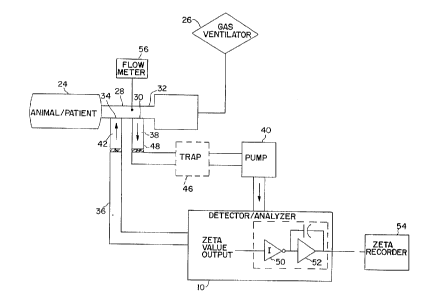
Fig. 12 shows an in vivo sch~m~tir diagram of a PFC elimin~fion
10 analysis system for sampling respifatoly gas in a partial liquid ventilation
process. The trachea of a living being, such as an animal or patient 24, is
conn~octed to a r~ oly gas ventilator 26 through an endotracheal tube 28
such as a HI-LO JET~ tracheal tube, m~nllf~ctured by M~11in~krodt Medical,
Inc., St. Louis, MO. The open end of this tube 28 is conn~oct~d to the
15 ventilator 26 which m~int~in~ the function of breathing by pushing ventil~tin~
gases in and out of the patient's lungs. One version of this tube 28 has two
different side ports. A first port 30 is proximal to the tube's median 32 and the
second port 34 is distal to the median 32. The first port 30 is in fluid
co,.""ll~ tion with the input of a sampling path 36 and the second port 34 is
20 in fluid co~,l"l~"iration with the output of the sampling path 36. The sampling
path 36 thus is a closed loop, continuous flow path for continuously sampling
the respiratory gas flowing through the tube 28 and leL~. "i~ the sampled gas
back into the tube 28. Accordingly, no net gas is added or removed from the
patient 24. This scheme also does not cause any physiologic pellull~ations such
25 as a lowered partial pressure of oxygen or an elevated partial ~res~,ure of carbon
dioxide.
The sampling path 36 comprises, in series order, inflow tube 38,
circulating pump 40, thermal conductivity detector/analyzer 10 and return tube
42. The inflow tube 38 allows for fluid col"",~ tion between the circ~ ting
pump 40 and the first port 30, whereas the return tube 42 allows for fluid
CA 02207871 1997-0~-30
W 096/22052 PCTrUS95/16703
- 20 -
co~ tion between the detector/analyzer 10 and the second port 34. The
cletector/analyzer 10 outputs a zeta value, as described above. A recorder 44
is connPcte~l in parallel to the detector/analyzer 10 to record the zeta value
calculated in the detector/analyzer 10 at discrete time periods.
The sampling path 36 may optionally include a fluid trap 46
between distal end 48 of the inflow tube 38 and the input end of the pump 40
for preventing lung fluid, mucus or other liquid or solid substances from
entering the pump 40 and the detector/analyzer 10. The detector/analyzer 10
may also optionally include a series connection of an inverter 50 and high pass
f~ter 52 to provide positive signal deflection and clarification of the output
signal before it's output is sent to zeta recorder 54.
Fig. 12 also shows flowmeter 56 for continuously measuring the
in~t~3nt~n.-ous flow rate of expired l~hd~ory gas. The i~ ous flow rate
the % saturation PFC value at each instant is sent to a co~ uLer to generate theil~ -lPous rate of loss, as well as total volume loss of PFC liquid from the
lungs. Subsequent figures show applications of the flowmeter 56.
Figs. 13-15 show how the digital zeta value output of the
detector/analyzer 10 is employed to alarm an operator and provide selected
feedback control functions. In Fig. 13, the zeta value controls the physical
position of the patient 24. In Fig. 14, the zeta value del~ es whether the
return tube 42 should draw from a reservoir of PFC liquid to replenish the PFC
in the patient's lungs. In Fig. 15, the zeta value controls the operation of theventilator 26.
Turning to Fig. 13, the zeta value is conn~octecl to the input of
central processing unit (CPU) 58. The CPU 58 is preprogrammed with
information for determines whether the operator should be alerted via audible
or visual alarm/display 60 that the zeta value, or the time rate of change of the
zeta value (d~/dt), is outside of a given range. The CPU 58 is also
preprogrammed with instructions on how to respond to out of range conditions
30 and to output applopliate control signals to feedback controller 62. One
CA 02207871 1997-0~-30
W O 96t22052 PCTAUS9~16703
possible response is to alert the operator via the alarm/display 60 to reposition
the patient's bed 64 or to autom~tir~lly control a bed positioning motor 66 to
perform that function.
Fig. 14 shows the feedb~c1~ controller 62 connected to a reservoir
S 68 of PFC. If ~e bed repositioning does not improve the PFC-gas interaction,
the CPU ~8 instructs the feedback controller 62 to release more PFC into a
patient's lungs by allowing PFC from the reservoir 68 to enter the return tube
42 of the sampling pa~ 36.
Fig. 15 shows the feedback controller 62 connected to the
10 ventilator 26 to cause the ventilator 26 to increase its respiration rate or
inspiratory pressures.
The feeAb~ck control functions shown in Figs. 13 and 14 are
most a~lopliate during partial liquid ventilation, whereas the function shown
in Fig. 15 is most a~r~liate while weaning a patient from total liquid
15 ventilation to conventional gas ventil~tion. When a patient is weaned from total
liquid ventilation, a residual amount of PFC liquid will remain in the lungs.
Eventually, the residual PFC will completely volatilize. However, if the
residual PFC is taking too long to volatilize (as intlir~trd by a zeta value that
is taking too long to reach the value for unsaturated pure ventilation gas), the20 feedb~ck controller 62 may cause the ventilator 26 to increase its respiration
rate or inspiratory pressures. This will increase the arnount of alveolar
ventilation, and thus will more rapidly promote PFC vol~tili7~ti~ n.
The application of the control function in Fig. 15 is best
understood with respect to Fig. 16 which gr~phir~lly depicts zeta values during
25 the weaning process. At zero minlltrs, liquid ventilation with FC-75 type PFCliquid has ceased and conventional gas ventilation with room air has begun.
The residual PFC in the patient's lungs vol~tili7rs and partially SdLUld~S the
expired respiratory gas. This results in a zeta value of about 7.0 ~ which is
between the ~.4 ~ value of room air and the 6.2 ~ value of air fully saturated
30 with FC-75. As time progresses, the amount of liquid PFC in the lungs slowly
CA 02207871 1997-0~-30
W O 96/22052 PCTrUS95/16703
depletes as it volatilizes and evaporates thelcrlulll. Since no PFC is being
added, the zeta value trends upward to 8.4 ~ (the value for unsaluldled, room
air). Since the weaning process from total liquid ventilation to conventional gas
ventilation should be relatively quick (e.g., about 30-60 mimlt~s), the solid
S trend line shows the desired progression of the zeta value. However, if
vol~tili~tion is occurring too slowly, due to insufficient alveolar ventilation, the
trend line will appear as shown in the dashed line. Mathem~tir~lly speaking,
this occurs when the slope of the trend line, d~/dt, is below a predesired value.
The CPU 58 is programmed to detect this condition and to increase the
10 respiratory rate or inspilatoly ~res~ule of the ventilator 26. In the weaningprocess shown in Fig. 16, the CPU 58 determines after about 12 mimltes that
the vol~tili7~tion is procee-ling too slowly. Corrective action is taken and
shortly thereafter, the dashed trend line merges with the desired solid trend line.
From the information in the Fig. 16 trend line, the amount of
15 PFC left in the lungs and the time course of vol~tili7~tion is easily derived(using the algorithm in Equation 7). Heretofore, there was no accurate or even
theoretical means of coll~c~ly ~sessing these parameters.
The application of the control functions in Figs. 13 and 14 are
best understood with respect to Fig. 17 which graphically depicts a hypothetical20 partial liquid ventilation session. In the scenario depicted in Fig. 17, the
respirator supplies air and the patient's lungs are filled with FC-75 type PFC
liquid. The zeta value begins at 6.2 ~ since the air in the expired lc~hdlory
gas is fully salul~t~d with FC-75 vapor. As time progresses, the amount of
liquid PFC in the lungs slowly depletes as it volatilizes and evaporates
25 thercrlolll. If the ventilation set-up is left alone as in the set-up depicted in Fig.
10, the zeta value would eventually approach and stabilize at 8.4 ~ (the value
for unsaturated, room air). However, unlike the test set-up in Fig. 10,
measures are continuously taken to m~int~in the zeta value at, or near, the fully
saturated value of 6.2 ~. This is because when the air is fully sa~ulaLed,
30 maximum gas-PFC interaction, and thus m~ximllm alveolar ventilation occurs.
CA 02207871 1997-05-30
WO 96/22052 PCT~JS9S~6703
- 23 -
Accordingly, the trend line will be relatively flat (average dg/dt = 0) during the
ventilation session. (For illustration purposes, the zeta value scale in Fig. 17is greatly exaggerated, thereby causing the slope of the trend line to appear
steeper than it really is.)
During the first 80 mimites of the session, the zeta value
gradually rises from 6.2 ~. The CPU 58 in Fig. 13 is set to take corrective
measures once the zeta value exceeds about 6% of its desired value. Thus,
when the zeta value reaches 6.6 ~ at 80 minutes, the CPU 58 alerts the operator
via alarm/display 60 with a prompt such as, "PFC LEVEL OUT OF RANGE! !
REPOSITION PATIENT OR ADD ADDITIONAL PFC." Alternatively, the
feeclb~rk controller 62 will autom~ti~lly send a signal to the bed positioning
motor 66 to reposition the patient. In the hypothetical session depicted in Fig.17, varying the bed position returned the zeta value back to an acceptable
amount.
At 160 mimlt~s, the zeta value is again out of range. Bed
repositioning is dL~ Led, but this time it fails to bring the value back into
range. The CPU ~;8 detects that the zeta value is not declining and determines
that it is nPces~ry to add additional PFC. The operator is alerted to perform
this function, or the fee~lk~rk controller 62 ~lltom~ti~lly releases more PFC
into a patient's lungs, as described in Fig. 14.
Although the systems in Figs. 13-15 are illustrated separately, it
should be understood that a single system may include more than one type of
fee~lb~ck control.
PFC recovery
Breathable liquid such as PFC volatilizes from a
diffuser/condenser circuit during total liquid ventilation. Expired breathable
liquid is scrubbed to remove the carbon dioxide, reoxygenated and returned to
the lungs during a subsequent inspiratory bl~edLllillg stage. Currently, the
vaporized breathable liquid in the e~hdlol~y liquid is not recovered during this
CA Oi207871 1997-0~-30
WO 96122052 PCT/US95/16703
- 24 -
process. Tn~te~, it is vented to the el-vh~ol.lllent. Accordingly, the system
must periodically add more breathable liquid from a storage reservoir. As noted
above, the loss of breathable liquid in this process is costly.
Fig. 18 shows a total liquid ventilation (TLV) system 70
5 employing PFC as the breathable liquid. The system 70 recovers PFC from
vol~tili7e~1 breathable liquid which may potentially escape to the environment.
Furthermore, the system 70 employs the thermal conductivity of the PFC, as
measured in zeta units, to monitor and control the efficiency of the recovery
process.
10The TLV system 70 includes condenser circuit 72 connected in
parallel to oxygenator/diffuser 74. The oxygenator/diffuser 74 includes an ~2-
CO2 Membrane 76, as is well-known in the art, for removing dissolved gas
from expiratory liquid flowing th~ lhlough. As PFC is pumped through the
oxygenator/diffuser 74 by pump 78, the corresponding PFC vapor travels to the
15condenser circuit 72 via path 80. The condenser circuit 72 includes a condenser
82 to capture the PFC vapor via cold condensation and a condenser thermostat
84. The recovered PFC fluid is then re-introduced into PFC expiratory
reservoir 86.
Two important factors determine the amount of PFC vapor lost
20 from the TLV system 70 (and thus determine the efficiency of the TLV system
70 in recovering PFC). One important factor is pump flow through the
oxygenator/diffuser 74. Another important factor is the operating condition of
the elements of the condenser circuit 72. For example, PFC vapor loss is
proportional to the oxygenator/diffuser 74 pump flow. The TLV system 70
25 employs thermal conductivity detector/analyzer 10' to track the vapor recovery
process. A pump 88 draws off gas samples from the output path of the
condenser 82 and flows them through the detector/analyzer 10' to obtain a
voltage level correlated to a zeta value. The zeta value is sent to CPU 90 for
analysis. If the CPU 90 determines that the zeta value is outside of a
30 predetermined range, it sends a signal to feedback controller 92 to take
CA 02207871 1997-05-30
W 096/22052 rCT~US95~f6703
- 25 -
~pl~liate rem~di~l action. One type of action is to increase or decrease the
pump flow in the pump 78 of the oxygenator/diffuser 74. Another type of
action is to modify operating conditions of the condenser circuit 72 elem~nt~.
Continuous feedbar~ control determines the most efficient pump flow amount
5 and condenser operating conditions. Of course, the goal of the feedback loop
is to minimi7ç the amount of PFC vapor in the drawn off gas sample (as
determined by the zeta value) without con.~r~...ieing other functions of the TLV system 70.
In the embodiment of the invention shown in Fig. 18, the
condenser circuit 72 includes a condenser thermostat 84 with a variable set
point. Thus, the operating condition modified in this embodiment is the set
point of the condenser thlormost~t 84. It is raised or lowered to achieve
~liullunl vapor recovery. Other known ways to improve the efficiency of the
con~len.eer 82 include applying ultrasound or vibrations thereto. Although the
disclosed embodiment adjusts only the condenser thermostat 84, the scope of the
invention includes all known methods for varying the o~ Lhlg conditions of the
condenser circuit elements. Thus, instead of, or in addition to, adjusting the
condenser thermostat 84 in response to a feeAb~clr control signal, the ultrasound
or vibration level may be adjusted.
The recovery of PFC is easy to monitor with this system. The
total amount of liquid PFC in e~ alol~ reservoir 86 and inspiratory reservoir
94 will remain constant if PFC recovery is 100% efficient. The output from
reservoir level int1i~tors (not shown) are connected to the CPU 90 to monitor
recovery amounts. If recovery efficiency drops si~nifir~ntly below 100%, the
reservoirs will need regular refilling. The refill rate will be proportional to the
recovery efficiency.
Fig. 19 shows a partial liquid ventilation (PLV) system 96
employing PFC as the breathable liquid. The system 96 employs the same
condenser circuit 72 of Fig. 18 to recover vol~tili7fd PFC from expired
respiratory gas. Likewise, the system 96 employs the thermal conductivity of
CA 02207871 1997-0~-30
W O 961220S2 PCTrUS95/16703
- 26 -
the PFC vapor to monitor the efficiency of the PFC recovery process and adjust
the operating conditions of the condenser circuit 72 elements.
The PFC recovery efficiency is measured in one of two ways.
In one scheme, the PFC vapor is ~ttoctç~ in the sampling path 36 and in the
5 output path of the condenser 82. The zeta values of the two samples are
compared to determine how well the con~leIl~er circuit 72 is recovering the PFC
vapor. In another scheme, the zeta values of the samples detected in the output
path of the condenser 82 are employed (see the calculation method described
above with respect to Fig. 7) to determine the total amount of liquid PFC not
10 being recovered. The unrecovered liquid amount is compared to the recovered
amount (i.e., the liquid amount of PFC condensed by the condenser 82) to
determine the recovery efficiency. The output from reservoir level in~ tor 98
is conn~cted to the CPU 90 to monitor recovery amounts. Of course, the CPU
90 employs the zeta value from detector/analyzer 10' to continuously adjust the
15 operating conditions of the condenser circuit 82 for m~ximllm achievable
efficiency.
End-expiratory ,~as samplin~
Fig. 20 shows how the thermal conductivity detector/analyzer 10
20 is utilized for end-expiratory gas sampling during partial liquid ventilation. At
the end of expiration, a volumetric syringe 100 with hP~ p~re draws off
expiratory gas from inflow tube 102 conn~ct~d to endotracheal tube 104.
(Endotracheal return tube 106 is left unconn~ctçcl.) The syringe contents are
then injected at a constant flow rate into the detector/analyzer 10 and the zeta25 value is determined. The zeta value is then extrapolated to determine PFC-gasinteraction or used for respiratory gas measurement. In cases of respiratory
co~ rolllise, this may be the preferred method of analysis because sampling
time is minim~l.
30 Solenoid l~cblea~
CA 02207871 1997-05-30
WO 96122052 PCT/lJS9~J16703
Fig. 21 shows how PFC-gas interaction is assessed using a
solenoid ~bl~LLhillg a~a~aLus set-up 108 during a partial liquid ventilation
session. The apparatus 108 includes ventilator 110, rebreather 112 and three-
way solenoid valve 114 conn~cte~l therebetween. Animal or patient 116 inspires
and expires ventilation gas through the solenoid valve 114. The solenoid valve
then opens to the rebreather 112 and simlllt~n~ously closes the pathway to and
from the ventilator 110. PFC vapor from PFC vol~tili7~tion flows into the gas
in the rcbled~ler 112. Eventually, that gas becomes sa~uldted by PFC vapor.
After saturation, the solenoid valve 114 switches back to the ventilator 110
mode. Thelmal conductivity detector/analyzer 10 measures the gas and sends
a zeta value output to CPU 117. The CPU 117 calculates the rate of change of
the zeta value over time, d~/d~, which provides an indirect measure of the PFC-
gas interaction. The i"~ 0us slope is a function of the PFC-gas
interaction. The faster that the rate reaches equilibrium, the greater the PFC-
gas interaction. Thus, a large slope inrlieAtes significant interaction, whereasa small slope indicates relatively little interaction. This interaction efficiency
measurement may be used in place of the in-line system depicted in Fig. 12.
Fig. 21 shows two trend graphs A and B which lcplese
dirrerclll partial liquid ventil~tion sessions. The breathable liquid is PFOB. In
trend graph A, the trend line slope (d~/dt) between the time that the solenoid
valve 114 switches between the ventilator 110 and the lchlca~ler 112, to~ and
the time of full saturation, *, is si~nifi~ntly less than the trend line slope
between those same points in time in trend graph B. Thus, the PFC-gas
interaction in session B is greater than in session A. In the system shown in
Fig. 21, the trend graph begins at a zeta value for air (8.4 0 and stabilizes atthe value for air fully saturated with PFOB vapor (7.8 ~).
CA Oi20787l l997-0~-30
W 096/22052 PCTrUS95/16703
PEC evaporative loss
Fig. 22A shows how PFC vapor levels are employed to quantify
PFC evaporative loss during partial liquid ventil~ti~-n. As described in the
Background section above, a portion of breathable liquid (e.g., PFC) is lost due5 to evaporation into the lungs. This evaporated liquid becomes absorbed by the
lungs by diffusing into the blood near the lungs and around the alveoli. It
eventually leaves the patient's body by Lld.~h~lion through the skin. A gas
collection device 118isatt~chPCl to and/or pressed against the skin of an animalor patient 120. The device 118 may be a skin patch or collection vial.
Fig. 22B shows an exploded view of the collection region and a
simplified illustration of device 118 against the outer surface of the skin 119.A small gas stream flows through a sampled region associated with the area of
the device 118. The gas flow into the region is pure (i.e., unsaturated by PFC).If there is a measurable PFC evaporative loss from the skin, the gas flow out
of the region will have a ~letect~ble saturation level. This gas flow output of the
device 118isconnPcted directly to thermal conductivity detector/analyzer 10 to
detect the saturation level. To ~luanLiry the PFC evaporative loss from the
sampled region, Equation 7 is employed, wherein VR is the gas flow from the
sampled region.
Knowing the amount of evaporative loss is important because it
improves the accuracy of the amount of PFC liquid known to be in the lungs.
The amount in the lungs is equal to the amount originally input minus the
amount vol~tili7~ minus the amount evaporated. The amount vol~tili7~d is
calculated from zeta values sampled from the endotracheal tube. The amount
evaporated will be functionally related to the zeta value determined from the
scheme in Fig. 22A.
To quantify the total PFC evaporative loss from the entire body,
the evaporation determined from the sampled region is extrapolated. For
example, if the device 118 is a skin patch, the skin patch will cover a known
percentage of the skin's total surface area. The evaporation from the surface
CA 02207871 1997-0~-30
WO g6/22052 PC'rJUS95/1670:~
- 29 -
area of the skin patch will thus be a known percentage of the total evaporation
from the entire body. Standard skin surface area values are known for hllm~n~
of a given age, size, weight and the like.
During partial liquid ventilation, evaporative losses are very small
5 compared to losses from the lc~h~tol~y system. For example, evaporative
losses may be 1/50 of the amount lost from the l~sL,il~loly system. However,
an accurate qll~ntif;r~tion of the total PFC loss during partial liquid ventilation
should preferably include the amount lost from evaporation.
10 PFC-Blood Substitute
Emulsions of PFC have been found to be a suitable blood
substitute, capable of dissolving oxygen and carbon dioxide. However, when
PFC is employed for this purpose, evaporative loss occurs via the skin and the
respiratory system (e.g., the lungs). There is a need to quantify this
15 evaporative loss. The level of PFC vapor in the lungs is related to the
evaporative loss, and thus in~1ir~t~ when the PFC in the bloodstream must be
replenished.
Fig. 23 shows a set-up for moni~o,illg and controlling PFC blood
levels after the blood of patient 24 is injected with PFC. Expired re~h~Lc,ly
20 gas cont~inin~ PFC vapor flows through the endotracheal tube 28, is pumped
through the sampling path 36 and is measured by the thermal conductivity
detector/analyzer 10. The ~letector/analyzer 10 outputs a signal to the CPU 58.
The CPU 58 is programmed to signal when the PFC vapor in the lungs reaches
a preset level and to either alert an ~ ~elalor or autom~ti~lly add PFC to the
25 patient's bloodstream through feedback controller 62. The fee~lb~ck controller
62 causes PFC from the reservoir 68 to flow into intravenous tube 122
connected to the patient's vein. Alteratively, end expiratory sampling of PFC
vapor in the lungs can be employed, as shown in Fig. 20, in place of the
sampling path 36 and the results fed into the CPU 58 of Fig. 23.
CA oi207s7l 1997-0~-30
WO 96/22052 PCT/USg5/16703
- 30 -
In the set-up shown in Fig. 23, the patient 24 breathes through
gas ventilator 26. However, the patient 24 need not be ~tt~ch~d to an ~si~ttod
l),catllillg device. It is only n.-cçss~ry that there be a means, such as, but not
limited to, the endotracheal tube 28, a nasal CPAP (continuous positive airway
5 pressure), a mask, or the like, to collect a sample of expired respiratory gas for
analysis. Equation 7 is employed to detect PFC loss.
Again, the set-up in Fig. 22A may be employed to detect the
amount of Llanspiled PFC during PFC blood substitution. This amount is then
added to the arnount leaving the patient through the lungs to determine the total
10 amount lost from the bloodstream.
Funçtional Residual Capacit,v
The thermal conductivity detector/analyzer 10 can also be
employed for correcting conventional Functional Residual Capacity (FRC)
15 patient lung measurements and for measuring FRC in a new way.
FRC is the volume of gas left in the lung (i.e., lung volume) at
the end of normal exhalation or expiration. Traditionally, a helium dilution test
is employed when making FRC measurements. This test employs thermal
detectors. The output of the detectors will be in error if a breathable liquid
20 such as PFC is present in the expired gas. When a patient undergoes partial
liquid ventilation, PFC vapor will be present in the expired gas due to
vol~tili7~tion of the PFC liquid. Thus, the FRC measurement will be in error.
To correct for this error, the output value of the detector/analyzer 10 is
employed to detect the amount of PFC vapor in the lung. This value is then
25 used to offset and normali_e the conventionally measured FRC value.
Furthermore, PFC vapor can be employed, instead of helium, as
the diluent or tracer gas to make an FRC measurement. Since helium is soluble
in blood, large ql-~ntiti~s of helium become absorbed during this measurement.
PFC vapor is an ideal gas for making such a measurement because it is inert
30 and minim~lly absorbed into circulation (FRC < < Helium).
CA 02207871 1997-0~-30
Wo 96/220S2 PCTJIJS95~16703
- 31 -
To pelr~ this mea~ulelllcnt, the patient breathes from a bag of
known volume and cont~ining a known quantity of PFC vapor.
The FRC is calculated using the following equation which is a
modification or rearrangement o~ Fick's law:
s
FRC = Vl [(C,/Cf) - 1~ (Equation 9)
where Vj is the system volume (i.e., the volume of the bag), Ci is the initial
concentration of PFC vapor in the bag, and CfiS the final concentration of PFC
10 vapor in the bag. System volume, V;, was also ~letermin~ by rearrangement
of Fick's Law. Various known syringe volumes can be accurately assessed in
this fashion.
The final concentration of PFC vapor in the bag, Cf, iS
~lete~ninecl by talcing a syringe sample and employing the set-up shown in Fig.
15 20. The resultant zeta value is then correlated to the percent concentration
using i,~ol",ation from the graph of Fig. 7.
PFC vapor is also suitable for use as a tracer gas for other types
of pulmonary function evaluations including the determination of static lung
volumes, including residual volume and total lung capacity. PFC vapor is less
20 expensive than current diluent gases currently employed in these tests. PFC
vapor may be used in the standard equipment employed for washout and single
breath techniques (either closed or open circuit) present in most hospitals. PFCvapor may also be applied in gas mixing analysis for distribution of ventilationin obstructive lung disease.
Radiolo~ic Dia~nosis
Certain breathable liquids such as PFC are radiopaque and make
ideal contrast agents for high resolution computed tomography (CT). Thus, a
CT scan of the lungs made during partial liquid ventilation provides an image
30 of the PFC in the lungs. The image is used to assess distribution of the PFC
CA 02207871 1997-0~-30
W 096122052 PCTrUS95/16703
in the lungs. However, the scan can be misleading because it does not
distinguish between PFC in the alveolar spaces and PFC in the pulmonary
inle~ l,. The thermal conductivity detector/analyzer 10 can be employed
in conjul~;Lion with CT to alleviate this problem. The level of PFC-gas
S interaction is ~ses~e~l either before or after the scan, thus providing correlation
with the physician's diagnosis. A high level of interaction in~ tes a signifie~nt
llily of PFC appearing in the CT scan is in the alveolar spaces, whereas a
low level of interaction in~lir~ttos that the PFC is primarily in the pulmonary
hllel~liliu~ll.
PFC Delivered A~ents and Therapies
The thermal conductivity detector/analyzer 10 can be employed
during pulmonary ~lmini~tration of drugs (PAD). During PAD, the level of
PFC vapor in the lungs can be used to estimate bioavailability of the relevant
ph~ ologic agent or ~n~sth~tic. Accurate a~ses~mtont of PFC-gas interaction
and relative amount of PFC in the lungs is also important during intratracheal
in~till~tion of PFC for tre~tment of meconium aspiration syndrome (MAS),
congenital diaphragmatic hernia (CDH), neonatal respiratory distress syndrome
(NRDS), and other pulmonary pathologies.
Fig. 3 shows one type of thermal conductivity detector suitable
the invention. However, other types of thermal conductivity detectors which
measure the conductivity of re~pi,aLoly gases are also within the scope of the
invention.
The systems and methods described above employ a thermal
conductivity measurement device for ascertaining the PFC-gas interaction.
However, other types of analyzers, including a spectrophotometer or a gas
chromatograph may be employed in place of the thermal conductivity
detector/analyzer 10. These devices are equally able to distinguish between
PFC vapor and other types of gases (e.g., air, oxygen) due to differences in
CA 02207X71 1997-05-30
WO 96/220S2 PCT/~IS95/16703
electron density. However, they are less cost-effective than the ~:ullclllly
described measurement device.
Fu~lhellllore, as is well-known in the art, the elements of the
thermal conductivity detector/analyzer 10 can be employed to measure other
S properties of the gas flowing th~ rough, including mass or pressure. For
example, if the thermistors are employed in a mass flow detector, the zeta valuewill vary with the mass of the sample. The zeta value would then be
precalibrated with gases of known PFC percent saturation amounts in the same
manner as the thPrm~l conductivity detector/analyzer described herein. That is,
the second y-axis in Figs. 10, 11, 16 and 17 labelled "Percent saturation with
PFC " will be shifted up or down so that it properly correlates with the
ap~l~opliate zeta values. Thus, the zeta value of the detector/analyzer 10 need
not n~ces~rily be the result of a thermal conductivity meas.~ .enl. The scope
of the invention includes any type of measurement detector/analyzer which
15 outputs signal levels (e.g, discrete zeta values) that may be correlated with percent saturatiorl of the gas sample.
When it is desired to determine the quantity of PFC liquid in
expired respiratory gas, the flowmeter 56 is employed. T~ ntsous flow rate
measurements are taken and sent to the CPU 58. The flow rate measurements
20 are correlated with liquid volume amounts associated with a zeta value
measurement taken at the same in.ct~n~.e. These values, along with the
liquid/vapor conversion factor from Equation 7, are then employed to (letermin~
the totall amount of lost liquid PFC.
One example of an expelullelllal volume loss c~le~ tion is as
25 follows:
Gas Flow into TLV diffuser = 8 L/min.
% saturation (zeta) = 100%
Te~ cldlule = 37 degrees Celsius
Time course = 30 mimltes
PFC volume loss = (8000 ml/min) x
[(1.447 ml PFOB/100 ml air) x 100%] x
(1 ml fluid/86 ml vapor) x 30 minutes = 40.3 ml
CA 02207871 1997-0~-30
W 096/22052 PCTrUS95/16703
- 34 -
Thus, the PFC volume loss in 30 minutes is 40.3 ml.
The invention disclosed above allow for ~ignifir~ntly improved
control of liquid ventilation processes. No longer does an operator have to
guess if, and how much, PFC must be added to a patient's lungs to optimize
5 PFC-gas interaction and to replace vol~tili7ed and evaporated PFC liquid. The
invention also describes simple and cost-effective techniques to add a PFC
recovery to total liquid ventilation systems and to m~ximi7,o the efficiency of the
recovery system. Furthermore, the invention describes how PFC saturation
values are useful in a wide variety of other biom~ l applications.
The present invention may be embodied in other specific forms
without departing from the spirit or essential attributes thereof and, accordingly,
ref~lc;llce should be made to the appended claims, rather than to the foregoing
specification, as inl1ic~tin~ the scope of the invention.