Note: Descriptions are shown in the official language in which they were submitted.
CA 02208334 1997-06-19
WO 96/25670 PCT/US96/02171
T I TT ,~
A Cell Enumeration Immunoassay
FI~T~n OF T~ TION
This invention relates to an immunoassay and, more
particularly, to a cell enumeration immunoassay as an
efficient alternative to flow cytometry.
10BACKGROUND OF T~F INV~TION
With the development of Kohler and Milstein's
hybridoma technology around 1975, monoclonal antibodies
were utilized in the discovery and identification of
cell surface molecules. Cell marker analyses are
important in the prognosis, classification of state of
disease, treatment decisions, and monitoring of
therapy. For example, in human immunodeficiency virus
(HIV) infection, both the CD~+/CD8+ T-cell ratio and
the absolute number of CD4+ T lymphocytes are important
20 for the reasons noted above. The functions of cell
membrane molecules and the consequences of their
quantitative changes in several disorders (e.g.,
septicemia, burns, autoimmune diseases, graft
rejection) are better understood because of advances in
25 techniques in molecular biology and in the ability to
assess the state of the immune system and to give more
accurate prognoses.
Cell membrane markers are usually assayed by flow
cytometry using a fluorescence-activated cell sorter
30 (FACS) and a fluorescently labeled monoclonal antibody
specific for the cell marker to be assayed. A FACS
analysis measures cells as they flow through a flow
cytometer in single file, or the best approximation
thereof that can be achieved, in a fluid stream.
35 Standardization and reproducibility of tests for
clinical application is difficult, especially when
measuring quantitative cellular fluorescence intensity,
because different flow cytometers differ in
SUBSTITUTE SHEET (RULE 26)
CA 02208334 l997-06-l9
WO 96/25670 PCT/US96/02171
sensitivity. In addition, the instrument is expensive
(frequently >$200,000) and is labor intensive,
requiring highly trained personnel to run the FACS.
FACS analysis is also unreliable due to instrument
variables and due to the way results are expressed.
Usually, the results are expressed in units which
require a separate determination of the number of white
blood cells per ~L of blood and a differential count.
This combines the variability of three tests into one
clinical result. Consequently, it would not be unusual
for the same sample to give results differing by a
factor of two in two separate determinations.
Given the foregoing, researchers have endeavored
unsuccessfully to reliably determine markers in or on
cells using enzyme immunoassays as an alternative to
flow cytometry.
Franke et al., Clin. Chem., 40(1): 38-42 (1994)
and AIDSLINE, December 1993 reporting on the Int-Conf-
AIDS, page 259 (June 16-21, 1991) describe a cell
marker ELISA, Capcellia CD4/CD8, to quantitatively
determine CD4/CD8 molecules. The assay is performed in
a single step on microtiter plates, specifically, cells
are immobilized on the solid phase using pan-T
monoclonal antibodies adsorbed on the solid phase
surface along with simultaneous labeling of CD4 or CD8
by peroxidase-labeled immunoconjugates. The results
are expressed in molar concentrations of CD4 or CD8
molecules calculated from standard curves. The factors
used to convert concentrations of CD4 molecules into
cells per liter were only relative and were used as
guides. Thus, this assay falls woefully short in being
an efficient alternative to flow cytometry.
Other approaches include the following:
Baumgarten, J. Immunological Methods, 94: 91-98
(1986) describes the requirements for calibration of a
cell ELISA for the quantitation of leukocyte antigens
using air-dried and methanol-fixed cells which were
attached to microplate wells. The test was
SUBSTITUTE SHEET (RULE 26)
CA 02208334 1997-06-l9
W096/25670 PCT~S96/02171
standardized by measuring both the ~c~ a~t~g~ and
the amount of cellular protein in each single sample
and was calibrated either with intact cells or isolated
plasma membranes prepared from the cells under study.
Hessian et al., J. Immunological Methods, 91: 29-
34 (1986) describes a cell-associated enzyme
immunoassay as an alternative to FACS analysis by
employing immunofiltration methodology and soluble
complexes of alkaline phosphatase and monoclonal anti-
alkaline phosphatase.
U.S. Patent No. 4,661,466, issued to Schlossman et
al. on April 28, 1987, describes the use of a
monoclonal antibody to distinguish subsets of cells on
the basis of different degrees of reactivity with the
monoclonal antibody.
Similarly, U.S. Patent No. 4,677,061, issued to
Rose et al. on June 30, 1987, describes T-cell
lymphocyte subset monitoring of immunologic disease.
T-cell subsets are monitored for a designated pattern
of epitopic sites associated with specific surface
membrane proteins where ratios of cells having
different patterns are determined by multi-parameter
flow cytometric analysis, the ratios being indicative
of a probable change in the immunologic disease.
WO 90/04180, published April 19, 1990, describes a
method for measuring soluble CD4 antigens to diagnose a
state of immune activation.
WO 92/08981, published May 29, 1992, describes the
measurement of total leukocyte antigens and the use of
such measurements to enumerate cells.
None of these references describes a standardized
and reproducible cell enumeration immunoassay which is
an efficient alternative to flow cytometry.
SUMMARY OF THE INV~NTION
The present invention concerns a cell enumeration
immunoassay for quantitating the number of cells in a
gUBSTlTUTE SHEET (RULE 26~
CA 02208334 1997-06-l9
WO 96125675) PCT/US96/02171
subpopulation or a subset of the subpopulation of the
total cell population in a sample which comprises:
(a) contacting a sample simultaneously with a
modified solid phase, a first labeled antibody specific
for the subpopulation and a second detectably labeled
antibody specific for the subpopulation or the subset
of the subpopulation wherein the label on the first
antibody is used for immobilization onto the modified
solid phase, the label on the second antibody is used
for detection and the label on the first antibody is
different from the label on the second antibody and
further wherein the first antibody and the second
antibody can have the same or different specificities
and bind to different sites on the cells in the
subpopulation;
(b) contacting separately a calibrated standard
which can be labeled or unlabeled with the modified
solid phase and (i) no other reactants if the
calibrated standard is doubly labeled with the same
labels attached to the first antibody and the second
antibody of step (a), (ii) the first and second
antibodies if the calibrated standard is unlabeled and
capable of binding to the ~irst and second antibodies,
(iii) the first antibody only if the calibrated
standard is singly labeled with the same label as the
second antibody and is capable of binding with the
first antibody, or (iv) the second antibody only if the
calibrated standard is singly labeled with the same
label as the first antibody and is capable of binding
with the second antibody, provided that if the
calibrated standard is a cell then it is labeled either
singly or doubly with the same label or labels attached
to the first and second antibodies;
(c) measuring separately a signal generated by
step (a) and a signal generated by step (b); and
(d) quantitating the number of cells in the
subpopulation or subset of the subpopulation in the
sample by comparing the results from the measurement of
SUBSTITUTE SHEET (RULE 26)
CA 02208334 1997-06-19
WO 96/25670 PCT/US96/02171
step (a) with the results obtained from the measurement
of step (b).
In another embodiment the invention concerns a
cell enumeration immunoassay for quantitating the
number of T cells in a subset of the total T cell
population in a sample which comprises:
(a) contacting the sample simultaneously with a
modified solid phase, a labeled anti-pan T cell
antibody wherein said label is used for immobilization
onto the modified solid phase and a detectably labeled
anti-subset specific antibody wherein the label on the
anti-subset specific antibody is used for detection and
is different from the label on the anti-pan T cell
antibody and further wherein the anti-pan T cell
antibody and the anti-subset specific antibody can have
the same or different specificities and bind to
different sites on the cells in the subpopulation;
(b) contacting separately a calibrated standard
which can be labeled or unlabeled with the modified
solid phase and; (i) no other reactants if the
calibrated standard is doubly labeled with the same
labels attached to the anti-pan T cell antibody and the
anti-subset specific antibody o~ step (a), (ii) the
anti-pan T cell antibody and the anti-subset specific
antibody if the calibrated standard is unlabeled and
capable of binding to both antibodies, (iii) the first
antibody only if the calibrated standard is singly
labeled with the same label as the anti-subset specific
antibody and is capable of binding to the anti-pan T
cell antibody, or (iv) the anti-subset specific
antibody only if the calibrated standard is singly
labeled with the same label as the anti-pan T cell
antibody and is capable of binding with the anti-subset
specific antibody, provided that if the calibrated
standard is a cell then it is labeled either singly or
doubly with the same label or labels attached to the
antibodies;
SUBSTITUTE SHEET (RULE 26)
CA 02208334 1997-06-19
W~6/2S~70 PCT~S96102171
(c) measuring separately a slgnal genera~c~y
step (a) and a signal generated by step (b); and
(d) quantitating the number of T cells in the
subset of the total T cell population in the sample by
comparing the results from the measurement of step (a)
with the results obtained from the measurement of step
(b).
BRIF.F ~)T'..'~CRIPTION OF FIGU~S
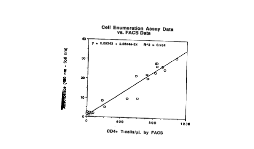
Figure l is a standard curve of absorbance of the
samples versus CD3+/CD4+ cells per ~l of blood obtained
by FACS analysis of the samples.
Figure 2 presents the absorbance of the standard
dilutions, i.e., the concentration of standard required
to produce a given amount of absorbance.
Figure 3 shows the combination of the data
presented in Figures l and 2 to calibrate the standard
concentration in pg/m~ versus the concentration of
CD3+/CD4+ cells per ~L of blood.
PETAITl~n DT~'.sCRIPTION OF THE INVENTION
The term "calibrated standard" as used herein
means a substance that behaves like the sample under
study and the concentration of which can be correlated
to the concentration of the cells. The substance can
be a cell which is singly or doubly labeled, a
particle or any other material which can be designed to
function like the sample and the concentration of which
can be correlated to the concentration of the cells in
the sample.
The calibrated standard can be labeled or
unlabeled provided that if the calibrated standard is a
cell then it is labeled either singly or doubly. The
calibrated standard reacts with the modified solid
phase and no other reactants if the calibrated standard
is doubly labeled with the same labels attached to the
first and second antibodies. It reacts with the
modified solid phase and the first and second
SUBSTITUTE SHEET (RULE 26)
CA 02208334 1997-06-19
WO 96/25670 PCTIUS96/02171
antibodies if the calibrated standard is unlabeled and
capable of binding to the first and second antibodies.
It reacts with the modified solid phase and the first
antibody only if the calibrated standard is singly
labeled with the same label as the second antibody and
is capable of binding with the first antibody. It
reacts with the modified solid phase and the second
antibody only if the calibrated standard is singly
labeled with the same label as the first antibody and
is capable of binding with the second antibody.
The standard can be calibrated using flow
cytometry, a technique well known to those skilled in
the art. Other methods of calibration include using
immunocytochemistry or a cell counter.
Examples of substances which can be used as a
calibrated standard include, but are not limited to,
biotin and fluorescein labeled dextran, biotin and
fluorescein labeled bovine serum albumin and
biotinylated fluorescein. Biotin and fluorescein
labeled dextran and biotinylated fluorescein are
commercially available. Biotin and fluorescein labeled
bovine serum albumin can be made using standard
techniques as illustrated in the example below.
The cell enumeration immunoassay of the instant
invention provides an accurate and efficient
alternative to flow cytometry. It enables one to
quantitate the number of cells in a subpopulation or a
subset of the subpopulation of the total cell
population in a sample such as blood or other cell
containing specimen without the need for expensive
instrumentation.
The cell enumeration immunoassay of the instant
invention is a simultaneous solid phase or
heterogeneous sandwich immunoassay in which the first
labeled antibody, second detectably labeled antibody,
modified solid phase and a sample are reacted together.
The solid phase or support is modified so that it will
bind with the first labeled antibody or capture
SUBSTITUTE SHEET (RULE 26)
-
CA 02208334 1997-06-l9
WO 96125670 PCT/US96/02171
reagent. Thus, the label on the firs~ antibody is not
used for detection but for immobili~ation onto the
solid phase. Typically, the solid phase and first
antibody are modified directly or indirectly with
members of a specific binding pair which can be immune
or non-immune. For example, if the solid phase is
modified with streptavidin then the first antibody
would be labeled with biotin. Modification of the
solid phase is accomplished using techniques well known
to those skilled in the art. Similarly, labeling of
the first antibody involves techniques well known to
those skilled in the art.
Immune specific binding pairs are exemplified by
antigen/antibody systems or hapten/anti-hapten systems.
There can be mentioned fluorescein/anti-fluorescein,
biotin/anti-biotin, dinitrophenol/anti-dinitrophenol,
etc. The antibody member, whether polyclonal,
monoclonal or an immunoreactive fragment thereof, of
the binding pair can be produced by customary methods
familiar to those skilled in the art. The terms
immunoreactive antibody fragment or immunoreactive
fragment mean fragments which contain the binding
region of the antibody. Such fragments may be Fab-type
fragments which are defined as fragments devoid of the
Fc portion, e.g., Fab, Fab' and F(ab')2 obtained by
reductive cleavage of the disulfide bonds connecting
the heavy chain components of the intact antibody. If
the antigen member of the specific binding pair is not
immunogenic, e.g., a hapten, it can be covalently
coupled to a carrier protein to render it immunogenic.
Non-immune binding pairs include systems wherein
the two components share a natural affinity for each
other but are not antibodies. Exemplary non-immune
binding pairs are biotin-avidin or biotin-streptavidin,
folic acid-folate binding protein, complementary probe
nucleic acids, etc. - --
Suitable supports include synthetic polymer
supports, such as polystyrene, polypropylene,
SUBSTITUTE SHEET (RULE 26)
CA 02208334 1997-06-19
WO 96/25670 PCT/US96/02171
substituted polystyrene, e.g., aminated or carboxylated
polystyrene, polyacrylamides; polyamides;
polyvinylchloride, etc.; glass beads; agarose;
nitrocellulose; nylon; polyvinylidenedifluoride;
surface-modified nylon, etc. The preferred support is
a polystyrene microplate.
The second or detector antibody is detectably
labeled with a label different from that on the first
antibody. It can be labeled directly or indirectly
with a member of an immune or non-immune specific
binding pair as discussed above using conventional
techniques. In order to facilitate signal detection,
either the antibody or a member of a specific binding
pair is labeled with a component of a reporting system.
For example, the second antibody could be conjugated to
fluorescein. Fluorescein can be detected directly or
indirectly by using an enzyme-labeled anti-fluorescein
antibody.
The term "reporting system" refers to the reporter
selected and any means of linking the reporter to the
antibody or to a component of a specific binding pair.
Thus, a reporter can be linked directly or indirectly,
covalently or non-covalently to an antibody or a member
of a specific binding pair. Reporters may be
radioactive isotopes, enzymes, fluorogenic, magnetic,
chemiluminescent or electrochemical materials. Two
commonly used radioisotopes are 125I and 3H. Standard
radioactive isotopic labeling procedures include
choramine T, lactoperoxidase and Bolton-Hunter methods
for 125I and reductive methylation for 3H.
Enzymes are the preferred reporters. These
include horseradish peroxidase, alkaline phosphatase, t3
-galactosidase, glucose oxidase, luciferase, t3-
lactamase, urease, and lysozyme. Labeling with enzymes
is facilitated using dialdehyde, carbodiimide coupling,
~ homobifunctional crosslinkers, etc. The labeling
method chosen depends on the functional groups
available on the enzyme and the material to be labeled,
SUBSTtTUTE SHEET (RULE 26)
CA 02208334 1997-06-19
WO 96/2~;670 PCT/US96/Otl71
and the tolerance of both to the conjugat~h
conditions. Labeling can be accomplished using any
conventional methods including those described by
Engvall and Pearlmann, Immunochemistry 8, 871 (1971),
Avrameas and Ternynck, Immunochemistry 8, 1175 (1971),
Ishikawa et al., J. Immunoassay 4(3): 209-327 ~1983)
and Jablonski, Anal. Biochem. 148: 199 (1985).
Labeling can be accomplished by indirect methods such
as using spacers or other members of specific binding
pairs. Detection of enzyme activity can be facilitated
by measuring chromogenic, fluorogenic, magnetic,
chemiluminescent or electrochemical changes by commonly
known methods.
The first and second antibodies can have the same
or different specificities and bind to different sites
on the cells in the subpopulation.
The first and second antibodies can be polyclonal,
monoclonal or an immunoreactive antibody fragment
thereof as discussed above. Such antibodies and/or
immunoreactive antibody fragments can be made using
standard techniques well known to those skilled in the
art.
After the sample has reacted with the modified
solid phase, first labeled antibody and second
detectably labeled antibody, it can be further
subjected to treatment with an inactivation reagent, as
described in the examples below, which serves to
inactivate any endogenous peroxidases which may be
present, fix the cells, and inactivate human
immunodeficiency virus which might be present if the
sample was obtained from a subject infected with HIV.
Any marker in or on a cell can be quantitatively
determined using the instant cell enumeration
immunoassay. Examples of cells which can be
quantitatively determined using the cell enumeration
immunoassay include CD4+ T cells, CD8+ T cells, B-
cells, activated B cells, activated T cells, CMV-
SUBSTITUTE SHEET (RULE 26)
CA 02208334 1997-06-19
WO 96/25670 PCT/US96/02171
in~ected granulocytes, E3v in~ected s-cells, H~-
infected monocytes, etc.
In a preferred embodiment, the instant invention
concerns a T-Cell enumeration immunoassay for
quantitating the number of T cells in a subset of the
total T cell population in a sample which comprises:
(a) contacting the sample simultaneously with a
modified solid phase, a labeled anti-pan T cell
antibody wherein said label is used for immobilization
onto the modified solid phase and a detectably labeled
anti-subset specific antibody wherein the label on the
anti-subset specific antibody is used for detection and
is different from the label on the anti-pan T cell
antibody and further wherein the anti-pan T cell
antibody and the anti-subset specific antibody can have
the same or different specificities and bind to
di~ferent sites on the cells in the subpopulation;
(b) contacting separately a calibrated standard
which can be labeled or unlabeled with the modified
solid phase and; (i) no other reactants if the
calibrated standard is doubly labeled with the same
labels attached to the anti-pan T cell antibody and the
anti-subset speci~ic antibody o~ step (a), (ii) the
anti-pan T cell antibody and the anti-subset specific
antibody if the calibrated standard is unlabeled and
capable of binding to both antibodies, (iii) the first
antibody only if the calibrated standard is singly
labeled with the same label as the anti-subset specific
antibody and is capable of binding to the anti-pan T
cell antibody, or (iv) the anti-subset specific
antibody only if the calibrated standard is singly
labeled with the same label as the anti-pan T cell
antibody and is capable of binding with the anti-subset
specific antibody, provided that if the calibrated
standard is a cell then it is labeled either singly or
doubly with the same label or labels attached to the
antibodies..
SUBSTITUTE SHEET (RULE 26)
CA 02208334 1997-06-19
WO 96/25~70 PCT/US96/02171
(c) measuring separately the signal generated by
step (a) and the signal generated by step (b); and
(d) quantitating the number of T cells in the
subset of the total T cell population in the sample by
comparing the results from the measurement of step (a)
with the results obtained from the measurement of step
(b).
The anti-pan T cell antibody can be, by way of
illustration, an anti-CD2 antibody or an anti-CD3
antibody. The preferred anti-pan T cell antibody is an
anti-CD3 antibody. The CD3 antigen is called the T
cell receptor (TcR). It has a complex multistranded
structure and is involved in T cell activation in the
immune response. It is found on virtually all mature T
cells in the peripheral circulation as well as in
precursor cells present in the thymus (thymocytes).
The anti-subset specific antibody can be an anti-
CD4 antibody, anti-CD8 antibody, anti-CD25 antibody,
etc. The pre~erred anti-subset specific antibody is an
anti-CD4 antibody.
The CD4 antigen is found on about 2/3 o~
peripheral T-Cells (those which are CD8 negative) and
also on monocytes and most thymocytes. Functionally,
CD4 is an accessory molecule involved in the
recognition of foreign antigens by T cells. The
population of T cells which expresses CD4 is considered
to represent the "helper" T cells. CD4 has been shown
to be the cellular receptor necessary for binding of
HIV to a cell during infection. As the disease
progresses, the fraction of CD4+ T lymphocytes in the
blood decreases. The Center for Disease Control
recommends that a level of less than 200 CD4+ T cells
per ~L of blood be used in the case definition for
AIDS. It is recommended that AIDS patients have their
CD4+ T cell levels measured 2-3 times per year. A
level of less than 500 CD4+ cells per ~L of blood is
frequently used as an indication for beginning
antiviral therapy. The number of CD4+ T cells is also
SUBSTITUTE SHEET (RULE 26)
CA 02208334 1997-06-19
WO 96/25670 PCTIUS96/02171
commonly used as a criterion ~or admission of patients
to clinical trials. The absolute number of CD4+ T
lymphocytes is important for prognosis, classification
of state of disease, treatment decisions, and
monitoring of therapy in human immunode~iciency virus
(HIV) infection.
The following examples are intended to illustrate
the invention and should not be construed as a
limitation thereon.
Example 1
Part A. Synthesis of ~ntibody Reagents
1. ~nti-C~3-Biot;n ~ntihody
An anti-CD3 monoclonal antibody solution
(BioDesign) was dialyzed against O.lM NaHCO3 and then
adjusted to 1 mg/ml in O.lM NaHCO3.
Biotinamidocaproate, N-hydroxysuccinimide ester
(Molecular Probes) was dissolved in dimethyl sulfoxide
at 0.75 mg/ml. To 4 ml of antibody solution, 400~1 of
biotin solution was added and mixed ~or 2 hours at room
temperature, and then 400~1 of 2M tris-HCl, pH 8.0 was
added and mixed for 15 minutes. The solution was
dialyzed against phosphate buffered saline (PBS) at 4~C
and then 4 ml of 2~ bovine serum albumin in PBS with
0.1~ chloroacetamide was added.
2. Anti-CD4-Fluorescein ~ntibody
An anti-CD4 monoclonal antibody solution (Ancell)
was adjusted to 1 mg/ml in O.lM NaHCO3 and then
dialyzed against O.lM NaHCO3 6-(fluorescein-5-(and-6)
carboxamido) hexanoic acid, succinimidyl ester
(Molecular Probes) was dissolved in dimethyl sulfoxide
at 1 mg/ml. To 1 ml of antibody solution, 100~1 of
fluorescein solution was added and mixed for 2 hours at
room temperature, and then 100~1 of 2M tris-HCl, pH 8.0
was added and mixed for 30 minutes. The solution was
dialyzed against phosphate buffered saline (PBS) at 4~C
SUBSTITUTE SHEET (RULE 26)
CA 02208334 1997-06-l9
W096l25670 PCT~S96/02l71
and then 1 ml of 2~ bovine serum albumin in PBS with
0.1~ chloroacetamide was added.
Part B.
Three tubes of EDTA blood were collected from 10
normal persons and 10 AIDS patients. One tube was
analyzed for CD3+/CD4+ lymphocytes content by
fluorescence-activated cell sorter (FACS) according to
established procedures (Morbidity and Mortality Weekly
Report, 1994 Revised Guidelines for the Performance of
CD4+ T-Cell Determinations in Persons with Human
Immunodeficiency Virus (HIV) Infection, U.S. Department
of Health and Human Services, Public Health Service,
Centers for Disease Control, Vol. 43, No. R, March 4,
1994). In brief, the cells are reacted with two
fluorescently-labeled monoclonal antibodies that react
with CD3 antigen and ~he CD4 antigen respectively if
present on the cell surfaces. By definition, T cells
have the CD3 antigen on their surface. The helper T
cells (a subset of the total T cells that can be
in~ected by HIV) have both CD4 and CD3. The FACS
results are reported as the ~ of the total lymphocytes
labeled with both fluorescent anti-CD3 monoclonal
antibody and labeled with fluorescent anti-CD4
monoclonal antibody.
The second tube of EDTA blood was used for
hematology analysis (total white cell count and
differential count). The total white cell count was
performed using an automated cell counter (Coulter) and
is reported as total white cells/~L of blood. From
microscopic ex~m-n~tion of a stained slide, a manual
differential count reports the ~ of the white cells
that are morphologically lymphocytes. The combination
of these two analyses yields the number of
lymphocytes/~L of blood. Combining this result with
the ~ of the lymphocytes that express CD3 and CD4 on
their surfaces (the FACS result) allows the calculation
of the number CD3+/CD4+ lymphocytes/~L of blood.
14
SUBSTITUTE SHEET (RULE 26)
CA 02208334 1997-06-19
WO 9612~670 PCTIUS96/02171
The third tube was tested using the cell
enumeration immunoassay of the present invention as
described below.
o.l mL of blood was added to 1 mL of Lysing
Reagent (1.68 M ammonium chloride, o.l M potassium
bicarbonate, lmM EDTA, pH 7.4). After 5 minutes, the
white blood cells were separated from the lysed red
blood cells by a 10 second centrifugation at maximum
speed in a microcentrifuge (Sorvall). A 1:5 dilution
o~ the original white cells was prepared by
resuspending the white cell pellet in 0.5 mL of
Antibody Reagent (UCHT1 anti-CD3 monoclonal antibody
[BioDesign] was labeled with biotin and QS4120 anti-CD4
monoclonal antibody [Ancell] was labeled with
fluorescein. They were used at 5 ~g/mL and 20 ng/mL,
respectively in PBS containing 4% BSA and o.o9~
chloroacetamide). A 1:25 dilution of the original
white cells was also prepared by adding 0.1 mL of the
1:5 dilution of white cells to 0.4 mL of Antibody
Reagent. For each of the dilutions (1:5 and 1:25) 0.1
mL samples were added to duplicate wells of a
streptavidin-coated microplate (DuPont). Dilutions of
a biotinylated and fluoresceinated dextran (the
calibrated standard) (Molecular Probes) were also added
to duplicate wells.
After 2 hours at room temperature, 0.05 mL of
Inactivation Reagent (0.1% sodium azide, 0.002% sodium
stannate, 0.01% hydrogen peroxide in 75% ethanol) were
added to each well. During a 30 minute room
temperature incubation, the Inactivation Reagent
inactivated endogenous cellular peroxidases,
inactivated any HIV particles from the sample, and
fixed the cells. The microplate was then washed 6X
with Plate Wash (DuPont) to remove unreacted
antibodies.
To each well was added 0.1 mL of Detector Reagent
(1:200 affinity-purified sheep antifluorescein
polyclonal antibody labeled with HRP [Boehringer-
SUBSTITUTE SHEET (RULE 263
-
CA 02208334 1997-06-19
WO 96/25670 PCI'rUS96/02171
Mannheim] in PBS containing 50~ normal rabbit serum, 1
casein and 0.09~ chloroacetamide). During a 30 minute
room temperature incubation, the anti-fluorescein-HRP
bound to any fluoresceinated anti-CD4 monoclonal
antibody on the surface of cells that are bound to the
streptavidin-coated microplate via the biotinylated
anti-CD3 monoclonal antibody. The microplate was then
washed 6X with Plate Wash Buffer to remove unreacted
Detector Reagent.
To each well, 0.1 mL of TMB (tetramethylbenzidene)
Substrate (Proteins International) was added. During a
1 hour room temperature incubation, any HRP in the
wells generated a colorimetric signal from the TMB. To
each well, 0.1 mL of TMB Stop (Proteins International)
was added to stop the enzymatic reaction. The
absorbance in each well was measured at 450 nm using a
Molecular Devices plate reader with a 650 nm reference
filter.
Part C. Calihrat;on o~ the Standard
FACS results were combined with hematology results
of the samples to generate a value for each sample in
units of CD3+/CD4+ cells ~L of blood.
The absorbance produced in the assay by a given
sample may vary due to fluctuations in the assay. This
can be controlled for by running the same
concentrations of standard in each assay and generating
a calibration curve (or standard curve).
Changes in the standard curve are used to correct
the sample absorbance for assay variations by
calculating the sample results in terms of the
concentration of standard needed in that assay to
produce the observed absorbance. So each sample result
is equal to the result that would be obtained with a
certain number of pg/mL of dextran standard.
Doing this experiment once and also testing a
number of samples by FACS permits determination of the
amount of pg/mL of dextran standard which corresponds
SUBSTITUTE SHEET (RU~E 26)
CA 02208334 1997-06-19
WO 96/25670 PCT/US96/02171
to the number of CD3+/CD4+ cells/~L of blood. This
does not change with assay varlation.
Since the amount of pg/mL of dextran standard
which corresponds to each sample is known, one can now
calculate the number CD3+/CD4+ cells/~L of blood which
corresponds to each sample.
~xample 2
Synthesls of biotin and fluorescein labeled bovine
serum albumin
10 mg of bovine serum albumin (Sigma) were
dissolved in 10 ml 0.1 M NaHC03. 0.5 mg each of
biotinamidocaproate, N-hydroxysuccinimide ester
(Molecular Probes) and 6-(fluorescein-5-(and-6)
carboxamido) hexanoic acid, succinimidyl ester
(Molecular Probes) were dissolved in 1 ml dimethyl
sulfoxide. The two solutions were mixed for 2 hours at
room temperature and then dialyzed against phosphate
buffered saline.
SUBSTITUTE SHEET (~UI E 26)