Note: Descriptions are shown in the official language in which they were submitted.
CA 02208345 l997-06-l9
WO 96/19941 PCT/US9~/16792
VENA CAVAL ~ILTER
Field of the Invention
This invention relates gene~lly to a device for filtPring blood in a blood
vessel, and more particularly, to a radially-Pxp~nd~hle filtering device for temporary use
in the vena cava, for example during orthopedic surgery and after long bone fractures.
Back~round of the Invention
During joint replacement :jUl~l,y such as total hip arthroplasty, emboli in
the form of lllallOW fat, m~ ry bony debris and cement are released into the patient's
bloodstream. These emboli may be released into the inferior vena cava (IVC) in large
qll~ntities during the relocation of the prosthetic femoral head into the acetabular
component, which sends a shower of embolic debris into the cardiopulmonary circulation.
The emboli have detrimental effects due to both mech~nic~l pulmonary arterial blockage
and potentially cardiosuppressive activity, thus requiring the emboli to be filtered.
Conventional blood filte.ring devices are designed primarily to filter
relatively large blood clots and to be perm~nPntly placed within the blood vessel. The
majority of these filters include an anchoring device to prevent migr~rion of the filter after
placement. Once the filtering device is positioned within the blood vessel, the device is
perrn~3nP,nt
An example of such a blood clot filtering device is disclosed in U.S. Pat.
No. 4,619,246 issued October 28, 1986 to Molgaard-Nielsen et al. This reference
Y discloses a collapsible filter receptacle adapted to be introduced into a blood vessel for the
purpose of ellLld~illg thrombi and emboli in the patient's blood. The Molgaard-Nielsen
device has a plurality of anchoring legs secured to the filter receptacle. The free end of
each anchoring leg is bent outwardly tO ~orm a hook. When the filter receptacle is
CA 0220834~ 1997-06-19
WO 96119941 PCT/US95~16792
positioned within the blood vessel, each hook penetrates into the wall of the vessel to hold
the filter receptacle in position, thus ~l~m~ing the vessel wall. Similar examples are
shown in U.S. Pat. No. 5,242,462 issued September 7, 1993 to El-Nounou et al. and
more recently U.S. Pat. No. 5,324,304 issued June 28, 1994 to l~m~ en
There are several disadvantages to a perm~n~ntly placed filter. First, some
types, if not correctly seated in the IVC, have a m~rk~lly decreased filtt-rin~ ab~lity, thus
neces~it~ting placement of a second filter. Second, the filter may ~ccllm~ te so much clot
as to occlude the entire vena cava, a circumstance which occurs in up to 19% of cases.
Third, the rate of rupture of the IVC may be as high as 15 % due to erosion of l:he vessel
wall by the anchoring hooks of the filter. Fourth, if removal becomes n~cesc~ry, it almost
always must be done surgically; moreover. removal may greatly damage the vessel
because over time the filter's struts become endoth( li~li7ecl by the interior wall oi the vena
cava. Fifth, a patient with a permanent filter must take anticoagulant me lic~tiott for life.
These disadvantages are regrettable since the placement of the filter in the vein is usually
only nPces~ry for a limited ti~ne, corresponding to the period during which there is a real
risk of embolism.
A technique has been developed for temporarily placing a removable vena
caval filter so that it can be withdrawn at a later time. For this, a c~thPt~r remains
inserted in the vein after the filter is deployed and projects from the body at the
introduction site. Unlike the prior art, this f~ter is capable of being flushed while
positioned within the blood vessel. Rec~llse the catheter projects from the skin, it
conctitllt~s a potential source of infection.
Another technique is disclosed in U.S. Pat No. 5,300,086 issued April 5,
1994 to Gory et al. In the Gory reference, the ç~theter is cut outside the jugular vein,
crimped to seal the proximal end and sutured just below the surface of the skin. This
technique significantly reduces the risk of infection. UnfiJlLun~Lely, the catheter, which
is relatively rigid is uncomfortable for the patient. Moreover, as with the prior art, the
filter is incapable of being flushed while positioned within the blood vessel.
Another technique is disclosed in U.S. Pat. No. 5,102,415 issued April 7,
1992 to Guenther et al. This reference discloses a triple c~theter having a rec~ptacle at t
the distal end of an intermediate catheter and a suction device at the proximal end for
ting in the removal of debris during placement of the receptacle within the blood
vessel. However, the receptacle is covered with an elastic coating or membrane and thus
CA 02208345 1997-06-19
WO 96119941 PCT/US95/16792
is in~p~hle of filtPring blood in the conventional sense. Thus, the receptacle is not a
f~ter for emboli but only a receptacle for relatively large blood clots. Furthermore, the
Guenther reference requires the use of an inflatable balloon attached to the distal end of
the inner c~thPtPr, which forces the clot into the receptacle. l~e intçrm~1i~t~ and inner
S c~th~ters must then be completely removed from the outer c~theter to remove the blood
clot. The receptacle of the Guenther reference does not filter the blood and must be
completely removed from the patient's body to remove the blood clot.
Sun~nary of the Invention
Accordingly, it is an object of the present invention to provide a temporary
vena caval filter for filt~Prin~ emboli and thrombi.
Another object of the present invention is to provide an improved vena
caval filter capable of being flushed while positioned within the blood vessel.
Still another object of the present învention is ~o provide a vena caval
filtering device which does not become endothPl;~li7ecl and thus ~tt~hP~ to the interior
vena cava wall.
A still further object of the invention is to provide a vena caval f~ter which
requires no hooks or the like that may tend to injure the vessel wall.
Yet another object is to provide a vena caval filter which is autom~ti~lly
seated correctly after it has been deployed.
In carrying out the above and other objects of the present invention, there
is provided a filtering device preferably comprising inner and outer catheters each having
proximal and distal ends, and a self-çxp~n-1in~ filter ~tt~chç~l at the distal end of the inner
catheter. The filter is made of a mesh-like m~t.ori~l through which blood can flow when
it is deployed and which can be collapsed by pressure so that it can be pulled through the
outer catheter when the filter is no longer n~edecl.
Brief Description of the Draw;n~
A FIG. 1 is a diagr~mm~tic view of a human body illustrating use of a vena
caval filter device in accordance with the preferred embodiment of the invention.
FIG. 2 is an enlarged view, partially in section, of the filter device in its
retracted state.
FIG. 3 is a view, partially in section. of the filter device in its exp~n~led
state.
CA oi20s34~ 1997-06-lg
wo 96/19941 PCT/US95/l6792
FIG. 4 is a view, partially in section, of the filter device in its exr~n~1e~
state showing particles being aspirated through the inner c~thPtPr into the SUCtiOIl device.
Detailed Description of the Preferred Embodiment
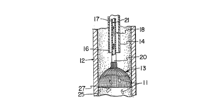
Referring to the drawings, a ~l~re;lrt;d venal caval filter device 10 is shown
in accordance with the present invention in its deployed condition wherein emboli and/or
thrombi 11 (Fig. 4) are filtered from the inferior venal caval 12 using a f~ter 13. The
filter device 10 comprises a sheath or outer c~thP.ter 14 having proximal and distal ends
15 and 16, respectively, and a coaxial inner c~thP.ter 17 which is slidable axially within
a through lumen 18 of the outer C~ rlP.I 14. The inner c~tl~P.ter 17, having proximal and
distal ends 19 and 20, respectively, in~ludes a through lumen 21 and is connec~P~ at its
proximal end 19 to a suction device, preferably a syringe 22, for reasons explained below.
The filter 13 comprises a mesh-like, collapsible basket connected to the
distal end 20 of the inner catheter 17 in such a way that when it is deployed (opened), the
patient's blood can flow past the open filter 13 to the patient's heart (see arrow 29). After
deployment, the m~teri~l 11 to be filtered is aspirated through the lumen 21 of lhe inner
catheter 17 by means of the syringe 22, which is connPctçd to a port 24 at the proximal
end 19 of the inner c~thPter 17.
The construction of the filter 13 is not critical and various dirrelclll
constructions may be used in accordance with the invention. What is necessary is that the
filter be self-exr~ntling so that when it is released from the outer ç~thPtP.r 14, it expands
into contact with the vena cava and, of course, that it be made of a m~teri~l which when
exr~ncle-l will block the particles of concern. The filter must also be capable of being
compressed by contact with the outer c~thPt~Pr so that it can be withdrawn after the need
for filtering no longer exists.
M~tP.ri~l~s which are suitable for use as a filter in accordance with this
invention have been proposed for use as self-exr~n~ing stents. One such construction is
shown in U.S. Patent No. 4,655,771 which is incorporated herein by reference. The stent A
disclosed in that patent is a radially and axially flexible, elastic tubular body made of a
multiplicity of thread elements defining a radially self-Pxr~n~ing helix. The thread
elements may be metallic or plastic as described in the patent. When radial pressure is
applied to such a self-expanding helix, the diameter is decreased which, in the case of the
CA 02208345 1997-06-19
WO 96119941 PCTJ~JS9SJ16792
s
invention, means that the ~i~meter of the filter can be decreased sllfficiently so that it will
fit within the outer c~ ler 14 when co~ t;ssed.
The proximal end 15 of the outer c~theter 14 is connPcted to a hub 30
which inclndes a side port 32 and a locking diaphram 34 through which the inner c~theter
5 17 extends. This type of hub is collve-~ n~l The proximal end of inner c~lllrlPI 17 may
be attached to a syringe 22 by a luer lock (not numbered) so that a negative pressure can
be applied to c~thPter 17 as descrihe(l below. When locking diaphram 34 is loosened, the
inner catheter 17 can be moved axially relative to the outer c~thPter 14. When diaphram
34 is locked, the inner ç~th~ter 17 is held in position within a fluid tight seal. The side
10 port 32 can be connected to a syr;nge 36 co.~ E a hepr~ni7~1 saline solution which can
be forced under pressure through the outer c~thPtPr 14 after the filter 13 has been
e~cr~n~1e~ ~1tPrn~tively, a pres~llri7eA IV bag may be conn~ctP~l to side port 32 to
provide a continuous flow of h~~ ed saline solution between the c~thPtPrs. This
prevents clots from acc~lm~ tin~ in the space between the two ç~thPters 17 and 14.
Preferably, the filter 13 and the inner c~thPtPr 17 are loaded into the outer
catheter 14 during m~mlf~c*lre so that the entire assembly can be positioned in the
patient's blood vessel. Referring to FIGS. 1 and 2, to position the f~ter 13 properly
within the blood vessel 12, the distal end 16 of the outer c~thPter 14 is inserted within the
right internal jugular vein of the patient preferably using a Seldinger needle and guidewire
20 as is well known in the art. The outer c~ll.rLe~ 14 may include a suitable marker (not
shown) at its distal end 16 to enable its position to be ~let~rminpd fluoroscopically so that
the operator knows when the distal end 16 of ç~thPter 14 has been advanced to the
i~lfld~ al portion of the inferior vena cava 12.
When the device is properly positioned, as detPrminPd fluoroscopically, the
25 diaphram 34 is released and the inner catheter 17 advanced. As the inner catheter 17 is
advanced, the filter 13 starts to exit from the distal end of the outer c~tllpter 14, causing
the filter to deploy autom~ti~lly as the ~ E pressure of the outer catheter is
removed. When the filter is fully deployed as shown in Figs. 3 and 4, the distal portion
27 of the filter expands into light contact with the inner surface 25 of the Vena Cava 12.
30 This self-exp~n~inE action causes the filter to be seated correctly and retains the filter in
position within the blood vessel without resort to hooks or the like which might tend to
injure the vessel. Once exr~n~lpd~ particles 11 in the blood which flows through the filter
13 will be entrapped by the filter.
CA 0220834~ 1997-06-19
WO 96/19941 PCT/US95l16792
Although, in the ~lt;r~ ed embo-liment7 the physician will receive the
device with the inner cAthPtPr 17 and filter 13 loaded within the outer cAthPter 14, it is
possible to provide the cAthPter~ as separate components in which case the inner cAth~ter
17 and filter 13 would be threaded through the outer cAthPter 14 after the outer çAt~Pter
5 has been positioned within the patient's blood vessel.
The filter may be deployed by pushing the inner CAI1-~'lPI 17 so as to expel
the filter from the stationary outer c~thPt~Pr 14 or it may be deployed by pulling the outer
catheter 14 while holding the inner catheter 17 stationary. In either case, once the filter
braid is free of the constraint of the outer cAthPter 14, it will expand into a funnel-like
10 shape, expansion of the proximal portion of the filter being prevented by its ~ttAchment
to the distal end of the inner catheter 17. In this condition, the filter 13 will prevent the
undesired emboli and thrombi from passing into the aorta.
If the filter is made by the braiding process described in Patent No.
4,655,771, particularly using metal filAment~, the distal end of the filter may be enclosed
15 in a protective rim to prevent injury to the walls of the blood vessel, and re~uce the
possibility of snagging as the filter is moved relative to the outer c~thPter 14.
Referring to FIG. 4, as the filter 13 collects emboli and thrombi 11, the
syringe 22 may be used to provide a suction force that aspirates the particles 11 from the
filter 13 through the lumen 21 of the inner catheter 17 and into the syringe 22, where they
20 may be safely disposed.
The suction provided by the syringe 22 within the lumen 21 has a greater
force than the force from the rate of blood flow. Thus, the blood is directed into the
lumen 21 with sufficient force to flush the particles 11 from the mesh of the filter 13 into
the lumen 21 and into the syringe 22. This process can be performed repeatedly during
25 the surgery when the buL~ of the particles 11 will be dislodged into the vena cava 12.
Thus, the filter 13 may be flushed repeatedly without the need to remove the filter 13
from the blood vessel 12 since the flllshing or suction force is provided by the syringe 22
located outside the patient.
The shape of the filter 13 is important. In the plerell~d embo-lirnent7 the
30 funnel-like or inverted umbrella shape enables the filter 13 to be compressed by pulling
on the inner catheter 17 causing the mesh-like material of the filter 13 to contact the distal
end 16 of the outer catheter 14. Continued pressure causes the filter 13 to collapse or
CA 02208345 l997-06-l9
WO 96/19941 PCT/US9~i~16792
compress to the condition shown in Figure 2. In this condition, the filter 13 can easily
be removed after there is no longer a need for the filter 13.
While the embodiment of the invention shown and described is fully capable
of achieving the results desired, it is to be understood that this embodiment has been
5 shown and described for purposes of illllstr~ti~n only and not for purposes of limit~ticn.
Therefore, the invention is limited only by the appended claims.