Note: Descriptions are shown in the official language in which they were submitted.
CA 02210142 1998-10-07
. WO 97!20497 PCT/US96/17675
RFI1~ABLE SENSOR ACCESSORY CONTAINING A
CONFORMABLE SPRING ACTIVATED RUBBER SLEEVED CLIP
BACKGROUND OF THE INVENTION
This invention relates to sensors for use with non-invasive pulse monitors
such as plethysmographs or pulse oximeters.
A plethysmograph is a pulse monitor. The plethysomograph sensor shines
light into the patient's tissue, and the light transmitted through the tissue
is received by a
photodetector. The photodetector generates electrical signals corresponding to
the
transmitted light levels and transmits the signals to a monitor for
processing. Arterial
blood will absorb some of the light, with more light being absorbed when there
is more
blood. Thus, changes in the amount of transmitted light are related to pulses
of arterial
blood in the illuminated tissue.
A pulse oximeter is a device for noninvasively determining the oxygen
saturation of arterial blood. The pulse oximeter sensor shines light at two
different
wavelengths (one in the red range, the other in the infrared range) through a
portion of the
patient's blood-perfused tissue. The red and infrared light scattered by
through the tissue
2 0 is detected by a photodetector. The amount of light absorbed varies with
the amount of
oxygen in the blood, and varies differently for red and infrared light. The
pulse oximeter
monitor computes blood oxygen saturation based on the changes in the two
detected light
levels between two points in time.
A key issue regarding the performance of the above discussed sensors is the
2 5 degree to which the surface of the sensor conforms to the surface of the
tissue being
measured. Poor sensor-tissue conformance provides a path for ambient light to
reach the
photodetector and to thereby distort the measurements of the sensor. In
addition, poor
sensor-tissue conformance interferes with retention of the light generated by
the
transmitter and facilitates shunting (i.e. the transmission of light from the
transmitter to the
3 0 detector via a path not going through the tissue to be measured) as well
as relative motion
between the sensor and tissue, all of which phenomena can distort the
measurements of the
sensor.
One type of sensor providing excellent sensor-tissue conformance is
sometimes referred to as an "adherent sensor". U.S. Patent No. 4,830,014
describes one
3 5 such sensor, having a flexible web onto which a transmitter and detector
are mounted. The
SUBSTITUTE SHEET (RULE 26)
CA 02210142 1998-10-07
WO 97120497 PCTlUS96/17675
web has an adhesive surface for attachment to a patient tissue, thereby
permitting close
sensor-tissue conformance. Adherent sensors, however, are typically restricted
to one
application due in part to a decrease in adhesive effectiveness with each
application and in
part to the difficulties in cleaning and sterilization for reuse. Thus, the
use of adherent
sensors can make pulse oximetry expensive.
On the other hand, many reusable sensors tend to employ rigid structures
(such as two legs of a clip or clothes-pin type arrangement, for ex. see U.S.
Pat. Nos.
3,152,587 and 3,810,460) for housing the detector and transmitter and as a
result often
conform poorly to a patient tissue. U.S. Patent No. 4,685,464 discloses a
similar type rigid
l0 structure with deformable pads which facilitate tissue-sensor conformance.
However, the
sensor heads (i.e. detector and transmitter) in this sensor are permanently
embedded in the
rigid structure. A recent trend in sensors involves the use of sensor heads
that can be
removably attached to a variety of sensor accessories (each of which is
tailored to different
applications, e.g. for attachment to an ear, finger etc.), thereby rendering
pulse oximetry
over many different tissue sites less expensive. One such sensor is sold under
the name
"Y-sensor" by Nellcor Incorporated of Pleasanton, California (the assignee of
the present
application) and depicted by reference numeral 201 of Fig. 2.
What is needed is a reusable sensor with removably attachable sensor heads
that is capable of providing a high degree of tissue-sensor conformance.
SUMMARY OF THE INVENTION
The present invention provides a reusable accessory for a sensor (such as a
pulse oximeter or a plethysmograph) that allows close conformance between the
sensor
heads and the patient tissue to be measured. The accessory thus provides both
a key
2 5 advantage of adherent sensors (i.e. close tissue-sensor conformance) and a
key advantage
of rigid structure sensors (reusability). The accessory provides a structure
for mounting
the sensor heads, whereby the sensor heads can pivot about their respective
points of
attachment to the mounting structure within first and second voids,
respectively, thereby
allowing the sensors to closely conform to the patient tissue. Sensor
accessories for other
3 0 applications (e.g. for attachment to an ear) could be modified to provide
a similar
mounting structure and resulting close tissue-sensor conformance.
In one embodiment, the mounting structure includes a spring containing
two coils, and two frames connected to the coils, each frame having an end
shaped to fit a
2
SUBSTITUTE SHEET (RULE 26)
CA 02210142 1998-10-07
WO 97/20497 PCT/US96/17675
portion of a sensor head and onto which a sensor head is mounted. When mounted
on the
frames, the sensor heads can pivot within voids located inside the frames. In
another
embodiment, the structure for mounting includes a stamped spring having two
ends, each
of which is shaped to fit a portion of a sensor head. In another embodiment,
the structure
for mounting includes two stamping forms, each of the stamping forms having an
end
shaped to fit a portion of one of the sensor heads.
Another aspect of the present invention concerns the use of flexible sleeves
to cover the sensor heads when mounted on a reusable sensor accessory. Such
flexible
sleeves can be used in conjunction with the mounting structures described
immediately
l0 above (which permit the sensor heads to pivot) and with prior art mounting
structures (not
permitting the sensor heads to pivot). The sleeves can be replaced or cleaned
between
patient applications, thereby reducing the risk of cross contamination between
patients. In
addition, the sleeves can be colored and shaped to reduce the amount of
shunting (i.e. light
that travels from one sensor head to the other without passing through the
patient tissue).
Also, the use of a variety of sleeves with different shapes may enable the
same mounting
structure to conform to a variety of tissue sites and patient types.
Yet another aspect of the present invention concerns sensory accessories
providing light shields that reduce shunting in non-flat site applications but
yet do not
interfere with tissue-sensor conformance in flat site applications (such as
where the tissue
2 o site is the web between the thumb and forefinger). In one embodiment, such
an accessory
contains two frames in each of which a sensor head is mounted, as well as
Iight shields
attached to the sides of the frames. The shields can be in one of two
positions: coplanar
with the frames (for flat site applications) and rotated to be perpendicular
to the frames
(for non-flat site applications). In another embodiment, the sensor accessory
contains two
2 5 frames in each of which a sensor head is mounted, and two flexible sleeves
for covering
the two frames. The flexible sleeves have sides that act as light shields in
non-flat site
applications but flatten under pressure in flat site applications, thereby
permitting close
conformance between the sensor and flat sites. In yet another embodiment, the
sensor
accessory consists of two wire frames that can fold to surround a non-flat
site. Each of the
3 0 wire frames is covered by a flexible sleeve. When the wire frames are
folded to surround a
non-flat site, the flexible sleeves act as light shields.
3
SUBSTITUTE SHEET (RULE 26)
CA 02210142 1998-10-07
WO 97/20497 PCT/US96/I7675
BRIEF DESCRIPTION OF THE DRAVViNGS
Fig. 1 is a diagram illustrating the application to a patient of the sensor
accessory of Fig. 2.
Fig. 2 is an exploded view of a partially assembled sensor using an
accessory permitting the sensor heads to pivot.
Fig. 3 is a diagram illustrating the close tissue-sensor conformance realized
with the sensor accessory of Fig. 2.
Fig. 4 is a diagram illustrating gaps between the tissue and the sensor heads
that can occur when using a prior art rigid structure sensor accessory.
Fig. 5 is a diagram illustrating the spring component 203 of the accessory
of Fig. 2
Fig. 6 is a diagram illustrating upper clip portion 207a of the sensor
accessory of Fig. 2.
Fig. 7 is a diagram illustrating lower clip portion 207b of the sensor
accessory of Fig. 2.
Fig. 8 is a diagram illustrating a sensory accessory containing a stamped
spnng.
Fig. 9 is a diagram illustrating a sensory accessory containing a stamped
form and a standard coil spring.
2 o Fig. 10 is a diagram illustrating a sensor accessory that permits the
sensor
heads to pivot.
Fig. l la is a diagram illustrating a sensor accessory with folding light
shields.
Fig. 1 lb is a diagram illustrating the sensor accessory of Fig. 1 la applied
to
2 5 a non-flat site.
Fig. 12 is a diagram illustrating a sensor accessory with rubber sleeves
having sides that act as light shields on flat sites and flatten under
pressure on non-flat
sites.
Fig. 13a is a diagram illustrating a sensor accessory with wire frames that
3 o can fold to surround a non-flat site.
Fig. 13b is a diagram illustrating the sensor accessory of Fig. 13a applied to
a non-flat site.
4
SUBSTITUTE SHEET (RULE 26)
CA 02210142 1998-10-07
WO 97/20497 PCT/US96/17675
Fig. 13c is a diagram illustrating the sensor accessory of Fig. 13a applied to
a flat site.
DESCRIPTION OF THE PREFERRED EMBODIMENT
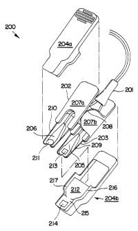
Fig.2 illustrates a partially assembled oximeter 200 according to the present
invention. Oximeter 200 consists of a sensor 201, a spring activated clip 202,
a spring 203
and rubber sleeves 204a and 204b (the latter four items are hereinafter
referred to as a
"sensory accessory"). (In alternate embodiments, sleeves 204a and 204b could
be made of
a flexible material other than rubber.) Sensor 201 contains a phototransmitter
205 and a
photodetector 206 (each of the latter two items is sometimes referred to as a
"sensor
head"). Clip 202 contains an upper portion 207a and a lower portion 207b,
which are
illustrated in more detail in Figs. 6 and 7, respectively. Spring 203, which
is illustrated in
more detail in Fig. 5, contains a left spring coil 208, a corresponding right
spring coil
(indicated by reference numeral 703 in Fig. 5), a lower spring frame 209 and
an upper
spring frame 210.
In order to assemble oximeter 200, transmitter 205 and detector 206 are
pulled through a cavity between clip upper portion 207a and clip lower portion
207b. A
button-shaped portion 211 of detector 206 and a similar button-shaped portion
(not shown)
of phototransmitter 205 are then mounted into ends (identified in Fig. 5 by
reference
2 o numerals 701 and 702, respectively) of upper and lower spring frames 210
and 209,
respectively. When so mounted, detector 206 and transmitter 205 rest within
voids 704
and 705, respectively. Ideally, the cavity between clip portions 207a and 207b
is
sufficiently small so as to prevent transmitter 205 and detector 206 from
slipping
backwards through the cavity (i.e. away from spring frames 209 and 210.) if
the grip on
2 5 transmitter 205 and detector 206 is lost while attempting to perform this
last fitting step.
Finally, sleeve 204b is placed over lower spring frame 209 and the flat part
of clip lower portion 207b (i.e. the part coplanar with lower spring frame
209). Lower
spring frame 209 and transmitter 205 are placed inside sleeve 204b underneath
a flap
portion 212 of sleeve 204b, in such a way so as to align a window 213 of
transmitter 205
3 0 with a window 214 of sleeve 204b. Sleeve 204a is similarly placed over
upper spring
frame 210 and the flat part of clip upper portion 207a, in such a way so as to
align a
window (not shown) of detector 206 with windows 213 and 214 and a window (not
shown) of sleeve 204a. Except for their respective windows, sleeves 204x-b are
largely
5
SUBSTITUTE SHEET (RULE 26)
CA 02210142 1998-10-07
WO 97120497 PCT/US96/17675
opaque to the light transmitted by transmitter 205 (i.e. the amount of light
transmitted
through sleeves 204x-b is comparable to the amount of light picked up by
detector 206
from noise sources). The above-mentioned windows should be thin, so as to
limit the
attenuation of the light transmitted from transmitter 205 to detector 206.
Window 214 of
sleeve 204b and the corresponding window of sleeve 204a should be shaped to
closely
match the respective shapes of transmitter 205 and detector 206, respectively.
Once oximeter 200 is assembled, the ends of upper and lower clip portions
207a-b are pressed together, thereby separating lower and upper spring frames
209 and
210 and permitting the patient's tissue to rest between a pad portion 215 of
sleeve 204b
and a corresponding pad portion (not shown) of sleeve 204a. Edge portions 216-
217 of
sleeve 204b flank pad portion 215 and, along with corresponding edge portions
of sleeve
204a (not shown), should be of sufficient thickness so as to inhibit the
rotation of oximeter
200 about the measured tissue at an angle perpendicular to edge portions 216-
217. Upon
release of upper and lower clip portions 207a-b, the tissue is held tightly in
place, as
illustrated in Fig. 1. In one embodiment, peel-off adhesives could be placed
on pad
portion 215 and the corresponding pad portion of sleeve 204a to minimize the
problem of
relative motion between the patient tissue and the sensor heads.
Upper clip portion 207a contains two cylinders 6002 and 6003 (Fig. 6),
each defining a hollow in which a respective one of discs 7001 and 7003 (Fig.
7) of lower
2 0 clip portion 207b rotates to separate or bring together lower and upper
frame portions 209-
210. A wedge shaped hollow 7002 is cut out of disc 7001. A similarly shaped
hollow (not
shown) is cut out of disc 7003. A wedge shaped piece 6001 is positioned inside
the
hollow defined by cylinder 6002. A similarly shaped piece (not shown) is
positioned
inside the hollow defined by cylinder 6003. When discs 7001 and 7003 are
positioned
2 5 within the hollows defined by cylinders 6003 and 6002, respectively, the
wedge shaped
pieces inside cylinders 6003 and 6002 sit within the wedge shaped hollows cut
out of disc
7001 and 7003, respectively.
Thus, the degree to which disc 7001 and 7043 can rotate within cylinders
6003 and 6002, respectively, is limited in both the clockwise and
counterclockwise
3 o directions. As a result, the degrees to which lower and upper frame
portions 209-210 can
be brought together and separated, respectively, are limited. The limitation
on the degree
to which frame portions 209-210 can be brought together may prevent sleeves
204a-b from
6
SUBSTITUTE SHEET (RULE 26)
CA 02210142 1998-10-07
WO 97120497 PCT/US96J17675
deforming under continuous pressured contact when the accessory of Fig. 2 is
not being
used.
A key advantage of the sensory accessory used in oximeter 200 is the
ability of detector 206 and transmitter 205 to pivot up and down within voids
704 and 705,
respectively, about their point of attachment to ends 701 and 702 of upper and
lower frame
portions 210 and 209, respectively. This pivoting ability permits detector 206
and
transmitter 205 to conform to a greater degree to the tissue to be measured
(particularly
irregular surfaces) over a broad range of patients and different tissue sites,
than is typically
possible with prior art oximeters in which the detector and transmitter are
embedded in a
l0 rigid structure accessory. This closer sensor to tissue conformance results
in less ambient
light reaching the detector and thus more accurate readings. Flap portion 212
of sleeve
204b and the corresponding flap portion (not shown) of sleeve 204a should be
sufficiently
thin so as to not interfere with the range of pivoting motion of detector 206
and transmitter
205.
For example, Fig. 3 illustrates a tissue 50 in place to be measured by
oximeter 200. As shown, the pivoting ability described above permits detector
206 and
transmitter 205 to conform closely to tissue S0, thereby reducing the problem
of ambient
light. By contrast, Fig. 4 illustrates a rigid structure oximeter 60. A tissue
61 determines
the angle at which the clip of oximeter 60 is opened. This angle determines
the angles at
2 0 which a detector 62 and a transmitter 63, both of which are embedded in
oximeter 60,
meet tissue 61, and thereby results in gaps 65 and 63 between tissue 61 and
detector 62
and between tissue 61 and transmitter 63, respectively. The presence of gaps
63 and 65
permits ambient light to distort the readings of oximeter 60.
The sensor accessory of Fig. 2 provides a new use for the existing "Y-
2 5 sensor", described above, in applications similar to those suited to the
rigid clothes-pin
structure sensor of U.S. Patent No. 4,685,464 (in which the sensor heads are
permanently
embedded). Existing sensor accessories for other applications (e.g. for
attachment to an
ear) could be modified to include a mounting structure for the sensor heads
similar to
spring 203.
3 0 Rubber sleeves 204a-b provide several advantages including the following:
7
SUBSTITUTE SHEET (RULE 26)
CA 02210142 1998-10-07
WO 97/20497 PCT/US96/17675
1 ) Due to their opacity, sleeves 204a-b act as light shields and thereby
reduce
shunting, i.e. the amount of light from transmitter 205 that reaches detector
206 through an
undesired path (i.e. not through the tissue whose oxygen level is to be
measured).
2) Sleeves 204a-b, which can be cleaned or replaced between applications,
protect
spring portions 209-210, detector 206 and transmitter 205 from contamination
which could
distort readings of oximeter 200 and result in cross-contamination from one
patient to
another. Ideally, sleeves 204a-b have no gaps or crevices, which could shelter
contaminants during cleaning.
IO
3) Different sleeves can be used to achieve good sensor to tissue conformance
for
different tissue sites and/or for different patient shapes. For example, pad
portion 215 of
sleeve 204b (and the corresponding pad portion of sleeve 204a) should have a
parabolic
shape when oximeter 200 is used to measure the oxygen level in a child's
finger. A sleeve
with a differently shaped pad portion 215 might be appropriate for adults. A
web-like
structure in a sleeve might be appropriate for measuring the tissue between
the thumb and
forefinger.
The above advantages can also be obtained by using rubber sleeves in
2 0 conjunction with prior art rigid structure sensor accessories in which the
sensor heads
cannot pivot.
Fig. 8 illustrates another type of sensor accessory that facilitates tissue-
sensor conformance by allowing the detector and transmitter to pivot. The
accessory
comprises a stamped spring 1000, a top left clip/lever section 1005, a bottom
left clip/lever
2 5 section 1006 and corresponding top and bottom right clip/lever sections
(not shown}. The
clip/lever sections are used to open stamped spring 1000 for tissue insertion.
The button shaped portions of transmitter 205 and detector 206 are mounted
onto bottom
and top ends 1002 and 1001 of stamped spring 1000, thereby permitting
transmitter 205
and detector 206 to pivot within voids 1004 and 1003, respectively. Stamped
spring 1000
3 0 is useful in applications requiring only a limited distance between the
sensor heads (i.e.
where the tissue is of limited thickness) so that the blending limits of
spring 1000 are not
exceeded.
8
SUBSTITUTE SHEET (RULE 26)
CA 02210142 1998-10-07
WO 97120497 PCT/US96/17675
When the sensory accessory of Fig. 8 is assembled, a circular portion 1007
of clip section 1006 is mounted onto a circular portion 1008 of clip section
1005. The
abutment of a notch 1009 of clip section 1006 against a notch 1 O 11 of clip
section 1005
(similar notches are present on the top and bottom right clip/lever sections)
limits the
extent to which ends 1001-1002 of stamped spring i 000 can be separated.
Similarly, the
abutment of a notch 1010 of clip section 1006 against a notch 1012 of clip
section I 005
(similar notches are present on the top and bottom right clip/lever sections)
limits the
extent to which ends 1001-1002 of stamped spring 1000 can be brought together.
This
latter limitation may prevent sleeves (not shown) placed on stamped spring
1000 over ends
1001 and 1002, from deforming under continuous pressured contact when the
accessory of
Fig. 8 is not being used.
Fig. 9 illustrates yet another type of sensor accessory that facilitates
tissue-
sensor conformance by allowing the detector and transmitter to pivot. The
sensor
accessory of fig. 9 is similar to that of Fig. 2, except that a standard
commercially
available coil spring 1101 (i.e. one without lower spring frame 209 and upper
spring frame
210) is used instead of spring 203. An upper stamping form I 102 with an end
1104 is
inserted into upper clip piece 1103. In one embodiment upper stamping form
1102 is
made of wire. (In another embodiment stamping form 1102 is made of a glass-
reinforced
thermoplastic material, in which case stamping form 1102 and upper clip piece
could be
2 0 formed as one piece.) Button-shaped portion 2I 1 of detector 206 is
inserted into end 1104,
thereby permitting detector 206 to pivot in void 1105. A similar lower
stamping form (not
shown) with an end similar to end 1104 (about which transmitter 205 pivots) is
inserted
into a lower clip piece (not shown). The upper and lower clip pieces of the
accessory of
Fig.9 may contain features similar to those described above with reference to
the accessory
2 5 of Fig. 2 for limiting the degrees to which the upper and lower stamping
forms can be
brought together and separated, respectively.
Fig. 10 illustrates yet another type of sensor accessory that facilitates
tissue-
sensor conformance by allowing the detector and transmitter to pivot. An upper
(lower)
clip piece 1201 (1202) defines a void 1205 (1206) in which detector 206
(transmitter 205)
3 0 is positioned. Button shaped portion 211 of detector 206 is fitted through
a hole 1204
inside a sleeve 1203 covering the part of upper clip piece 1201 defining void
1205. (In
other embodiments, sleeve 1203 has a structure for mounting button portion 21
l, other
than hole 1204, for example a button shaped pocket.) Detector 206 is thereby
secured in
9
SUBSTITUTE SHEET (RULE 26)
CA 02210142 1998-10-07
WO 97/20497 PCT/US96/17675
position and still free to pivot within void 1205 about button shaped portion
211.
Transmitter 205 is similarly secured in position. The upper and lower clip
pieces of the
accessory of Fig.lO may contain features similar to those described above with
reference
to the accessory of Fig. 2 for limiting the degrees to which voids 1205 and
1206 can be
brought together and separated, respectively.
Another aspect of the present invention will now be discussed, in particular
sensory accessories providing light shields that reduce shunting for non-flat
site
applications and yet do not interfere with sensor-tissue conformance in flat
site
applications. Figs. l la-b illustrate a sensory accessory 1300 suitable for
application to
l0 both flat sites (such as the web between the thumb and forefinger) and non-
flat sites {such
as a finger). A sensory accessory 1300 contains upper and lower frames 1301
and 1302,
respectively, which in one embodiment are made of polycarbonate. Button-shaped
portion
211 of detector 206 and button-shaped portion of transmitter (not shown) are
pushed
through respective holes in portions 1304 and 1305 of upper and Iower frames
1301 and
1302, thereby fixing in position detector 206 and transmitter 205 in
respective pockets in
portions 1304 and 1305. (In other embodiments, portions 1304-1305 have a
structure fox
mounting the button shaped portion of a sensor head, other than a hole, for
example a
button shaped pocket.) Light shields 1303a-b & 1303c-d flank portions 1304 and
1305,
respectively, and can be in an unrotated position in which light shields 1303a-
b & 1303c-d
2 0 are coplanar with portions 1304 and 1305 (as shown in Fig. 11 a), suitable
for flat site
application, or can be rotated downward and upward, respectively (as shown in
Fig. 1 lb
where accessory 1300 is applied to a non-flat site).
Fig. 12 illustrates a sensory accessory 1400 similar to that of Fig. 11 a-b,
with the rotatable light shields 1303a-d replaced with rubber sleeves 1401 and
1402, which
cover portion 1304 of upper frame 1301 and portion 1305 of lower frame 1302,
respectively. (In alternate embodiments, sleeves 1401 and 1402 could be made
of a
flexible material other than rubber.) Rubber sleeve 1401 (1402) has two sides
that extend
downward (upward) from portion 1304 (1305) and thereby reduce the amount of
light
shunted to detector 206 from transmitter 205 when sensory accessory 1400 is
applied to
3 0 non-flat sites. The sides of sleeves 1401 and 1402 flatten under the
pressure of the grip of
accessory 1400 on a flat site to become coplanar with portions 1304 and 1305.
Thus,
accessory 1400 is suitable for both flat and non-flat tissue sites.
SUBSTITUTE SHEET (RULE 26)
CA 02210142 1998-10-07
WO 97/20497 PCT/US96/17675
Figs. 13a-c illustrate a sensory accessory 1500 similar to that of Fig. 11 a-
b,
with the rotatable light shields 1303a-d and the portions 1304 and 1305 of
upper and lower
frames 1301 and 1302, respectively, replaced with wire frames 1501 and 1502.
Wire
frames 1501 and 1502 each surround a void in which a respective one of the
sensor heads
is positioned. Rubber sleeves 1503 and I 504 cover wire frames 1501 and 1502,
respectively. (In alternate embodiments, sleeves 1503 and 1504 could be made
of a
flexible material other than rubber.) The button shaped portion of each sensor
head is
fitted through a hole in a respective one of sleeves 1503-1504, thereby
securing each
sensor head in position but allowing each sensor head to pivot within a
respective one of
l0 the voids about the sensor's button shaped portion. (In other embodiments,
sleeves 1503-
1504 have a structure for mounting the button shaped portion of a sensor head,
other than a
hole, for example a button shaped pocket.) Sleeves 1503 and 1504 are attached
to the rest
of accessory 1500 at one or more hinge/attachment pins 1509.
Wire frames 1501 and 1502 are spring loaded such that the spring force
tends to bring together frames 1501 and 1502, which thereby grip patient
tissue and
conform to the tissue by folding at hinge points 1505-1508 to surround a non-
flat tissue.
The resulting folded sides of sleeves 1501 and 1502 serve as light shields as
shown in Fig.
13b. Wire frames 1501 and 1502 do not fold at internal hinge/break points 1505-
1508
when accessory 1500 is applied to a web site, as illustrated in Fig. 13c.
Thus, accessory
2 0 1500 is also suitable for both flat and non-flat tissue sites. In other
embodiments, means
other than hinge points 1505-1508 for facilitating the folding of wire frames
around a non-
flat tissue site can be employed such as:
a) A connection, made of an elastomeric material (e.g. epdm or silicone
rubber) or
an elastic thermoplastic (e.g. polypropylene), could replace the portion of
wire frames
1501 (1502) between the locations of hinge points 1505-1506 (1507-1508) in the
above
embodiment
b) The portion of wire frames 1501 (1502) between the locations of hinge
points
1505-1506 (1507-1508) in the above embodiment could be annealed and drawn
thinner.
In one embodiment, hinge points 1505-1508 could be spring loaded so as to
3 0 bias frames 1501 and 1502 in a folded position. Frames 1501 and 1502 would
flatten
under the pressure of the grip of accessory I 500 on a flat site.
While the above is a complete description of the preferred embodiments of
the invention, various alternatives, modifications, and equivalents may be
used. (For
11
SUBSTITUTE SHEET (RULE 26)
CA 02210142 1998-10-07
WO 97/20497 PCT/US96/17675
example, frames 1501-1502 of accessory 1500 (Fig. 13a) couid be made of a
glass-
reinforced thermoplastic material instead of wire.) Therefore, the above
description
should not be taken as limiting the scope of the invention which is defined by
the
appended claims.
12
SUBSTITUTE SHEET (RULE 26)