Note: Descriptions are shown in the official language in which they were submitted.
CA 02210~8~ 1997-07-16
P-3498 PATENT
p3498app.doc
Title:
Arterial Catheter and Catheter/Needle Assembly with Improved Flow
Characteristics and Method for Its Use
s
Field of Invention:
This invention relates to intravascular catheters and more particularly to
an over-the-needle arterial catheter with provisions to maintain fluid pathway
patency.
Background
An intravascular catheter is generally a flexible small diameter tube
inserted into a patient's blood vessel to allow withdrawal or addition of fluid.Typically, a practitioner places the catheter by locating a target blood vessel for
15 the placement, then pierces the patient's skin and the blood vessel wall with an
inserter needle, uses the needle to lead the catheter into the vessel and then
removes the needle, leaving the catheter in the vessel. Catheters may be
inserted into blood vessels either through the bore of the inserter needle or over
the outside of the inserter needle. In this disclosure, catheters that are inserted
20 over-the-needle are described. Additionally, a convention is followed in thisdisclosure using the term "proximal" to refer to the portion of the device closest
to the practitioner and the term "distal" for the portion of the device toward the
patient or away from the practitioner.
Over-the-needle catheters are generally supplied already mounted on an
25 inserter needle in a sterile, ready-to-use, unit package. In its simplest form, the
over-the-needle catheter generally resembles one tube slidably fit within another
CA 02210~8~ 1997-07-16
P-3498
tube, the flexible catheter being outermost with a sharp beveled point inserter
needle slidably fit within the catheter bore so that the sharp distal inserter needle
point projects beyond a gently tapered distal end of the catheter. In placement
of these over-the-needle catheters, the needle, with the catheter outside, is held
by the practitioner, generally with the point bevel face up, longitudinally aligned
with the target blood vessel, then placed into the vessel.
Catheters are placed both in veins and in arteries. When the target
blood vessel is a vein, the needle is then inserted at a shallow angle through the
patient's skin into the blood vessel. The practitioner then often determines that
10 the needle is properly positioned within the blood vessel by allowing a smallquantity of the patient's blood to flow through the hollow needle bore, impelledby the patient's blood pressure, so that the small quantity of blood can be seenat the rear of the needle. This practice of using the patient's blood to signal
proper placement of needle within the target vessel is termed "fl~hin~ or
15 flashback." The fl~hing step has the purpose of confirming that the catheter is
properly inserted into the blood vessel. Once the proper placement is
confirmed, the practitioner applies finger pressure to the vessel over the distal
tip of the needle and the catheter to occlude further blood flow, withdraws the
needle and attaches a fluid handling device to the catheter hub.
When an artery is the target blood vessel, the practitioner generally
inserts the needle at a steeper angle than is the usual practice for venous
placement, because arteries are deeper in the patient's tissue. The practitioneruses the flash to confirm that the needle point is in the target vessel. The
catheter is advanced into the vessel and the needle is withdrawn. The presence
25 of a second flash in the catheter hub is indicative of the catheter being in the
blood vessel. Often, a blood sample is analyzed to confirm that the catheter
CA 02210~8~ 1997-07-16
P-349~
needle is placed in the desired artery, not a vein. To obtain a blood sample,
several manipulative steps may be required. Alternatively, a practitioner may
choose to allow a sufficient amount of the patient's blood to escape to confirm
that the pulsatile blood flow characteristic of arterial blood is present.
An "ideal" arterial catheter has two conflicting physical property
requirements. A high degree of stiffness is desirable to facilitate placement ofthe catheter in the artery. However, once placed, since the placement angle is
relatively steep when compared to the placement angle of a venous catheter, a
stiff catheter is more likely to cause damage to the inside wall of the artery
10 opposite the penetration site. If an arterial catheter is formed from a compliant
soft material, it is less likely to cause damage to the patient's blood vessel Asoft arterial catheter is useful for administration of fluids to a patient, but if the
soft catheter is used for withdrawal of blood samples or for placement of a
pressure sensor, a compliant soft material may collapse at the tip when suction
15 is applied to withdraw a sample or become occluded where it bends, either at
the vessel or on the patient's skin.
Because of the likelihood of occlusion by bending or by collapse, most
commercially available arterial catheters are formed from
polytetrafluoroethylene (PTFE) and are somewhat stiff. There are also kink
20 resistant tubes used in the medical arts for oxygen tubes and certain types of
catheters. Some commercially available oxygen tube sets have formed
longit~ in~l ridges within the bore of the tubing, the ridges tend to interfere
with each other when the tube is bent or twisted and m~in~in a flow path
through the bore.
United States Patent No. 4,790,831 discloses a torque-control catheter
adapted to be inserted in to the cardiovascular system. The catheter has a body
CA 02210~8~ 1997-07-16
P-3498
formed from a soft outer sheath coextruded over a stiffer core or inner tube
having a multilobal cross section. The outer portion of the disclosed tube
defines a plurality of longitudinally extending ribs that protrude radially
outwardly at circulllrelelllially spaced-apart locations on the tube. The inner
5 bore of the disclosed tube is smooth. The patent is silent to the kinking and
fluid path occlusion, rather, the disclosure teaches achievement of desired
torque control properties for angiography and angioplasty procedures.
Another patent disclosing a catheter with internal structure is United
States Patent No. 4,~40,623. This patent discloses a medical catheter with a
10 splined internal wall. The patent teaches that the splined wall can be formed as
a coextrusion to provide a long catheter that is useful for angioplasty
procedures. The patent is silent to occlusion of the bore.
If an arterial catheter were available that had sufficient stiffness to
f~cilit~te placement, that softened after placement to substantially reduce
15 trauma to blood vessel walls and was resistant to occlusion from bending and
suction, the art of arterial catheterization would be advanced. Such a catheter
is disclosed below.
Summary
An arterial catheter of the present invention includes an elongate tube
20 that has a sidewall with an inside surface and an outside diameter. The catheter
has a proximal end, an open distal end with a tip portion, and a hollow bore
with an inside diameter. There is a hub attached to the proximal end of the
catheter that is in fluid communication with the hollow bore. The inside surfaceof the hollow bore has a plurality of inward projections disposed longit~ in~lly25 from the proximal end to the distal end. The tip portion has at least one hole
through the sidewall into the bore. The catheter sidewall outside diameter is
~ . CA 02210~8~ 1997-07-16
P-3498
tapered distally from the hole to the open distal end and the inward projectionson the inside surface of the bore are substantially (limini~hed in the tip portion.
The inward projections on the inside wall of the bore of the catheter of
the invention substantially reduce the possibility of occlusion of the bore whenthe catheter is bent, because as the bore is collapsed by bending, the inward
projections interfere with each other and maintain a pathway for fluid flow.
Additionally, the at least one hole through the sidewall provides a pathway for
fluid to enter the bore if the tip is occluded. Over-the-needle catheters generally
are formed from extruded tubing that has a uniform cross-section. The
extruded tubing is cut to the desired length and the distal tip of an over-the-
needle catheter is generally formed into a taper to facilitate the entrance of the
catheter tubing into a blood vessel. If a catheter is formed from a soft material,
the thin tip area is prone to collapse, either from suction or from bending, andsubstantially prevent fluid flow. Because of the need to avoid collapse and boreocclusion, many current arterial catheters are formed from substantially stiff
tubing. In the present invention, while formation of the tapered tip substantially
tlimini~hes the inward projections that keep the bore open, the at least one hole
in the sidewall of the present invention m~int~in~ fluid path patency. The
catheter of the invention is thus able to be formed from a softer material. The
use of the softer material in catheter of the invention thus substantially
elimin~tes problems associated with damage to the blood vessel opposite the
penetration site seen with catheters formed from stiffer materials.
Brief Description of the Drawings
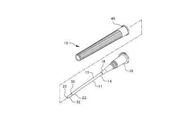
Fig. 1 is an exploded perspective view of the arterial catheter assembly
of the present invention;
CA 02210~8~ 1997-07-16
P-3498
Fig. la is an enlargement of the tip portion of the arterial catheter from
Fig. 1;
Fig 2 is a perspective view of the arterial catheter assembly from Fig. 1
as assembled and packaged;
Fig. 3 is a longitudinal cross-sectional view of the tip portion of the
arterial catheter from Fig. la;
Fig. 4 is a lateral cross-sectional view of the arterial catheter of Fig. 3
along the line 4-4;
Fig. S is a lateral cross-sectional view of the arterial catheter of Fig. 3
10 along the line 5-5;
Fig. 6 is a schematic partial longitudinal cross-sectional view illustrating
initial placement of the arterial catheter of the invention into a blood vessel;Fig. 7 is a schematic partial longitudinal cross-sectional view, sequential
to the view of Fig. 6, illustrating further placement of the arterial catheter of the
15 invention into a blood vessel;
Fig. 8 is a schematic partial longitu~in~l cross-sectional view illustrating
a common substantially rigid arterial catheter in a blood vessel;
Fig. 9 is a schematic partial longitudinal cross-sectional view illustrating
a common soft flexible catheter in a blood vessel;
Fig. 10 is a schematic partial longitudinal cross-sectional view of the
arterial catheter of the present invention positioned in a blood vessel;
Fig. 11 is a schematic partial cross-sectional view of the arterial catheter
of the invention positioned in a blood vessel;
Fig. 12 is a schematic side view of the arterial catheter of the invention
25 positioned in a blood vessel;
. . CA 02210=,8=, 1997-07-16
P-3498
Fig. 13 is a laid-open view of the exterior of the tip portion of the
arterial catheter of the present invention;
Fig. 14is a lateral cross-sectional view of an embodiment of the arterial
catheter of the present invention;
Fig. lS is a lateral cross-sectional view of another embodiment of the
arterial catheter of the present invention; and
Fig. 16 iS a perspective view of the arterial catheter assembly of the
present invention mounted on a hypodermic syringe.
Detailed Description
While this invention is satisfied by embodiments in many different forms,
there are shown in the drawings and herein described in detail, prerelled
embodiments of the invention with the understanding that the present disclosure
is to be considered exemplary of the principles of the invention and is not
considered to limit the invention to the embodiment illustrated. The scope of
the invention is measured by the appended claims and their equivalents.
Referring to Figs. 1-7 and 10-13, an arterial catheter assembly 10 of the
present invention includes an arterial catheter 11 that has an elongate tube 12
with a sidewall 14 that has an inside surface 16 and an outside diameter "D".
Arterial catheter 11 has a proximal end 18, an open distal end 20 with a tip
portion 22, and a hollow bore 24 with an inside diameter "n". Arterial catheter
11 has a hub 26 attached to proximal end 18 in fluid communication with
hollow bore 24. Inside surface 16 of the hollow bore of arterial catheter 1 1 has
a plurality of inward projections 28 disposed longitudinally from proximal end
18 to distal end 20. As shown in Fig. la, tip portion 22 has at least one hole 30
through sidewall 14 into bore 24 and also has a taper 32 of sidewall outside
CA 02210~8~ 1997-07-16
P-3498
diameter "D" distally from adjacent hole 30 to open distal end 20. Taper 32
facilitates entrance of catheter 11 into the patient's blood vessel. Inward
projections 28 in bore 24 are substantially diminished on inside surface 16 at tip
portion 22.
Assembly 10 also includes an elongate inserter needle 34 with a
proximal end 36, a distal end 38, and a passageway 40 therethrough. Inserter
needle 34 is sized to fit within inside diameter "n" of bore 24 of arterial
catheter 11. Inserter needle 34 has a hub 42 attached at proximal end 36 that issized to releasably fit within hub 26 of arterial catheter 11 and a sharpened point
10 44 on distal end 38 of needle 34. Needle 34 has a sufficient length so that
sharpened point 44 of needle 34 extends beyond tip portion 22 of catheter 11
when inserter needle 34 is disposed within bore 24 of catheter 11 and needle
hub 42 is positioned within catheter hub 26.
Preferably, assembly~ 10 is supplied with a shield 46 that releasably fits
15 hub 26 and protects arterial catheter 11 with projecting needle point 40.
Assembly 10 also preferably includes a porous plug 48 that releasably fits
needle hub 42 and allows blood flowing into needle hub 42 to displace air from
the needle and hub. As shown in Fig. 2, assembly 10 preferably supplied
assembled with shield 46 and porous plug 48 and sealed in a package 49
20 (shown in phantom). Package 49 is preferably formed from materials
substantially resistant to the passage of microorg~ni~m~. Package 49 with
assembly 10 therein is preferably exposed to conditions sufficient to render anymicroorg~ni~m~ therein nonviable and assembly 10 is then considered sterile as
long as package 49 is intact.
Referring to Figs. 3, 4 and 5, preferred arterial catheter 11 has
longitudinal inward projections 28 disposed at regular intervals about inside
. CA 02210~8~ 1997-07-16
. P-3498
surface 16 of hollow bore 24 of the catheter. When catheter 11 is flexed or
bent, inward projections 28 are of sufficient size, preferably extending inwardly
from inside surface 16 more than about 0.05mm, and shaped to engage each
other and m~int~in a fluid flow path through bore 24. Preferably, at least one of
5 inward projections 28 includes a radiopaque material 29 to provide the
practitioner the ability to identify the catheter by X-ray. Radiopaque materialssuch as barium sulfate and the like are suitable radiopacity agents. Arterial
catheter 11 may be formed from polymeric materials such as polyvinylchloride,
polyethylene, polytetrafluoroethylene, polyurethane and the like. Preferably,
10 arterial catheter 11 is formed by an extrusion process from a hydrophilic
polyurethane that softens when exposed to physiological conditions (37~/
aqueous saline or blood). A more plefelled arterial catheter 11 is formed by
incorporation of the radiopaque material into a flexible polyester matrix that is
co-extruded with the hydrophilic polyurethane so that one of projections 28
15 includes at least one stripe 29 of substantially radiopaque polyester
encapsulated in the polyurethane. The more prerelled co-extrusion provides a
catheter that softens with physiological exposure that retains dimensional
stability and is reinfol ced by the polyester.
When the radiopaque agent is incorporated as a stripe, several benefits
20 are provided to the catheter. Most common radiopaque agents, when
incorporated into a thermoplastic matrix, also render the thermoplastic
substantially opaque to light tr~n~mi~ion. By providing the prefelled
radiopacity agent as discrete stripe 29, a longit~l~lin~l space 31 between
projections 28 retains the optical properties of the polyurethane, and the density
25 of the x-ray image of the material is enhanced over the density of the x-ray
image provided by an equivalent amount of radiopacity agent uniformly
P-3498 CA 02210~8~ 1997-07-16
dispersed through the entire catheter. Preferably, the hydrophilic polyurethane
selected for the catheter is substantially transparent, or at least translucent, so
that the presence of blood or other fluids in the catheter is visible to the
practitioner through sidewall 14 of the catheter
Figs. 6 and 7 schematically illustrate placement of arterial
catheter/needle assembly 10 of the invention in an artery S0. Assembly 10 is
introduced at a steeper angle to the patient's skin surface than the angle used for
an intravenous catheter, i.e., about sixty-five degrees to about eighty degrees
for an arterial puncture compared to about fifteen degrees to about forty
degrees for a venipuncture. The practitioner longitudinally aligns assembly 10
with artery 50 and advances distal needle point 36 through the patient's tissue
52 until an artery wall 54 is penetrated. Blood from the artery enters
passageway 38 of the needle and is visible at hub 38 of the needle. The
practitioner then, as shown in Fig. 7, advances assembly 10 into the artery until
hole 30 in the catheter sidewall has entered the artery. The pl~re.led arterial
catheter 11 has longit~ldin~l spaces 31 between projections 28 and is formed
from a llanspa-enl, or at least translucent material. Thus, as soon as hole 30
passes through artery wall 54 blood enters spaces 31 between needle 34 and
inside surface 16 of the catheter and is visible through sidewall 14 of the
catheter. The ability to visualize the blood between the catherter and the needle
provides the practitioner with an indication of proper placement of the catheter.
The practitioner then withdraws the needle and continues with the planned
procedure.
Figs. 8 and 9 illustrate problems that may occur with arterial catheters
that are formed from a material that is too rigid or too soft. Fig. 8
schematically shows how a stiff arterial catheter 110 may cause trauma to artery
CA 02210~8~ 1997-07-16
P-3498
wall 54 opposite the penetration site. Trauma to the artery wall may lead to
phlebitis or development of other conditions with the artery. Fig. 9
schematically shows how the tip 112 of an arterial catheter 114 formed from a
soft material may be collapsed under suction or occluded by collapsing against
artery wall 54. When the flow path into the catheter is occluded, withdrawal of
samples or fluid communication with a transducer positioned in the catheter
may be compromised.
Fig. 10 illustrates arterial catheter 11 of the present invention. Since
preferred catheter 11 is formed from a hydrophilic polyurethane that softens
10 after exposure to physiological conditions, tip 22 may be occluded by collapsing
against wall 54 of the artery or under suction. When tip 22 is occluded, a fluidpath, as indicated by flow arrows, is still available into bore 24 through hole 30
in the catheter sidewall.
Figs. 11 and 12 show pl~relled catheter 11 in position in artery 50.
15 Since the preferred arterial catheter softens with exposure to physiological
conditions, it readily bends to conform to the artery wall and to be secured to
the patient's skin surface. When catheter 11 bends, fluid flow through bore 24
is substantially m~int~ined by contact between projections 28.
Fig. 13 illustrates plerelled catheter 11 that has three holes 30 through
20 sidewall 14 spaced about 120 degrees apart. Advantages provided by having
the plurality of holes in the sidewall at the tip include providing a higher flow
rate through the catheter than would available just through the open bore end,
because the total hole area is greater than the area of the open end at the tip
because the tip diameter is reduced by the tapering process. Additionally, if the
25 tip is partially or completely occluded, the flow pathway is still available. The
preferred plurality of holes also provides for a more dispersed flow of any
- CA 022l0~8~ l997-07-l6
P-3498
medicament being added through the catheter. This dispersed flow could
substantially reduce the effects of some toxic or highly concentrated
medicament upon the sidewalls of the blood vessel. Preferably, each of holes
30 are di~elelll distances, x, y, and z, from distal end 20 of the catheter.
5 Distances x, y, and z are preferably between about 2.8 to about 3.6 times
diameter "D" of the catheter. The preferred di~erenl distances x, y, and z
substantially reduce any weakening of catheter sidewall 14 at tip portion 22 that
could occur if all of the holes were the same distance from distal end 20. Taper32 of the tip portion of the arterial catheter of the invention preferably extends a
distance about 3 times diameter "D". Taper 32 at tip portion 22 iS preferably
formed by thermally softening catheter sidewall 14 with a mandrel positioned
into catheter bore 24. The tip portion is then advanced into a tapering and
llinlll~illg die to form taper 32. The sidewall is preferably then allowed to cool
and holes 30 are formed into sidewall 14 against the mandrel in the desired
15 locations. The tapering and llilllll~ing operation substantially riimin~hes interior
projections 28 on interior surface 16 ofthe catheter sidewall.
Table 1 presents the nominal outside diameter corresponding to
standard gauge sizes used for hypodermic needles and catheter tubing. The
most common catheter sizes used for arterial catheters are 18 gauge to 22
20 gauge with a length of about five centimeters. These catheters are supplied
fitted over needles 20 gauge to 24 gauge. A prerelled configuration for
assembly 10 of the present invention is a 20 gauge arterial catheter 11 suppliedover a 22 gauge inserter needle 34.
12
CA 02210~8~ 1997-07-16
. P-3498
Table 1
Hypodermic Tubin~ Size
Nominal Sizes Outside Diameter (mm)
(Gau~e!
0.30
29 0.33
28 0.36
27 0.40
26 0.46
0.51
24 0.56
23 0.64
22 0.71
21 0.82
0.90
19 1.08
18 1.27
17 1.50
16 1.65
Catheter hub 26 and needle hub 42 are preferably fitted to accept male
luer fittings. Catheter hub 26 further is sized and shaped to allow a portion 275 of the exterior of needle hub 42 to fit within it. Needle 34 is fit within arterial
catheter 11 to form assembly 10. Catheter hub 26 and needle hub 42 are
preferably formed from a thermoplastic material that is substantially llanspa-~;nl
or at least translucent, so that the presence of fluid, particularly blood, is visible
in the hub. Thermoplastic materials such as polycarbonate, polyamide and
10 polypropylene are suitable for forming catheter hub 26 and needle hub 42.
Hydrophilic polyurethanes that are substantially catalyst and additive free, andare extrudable are suitable for forming tubing for arterial catheter 11 of the
invention. Hydrophilic polyurethanes incorporating polytetramethylene
etherglycols that are sold under the tr~dçn~me Vialon~ by Becton, Dickinson
15 and Company, Franklin Lakes, NJ are preferred as the hydrophilic polyurethane for forming tubing for the arterial catheter of the invention.
13
CA 02210~8~ 1997-07-16
P-3498
Samples of 22 gauge catheter tubing were prepared from
polytetrafluorethylene (PTFE) and from the plerelled hydrophilic polyurethane.
Test sections of these tubings were subjected to co~--pa-~live physical tests atambient and physiologic (37~C./aqueous saline or blood) conditions. The
5 results of these comparative physical tests are shown in Tables 2 and 3.
Table 2
Co~pa-~live Physical Properties of 22 ~au~e PTFE and Hydrophilic tubin~
Hydrophilic Polyurethane
Time Tensile Tensile 5% mod. 5%mod. elon~. elon~.
37~C. amb. 37~C. amb. 37~C. amb.
Ohr 7507psi 8224psi 835psi 2337psi 398% 293%
4hr 6678psi 7668psi 554psi 1107psi 258% 303%
PTFE
Ohr 6737psi 6910psi 1841psi 2005psi 157% 142%
4hr 5063psi 6060psi 1560psi 1534psi 129% 130%
10 (In Table 2, 37~C. is indicative of physiological conditions)
Referring to Table 2, it is noteworthy that the tensile strength of the
hydrophilic polyurethane and the PTFE are similar and substantially unchanged
by the test conditions. The 5% modulus values, an indication of compliability,
are somewhat comparable for the prerelled hydrophilic polyurethane and the
15 PTFE at ambient conditions and before exposure to physiological conditions.
The somewhat comparable values for 5% modulus at ambient conditions are
indicative that the p-ere--ed arterial catheter would have relatively similar
behavior to the PTFE arterial catheter during the initial placement by the
practitioner. The results show the pl~r~--ed tubing formed from hydrophilic
20 polyurethane has a 5% modulus that is significantly lower after exposure to
14
P-3498 CA 02210~8~ 1997-07-16
physiological conditions and is further reduced by the 4 hour exposure to the
physiological conditions, while PTFE is substantially unchanged and not as
compliant. Thus, the plerelled arterial catheter of the invention is less likely to
cause trauma to the blood vessel wall during an extended duration placement
5 than the common PTFE arterial catheter.
Table 3
Bend force Softenin~
Bend Force (~ms)
Hydrophilic polyurethane
Time Dry Blood/37~C
Ohr 22.6 5.4
4hr 6.8
PTFE
Ohr 34.8 29.3
4hr 33.4
Referring to Table 3, the test results show the Bend force for the
10 hydrophilic polyurethane is originally somewhat lower than for PTFE and is
substantially reduced by the exposure to physiological conditions, while the
PTFE bend force is substantially unchanged by exposure to physiological
conditions. Again, these results suggest that the arterial catheter of the
invention is less likely to cause trauma to the patient's blood vessel. However,15 since the arterial catheter of the invention is much easier to bend and is more
compliant, particularly after exposure time to physiological conditions, were itnot for the presence of holes 30 and the interior projections 28 of the present
invention, the bore of an arterial catheter formed from such a compliant material
CA 02210=,8=, 1997-07-16
P-3498
would easily be occluded by bending or by tip contact with the interior wall of
the patient's blood vessel.
Fig. 4 illustrates a preferred cross-sectional configuration of the arterial
catheter of the invention. As shown in Fig. 4, arterial catheter 11 has six inward
projections 28, each projection being substantially rectangular and having a
generally circular radiopaque stripe 29. The precise cross-sectional shape of
projection 28 and the number and cross-sectional shape of radiopaque stripe 29
may be changed to fit requirements imposed by particular manufacturing and
procedure requirement. Thus other cross-sectional shapes and numbers of
10 projections 28 and radiopaque stripes 29 may be envisioned and are consideredto be within the scope of the present invention. Referring to Figs. 14 and 15,
alternative embodiments for the configuration of the cross-section of the arterial
catheter of the present invention is illustrated. In these alternative
embodiments, the structure of the arterial catheter is substantially similar to the
15 arterial catheter of Figs. 1-7 and 10-13. Accordingly, substantially similar
components that perform substantially similar functions are numbered
identically to those components of the embodiments of Figs. 1-6 except that
suffixes "a" and "b" are used to identify those components in Figs. 14 and 15
respectively.
As shown in Fig. 14, arterial catheter 1 la has elongate tube 12a with
sidewall 14a that has inside surface 16a. Arterial catheter 1 la has proximal end
18a, distal end 20a and hollow bore 24a therethrough with inside surface 16a.
Surface 16a has a plurality of inward projections 28a disposed longitudinally
from proximal end 18a to distal end 20a. Between projections 28a are spaces
25 31a. In this embodiment, projections 28a have a narrower base 60 where
16
P-3498 CA 02210~8~ 1997-07-16
projection 28a joins inside surface 16a than a top surface 62 Additionally,
radiopaque stripe 29a has a substantially triangular cross-section.
As shown in Fig. 15, arterial catheter 11b has elongate tube 12b with
sidewall 14b that has inside surface 16b. Arterial catheter llb has proximal end18b, distal end 20b and hollow bore 24b therethrough with inside surface 16b.
Surface 16b has a plurality of inward projections 28b disposed longitudinally
from proximal end 18b to distal end 20b. In this embodiment, projections 28b
have a more rounded configuration and the number of radiopaque stripes 29b
does not correspond to the projections 28b. Spaces 3 lb are seen between the
radiopaque stripes 29b.
In the embodiments illustrated in Figs. 14 and 15, as well of those of
Figs. 1-7 and 10-13, when the catheter is flexed or bent, the inward projectionsare of sufficient size, preferably extending inwardly from the inside surface
more than about 0.05mm, and shaped to engage each other and m~int~in a fluid
flow through the bore.
Referring to Fig. 16, for some applications, assembly 10 may include a
syringe 70. A practitioner may prefer to insert assembly 10 with syringe 70
mounted on needle hub 42, using the syringe chamber to confirm the presence
of arterial blood. The practitioner may then remove syringe 70 with the needle
attached, occlude catheter hub 26 and then mount either a three-way valve or a
PRN type device on the catheter hub.
The presence of inward projections on the inside wall of the bore of the
catheter of the invention substantially reduces the possibility of occlusion of the
bore when the catheter is bent, because as the bore is collapsed by bending, theinward projections interfere with each other and m~int~in a pathway for fluid
flow. Additionally, the at least one hole through the sidewall provides a
17
CA 02210~8~ 1997-07-16
P-3498
pathway for fluid to enter the bore if the tip is occluded. Prior arterial catheters
generally are not formed from a soft material because the thin tip area is proneto collapse, either from suction or from bending, and substantially prevent fluid
flow. In the present invention, while formation of the tapered tip substantially5 dimini~hes the inward projections that keep the bore open, the holes in the
sidewall of the present invention m~int~in fluid flow. The catheter of the
invention is thus able to be formed from a softer material. The catheter of the
invention thus substantially elimin~tes problems associated with damage to the
blood vessel opposite the penetration site seen with catheters formed from
10 stiffer materials and provides an advance to the art of arterial catheters.