Note: Descriptions are shown in the official language in which they were submitted.
CA 02211512 1997-07-25
ANEURYSM CLOSURF DEVICE ASSE1~'IBLY
FIELD OF THE INVENTION
This invention is an implantable medical device assembly for use in
surgical procedures. The invention includes an artificial occlusion kit that
uses a
S retaining device to prevent migration of artificial occlusion implants from
an
occlusion site, such as an aneurysm, and into an adjacent body space, such as
a
blood vessel.
BACKGROUND OF THE INVENTION
Different implantable medical devices have been developed for treating
various ailments associated with body lumens. such as ailments of body vessel
walls or other lumenal walls. One category of implantable medical device that
has
been developed for artificial occlusion of body spaces is the category of
"artificial
occlusion devices." Although artificial occlusion devices are useful in
occluding
body spaces, other applications include occluding body lumens. Examples of
lumens that have been identified as candidates for treatment with artificial
occlusion devices include, for example, the vas deferens or the fallopian
tubes.
Most commonly, however, artificial occlusion devices have been disclosed for
medical treatment of the vascular lumens and aneurysms in the walls of such
vessels. This treatment is commonly referred to as "artificial vaso-
occlusion."
Artificial Vaso-occlusion
Artificial vaso-occlusion is a medical treatment that has involved
techniques such as the delivery of various occlusive agents including
solidifying
suspensions, thrombogenic fluids, or emboli such as hog hair or suspensions of
metal particles. Delivery of such agents or emboli normally causes a
thrombogenic or other occlusive tissue response. Recent advancements in
CA 02211512 1997-07-25
artificial occlusion of vessels and aneurysms have included the delivery and
implantation of metal coils. Implantable metal coils that are useful as
artificial
occlusion devices in vascular lumens or aneurysms are herein referred to as
"vaso-
occlusion coils."
Vaso-occlusion coils generally are constructed of a wire, usually made of a
metal or metal alloy, that is wound into a helix. Vaso-occlusion coils are
normally delivered through microcatheters such as the type disclosed in U.S.
Patent No. 4,739,768 to Engelson. The microcatheter commonly tracks a guide
wire to a point just proximal of or within the desired site for occlusion. The
coil is
advanced through the microcatheter and out the distal end hole so to at least
partially fill the selected space and create an occlusion.
Once a vaso-occlusion coil is implanted at a desired site, occlusion results
either from the space-filling mechanism inherent in the coil itself, or from a
cellular response to the coil such as a thrombus formation, or both. The space-
filling mechanism of the vaso-occlusion coil may be either based upon a pre-
determined secondary geometry, or may be based upon random flow
characteristics of the coil as it is expelled from a delivery sheath lumen.
Vaso-occlusion coils have been disclosed that have a secondary geometry
or shape which dictates at least in part their space-filling occlusion
mechanism.
Such a secondary shape may include a secondary helical structure which
involves
the primary coil helix being itself wound into a second helix. In addition to
the
space-filling feature, another benefit to having a secondary coil shape is
that it
may allow the coil readily to anchor itself against the walls of a delivery
site. For
example, a vaso-occlusion coil having a secondary shape may be ejected from a
2~ sheath lumen where it was constrained in a stretched condition to have a
first outer
diameter equal to the sheath lumen inner diameter. When ejected, the coil
passively expands to its secondary shape, often having a larger, second outer
diameter to aid in space-filling the body cavity or lumen. This may be an
expansion to the coil's relaxed. unrestrained memory state--or at least until
the
;: ..
CA 02211512 1999-10-04
coil encounters a vessel wall against which it exerts a force to complete the
anchoring process.
One example of a type of vaso-occlusion coil having a pre-determined
secondary shape is described in US Patent No. 4,994,069 to Ritchart et al.
Ritchart
describes a vaso-occlusive wire having a memory imparted thereto by heating
the
wire at about 800°F for 24 hours after it is shaped. This memory is
effective to
return the wire from a stretched, linear condition in which it is advanced
through a
catheter to a space-filling relaxed condition as the wire is released from the
catheter. The diameter of the secondary shape is approximately equal to and
may
be larger than the vessel in which it is deployed.
In contrast to vaso-occlusion coils having pre-determined secondary shapes
that dictate in part their space-filling mechanism, other vaso-occlusion coils
have
been disclosed that take on random shapes when expelled from a delivery
sheath.
This type of vaso-occlusive coil is often referred to as the "liquid coil."
One
example of such a vaso-occlusive coil which takes on a random occlusive shape
when delivered into a body space is disclosed in pending U.S. Patent
Application
Serial No. 08/413,970, filed March 30, 1995 which corresponds in part to
Canadian
Patent Application No. 2,173, 023. This document describes very soft and
flexible
coils which are flow-injectable through the delivery catheter using, e.g.,
saline
solution.
In addition to the various types of space-filling mechanisms and geometries
of vaso-occlusion coils, other particularized features of coil designs, such
as
mechanisms for delivering vaso-occlusion coils through delivery catheters and
implanting them in a desired occlusion site, have also been described.
Examples of categories of vaso-occlusion coils based upon their delivery
mechanisms include pushable coils, mechanically detachable coils, and
electrolytically detachable coils.
One example of the type of vaso-occlusion coil referred to as the "pushable
coil" is disclosed in U.S. Patent No. 4,994,069 to Ritchart et al., introduced
above. Pushable coils are commonly provided in a cartridge and are pushed
or "plunged" from the cartridge into a delivery catheter lumen. A pusher
CA 02211512 1999-12-30
rod advances the pushable coil through and out of the delivery catheter lumen
and into the site for occlusion.
In contrast to pushable coils, mechanically detachable vaso-occlusion
coils are integrated with a pusher rod and mechanically detached from the
pusher after exiting a delivery chatheter. Examples of such mechanically
detachable vaso-occlusion coils are provided in U.S. Patent No. 5,261,916 to
Engelson, or U.S. Patent No. 5,250,071 to Palermo.
Further in contrast to the mechanically detachable type of vaso-occlusion
coil, the electrolytically detachable type is also integrated with a pusher
rod, but
is detached from the pusher by applying a direct current that dissolves a
sacrificial link between the pusher and the coil. Examples of such
electrolyrically detachable vaso-occlusion coils are disclosed in U.S. Patent
No.
5,122,136 to Guglielmi, et al. and U.S. Patent No. 5,354,295 to Guglielmi, et
al.
A further improvement upon the electrolytic detachment mechanisms just
previously referenced is disclosed in co-pending Canadian Application No.
2,162,117. This document describes superimposing an alternating current
signal over the direct current signal, wherein a sensing circuit monitors the
alternating current signal as an indicator of the progression of coil
detachment.
Improvements for enhancing the thrombogenic or other occlusive tissue
response to metal coils have also been disclosed. For example, vaso-occlusion
coils having vaso-occlusive fibers attached thereto have been described (see
for
example, U.S. Patent No. 5,226,911 to Chee et al.). A further type of vaso-
occlusion coil is used as a detachable dielectric electrode in .a radio-
frequency
artificial vaso-occlusion system, is disclosed in co-pending Canadian
Application
No. 2,182,738.
4
CA 02211512 1997-07-25
Vaso-occlusion Coils in Aneurysms
A wide variety of clinical abnormalities in body lumens may be treated
with artificial occlusion methods. For example, artificial occlusion methods
have
been disclosed for treating feeder vessels into tumors, arterio-venous
malformations, fistulas, and aneurysms of vessel walls. Among arterial
abnormalities, aneurysms present particular medical risk due to the dangers of
potential rupture of the thinned wall inherent in an aneurysm. Occlusion of
aneurysms with vaso-occlusion coils without occluding the adjacent artery is a
desirable method of reducing such risk.
In one disclosed method of treating aneurysms with vaso-occlusion coils, a
microcatheter is initially steered into or adjacent the entrance of an
aneurysm,
aided by a steerable wire. The wire is then withdrawn from the microcatheter
lumen and replaced by the vaso-occlusion coil. The vaso-occlusion coil is
advanced through and out of the microcatheter, desirably being completely
delivered into the aneurysm. After or during delivery of such a coil into the
aneurysm, a portion of the coil might then migrate out of the aneurysm
entrance
zone and into the feeding vessel. This may cause an undesirable response of
occluding the feeding vessel. Also, there is an additional risk that the blood
flow
may induce movement of the coil farther out of the aneurysm, resulting in a
more
developed embolus in the good vessel.
One type of aneurysm, commonly referred to as a "wide-neck aneurysm,"
is known to present particular difficulty in placing and retaining vaso-
occlusion
coils. Wide-neck aneurysms are herein referred to as aneurysms of vessel walls
having a neck or "entrance zone" from the adjacent vessel, which entrance zone
has a diameter that either: ( 1 ) is at least 80% of the largest diameter of
the
aneurysm; or (2) is clinically observed to be too wide to effectively retain
vaso-
occlusion coils that are deployed using conventional techniques.
In attempting to prevent potential migration of vaso-occlusion coils from
:0 aneurysms. catheter distal tip shapes may be formed on delivery
microcatheters to
CA 02211512 1997-07-25
help support the distal tip during deployment of vaso-occlusive agents.
However.
this may provide only a partial solution, particularly in the case of wide-
neck
aneurysms.
There is a need for a retaining device that is adapted to block an entrance
S zone to an aneurysm such that occlusion devices may be implanted in and
retained
within the aneurysm and are prevented from migrating through the entrance zone
of the aneurysm and into the adjacent vessel.
SUMMARY OF THE INV NTION
This invention is a novel artificial occlusion kit, which includes a novel
I 0 implantable medical device useful for retaining occlusion devices at an
occlusion
site, and related method for use. A particularly useful application of the
invention
is in the treatment of wide-neck aneurysms and aneurysms emanating from a
curving vessel..
An artificial occlusion kit is provided for implanting and retaining an
15 artificial occlusion device in a body space adjacent to and extending from
a bode
lumen in a mammal. The artificial occlusion kit has at least one occlusion
device
adapted for filling at least a portion of the body space, and a retaining
device
assembly that includes a retaining device.
The retaining device of the artificial occlusion kit is adapted to be
20 delivered and implanted at a retaining site in the body lumen adjacent to
the body
space to be occluded. This retaining device has a first shape that is radially
expandable to a diameter that is sufficient to engage the wall of the body
lumen at
a retaining site adjacent the body space to be occluded. V~~hen engaged with
the
body lumen wall, the retaining device forms a lumen having a diameter that is
2~ sufficient to allow flow therethrough, and also forms a barrier that
prevents
occlusion devices that are implanted in the body space from migrating out of
the
body space and into the adjacent body lumen.
In one retaining device variation. the first shape is formed when the
retaining device is radiallv constrained during delivery to the retaining
site. and a
(,
CA 02211512 1997-07-25
second shape with an expanded outer diameter is formed when the retaining
device is released from radial constraint at the retaining site. Either a
coaxial
delivery sheath or a coaxial delivery wire may provide this radial constraint.
Alternatively, the retaining device may be balloon expandable from the first
shape
S to the second shape.
In another retaining device variation, at least one semi-penetrable space is
provided in the barrier formed at the entrance zone into the body space to be
occluded. This retaining device may be delivered to the retaining site,
followed
by introduction of at least one occlusion device into the body space to be
occluded
through the semi-penetrable space.
In another aspect of the invention, the retaining device may be a metal
wire wound into a primary helix that has a secondary geometry which is also a
secondary helix. The adjacent windings of the secondary helix may be the semi-
penetrable space provided by the appropriate retaining device variation.
1 S In another variation, the semi-penetrable space is sized to allow at least
one occlusion device, when radially artificially constrained to a first
occlusion
device outer diameter, to be inserted therethrough. Subsequent release of the
radial constraint on the occlusion device allows it to reconfigure to a second
outer
diameter which prevents migration back through the semi-penetrable space.
In still a further variation, the semi-penetrable space of the retaining
device
is distendable. A delivery catheter with a tapered tip may be provided such
that
the semi-penetrable space is distendable by forcing the delivery catheter tip
therethrough and into the body space to be occluded. Alternatively to a
tapered-
tip delivery catheter, an introduces wire is provided to distend the semi-
penetrable
spaces of the retaining device. At least one occlusion device is introduced
into the
body space to be occluded either coaxially through the delivery catheter or
over
the introduces wire. Subsequent withdrawal of the delivery catheter or
introduces
wire allows the once distended semi-penetrable space to reform to its original
shape. forming a barrier-against migration of the occlusion devices out of the
~0 occlusion site and into the adjacent lumen.
7
CA 02211512 1997-07-25
In a further variation of the artificial occlusion kit, the retaining device
is a
wire wound into a primary helix over a core member. In a preferred variation
of
this embodiment, the core member is a metal, preferably a shape-memory alloy,
and most preferably a shape-memory alloy of nickel and titanium. The wire is
also preferably a metal, most preferably radiopaque.
Each of the variations discussed herein may further include a smaller
diameter "leading helix" in the retaining device to assist in the alignment
and
deployment of the retaining device as it exits the catheter.
An implantable medical device assembly is also provided, having the
structure described for the retaining device of the novel artificial occlusion
kit and
which is attached to an elongate pusher via a sacrificial link that is
electrolytically
dissolvable. In one variation, this implantable medical device may take the
form
of the "wire wound over core member" variation described for the artificial
vaso-
occlusion kit aspect of the invention.
1 S This invention includes methods for using the apparatus here described.
BRIEF DESCRIPTION OF THE DRAWINGS
Figure 1 A shows a side view of a vessel with an aneurysm in its wall,
wherein a vaso-occlusion coil component of an artificial occlusion kit is
shown
'_'0 being delivered into the aneurysm.
Figure I B shows a side view of the vessel and aneurysm of Figure 1 A,
wherein a retaining device assembly of the artificial occlusion kit is shown
being
delivered to a retaining site in the vessel adjacent the aneurysm which is
substantially filled with a plurality of vaso-occlusion coils.
2~ Figure IC shows a side view of the same vessel and aneurysm wherein the
retaining device is shown electrolytically detached from a pusher, the
retaining
device engaging the vessel wall adjacent the entrance zone of the aneurysm.
bridging across the entrance zone to form a barrier against vaso-occlusion
coil
CA 02211512 1997-07-25
migration into the vessel, and forming a lumen allowing for physiological flow
through the vessel.
Figures 2A-2C show in partial section, a side view of an electrolytically
deployed device.
Figure 3 shows in side view cross-section, a portion of the device shown in
figures 2A-2C emphasizing the section of the device employing an
electrolytically
erodible link.
Figures 4A, 4B, and 4C show in partial cut-away, cross-section, variations
of the shape of retainers made according to this invention.
Figure 5 shows a side view of a vessel having in its wall a wide-neck
aneurysm, showing a variation ~f the artificial occlusion kit where the
retaining
device has a semi-penetrable space through whic!~ an artificial occlusion
device is
being introduced into an aneurysm.
Figure 6 shows a side view of a vessel having in its wall a wide-neck
aneurysm, showing a variation of the artificial occlusion kit where the
retaining
device has a distensible semi-penetrable space that is shown distended by a
delivery catheter through which vaso-occlusion coils are being introduced into
the
aneun~sm.
Figure 7 shows a side view of the vessel and wide-neck aneurysm,
showing a further variation of the assembly shown in Figure 5, wherein the
distensible semi-penetrable space is shown distended by an introducer wire
over
which vaso-occlusion coils are being coaxially advanced into the aneun-sm.
Figure 8 shows a perspective view of a further variation of retaining device
wherein the retaining device is shown to be constructed of a wire wound into a
2~ helix over a core member, the helical wire and core member being formed
into a
secondary geometry.
Figures 9A and 9B show in schematic cross-section anatomically shaped
filler coils suitable for use in conjunction with the retention devices made
according to this invention.
9
CA 02211512 1997-07-25
The present invention provides a novel solution to the problem of vaso-
occlusion device migration out of aneurysms or other implantation sites and
into
the feeding vessels that are not the target of vaso-occlusion. A retaining
device is
used in a novel artificial occlusion assembly to prevent migration of one or
more
occlusion devices from a target occlusion site by forming a barrier at the
entrance
zone to the target site from a feeding vessel. Variations of a novel
implantable
medical device are provided as the retaining device, which novel implantable
medical device is included within the scope of the present invention.
Artificial Occlusion Kit wJ Retaining Device
Figures 1 A-C show sequential steps of a novel method of occluding a
body space -- here an aneurysm of a body lumen wall -- using one artificial
occlusion kit embodiment of the current invention. In this series of Figures,
a
retaining device is provided in a kit together with at least one vaso-
occlusion
device, which kit is also shown in use with at least one delivery catheter.
Figure lA shows the first of a plurality of vaso-occlusion coils is shown as
it is being implanted into an aneurysm. In Figure I B, a retaining device of a
retaining device assembly is shown being delivered to a retaining site in the
body
lumen adjacent the aneurysm after the aneurysm is substantially occluded with
vaso-occlusion coils. In Figure 1 C, the retaining device is completely
implanted
at the retaining site and detached from a pusher via electrolytic detachment
from a
pusher. The implanted retaining device shown forms a barrier against migration
of the vaso-occlusion coils from the aneurysm and into the body lumen, while
maintaining an open conduit for flow through the body lumen.
In Figure 1 A, a cut-away side view of a vessel (2) having an aneun~sm (4)
in its wall is shown. Vaso-occlusion coil (8) is shown being delivered into
aneurysm (4) out of the distal end of delivery catheter (10) in order to
occlude the
~0 aneuy sm (4).
CA 02211512 1997-07-25
Vaso-occlusion coil (8) for the purposes of this invention may be any one
of a wide variety of coils that are known in the art for occluding vessels or
aneurysms. For example, vaso-occlusion coil (8) may be a pushable coil of the
type described in US 4,994,069. Or, coil (8) may be a mechanically detachable
S coil such as that described in US 5,261,916 or US 5,250,071. Alternatively,
coil
(8) may be an electrolytically detachable coil such as that described in US
5,122, I 36 or US 5,354,294.
Still further, vaso-occlusion coil (8) may have a pre-formed secondary
shape that is constrained in a stretched orientation when being delivered
through
delivery catheter (10) but reconfigures when delivered beyond delivery
catheter
( I 0). Such reconfiguring often includes radial expansion to a relaxed memory
state having a desired, pre-determined shaped geometry. Alternatively, coil
(8)
may have highly flexible portions that ball up from random convolutions formed
while the coil flows distally during delivery, such as the coils described in
I S pending US Pat. Appl. No. 08/413,970, filed March 30, 1995.
The type and geometry of vaso-occlusion coil are normally chosen for the
particular delivery mechanism and space-filling characteristics, as may be
appropriate for a particular occlusion site. '
The appropriate design for delivery catheter (10) is defined by the ability
to reach the desired occlusion site atraumatically and to efficaciously
deliver the
vaso-occlusion coil into the site as an occlusion implant. One example of a
catheter that may be used in the present invention is described in US
4,739,768 to
Engelson.
Figure 1B shows a retaining device (19) being delivered through delivery
catheter (20) and into vessel (2) at the site of aneurysm (4). A plurality of
vaso-
occlusion coils (12) is also shown having been implanted into aneurysm (4)
prior
to delivery of the retaining device ( I 9). Retaining device ( 19) is shown as
a distal
segment of a retaining device assembly ( 15), wherein it is attached at its
proximal
end to a pusher ( I 6) which is relatively more stiff than the implantable
retaining
CA 02211512 1997-07-25
device ( 19). Pusher ( 16) is adapted for advancing the retaining device
percutaneously through the delivery catheter (20), even when in tortuous bends
of
the vasculature, into remote internal body spaces for occlusion.
In retaining device assembly (IS), retaining device (19) and pusher (16)
are shown to be coupled or attached via a joint or link (17). The scope of
this
invention contemplates that retaining device (19) and pusher (16) can be
either
electrolyically detachable at link (17) or mechanically detachable at link
(17). In
the electrol5~tically detachable embodiment, link ( 17) is electrolytically
dissolvable when current is applied thereto. Electrolytic detachment
mechanisms
of the types described in US 5,122,136 or US 5,354,295 may be suitable. In the
mechanically detachable embodiment, pusher (16) and retaining device (19) are
mechanically detachably engaged at link (17). In such an assembly, the
mechanical detachment mechanisms of the types described in US 5,261,916 or US
5,250,071 may be suitable.
It is further contemplated that the use of a retaining device to prevent
migration of vaso-occlusion devices from an occlusion site need not be limited
to
use with a "detachable" pusher-retaining device mechanism as is shown in
retaining device assembly ( 15). It may be equally efficacious, and perhaps
even
preferred in a given circumstance, to use separate, non-attached retaining
device
and pusher without the need for a detachable link such as Iink (17).
Where the pusher is separate and not attached to the retaining device,
pushers such as the type described in US 4,994,069 to Ritchart et al. may be
satisfactory. In use, the distal end of the pusher can be advanced axially
within a
delivery catheter lumen to abut a proximal end of the retaining device, also
0
disposed within the delivery lumen. With the distal pusher end in confronting
engagement with the retaining device proximal end, further advancement of the
pusher by the user will effectively push the retaining device distally through
the
12
CA 02211512 1997-07-25
lumen, out of the delivery catheter from a distal port thereof, and into a
vessel site
adjacent a body space where occlusion devices are deployed.
In the artificial occlusion kit variation of Figure 1 B, retaining device (
19)
has a memory in the form of a pre-determined, shaped, secondary geometry.
Retaining device (19) is shown to have a first shape with a first outer
diameter
"A" where it is positioned within delivery catheter (20). The delivery lumen
(22),
which ends distally in distal delivery port (23), radially constrains the
retaining
device such that the first outer diameter is defined by the delivery lumen
(22)
inner diameter. When released from a radially constraining condition,
retaining
device ( 19) also forms a second shape with a second outer diameter greater
than
the first outer diameter "A." and sufficient to engage the vessel wall. In
Figure
1B, retaining device (19) is shown extending beyond the distal delivery port
of the
delivery lumen (22) where it is radially artificially unconstrained and
expanded to
an outer diameter "B" larger than the first diameter "A," engaging the lumen
wall
at the retaining site adjacent the aneurysm.
It is contemplated that the completely relaxed, unconstrained second outer
diameter of the retaining device may be slightly greater than the diameter of
the
vessel. This may be necessary in order to maintain accurate placement of the
retaining device in the vessel lumen at the aneurysm site. However, the
purpose
of the retaining device is merely to form a barrier at the entrance zone of
the
aneurysm to prevent occlusion coil migration. Unnecessary trauma to the vessel
wall, such as from oversizing or coil designs that are too stiff to perform
the stated
purpose should be avoided.
Retaining device (19) is shown in Figure 1B to have a helical geometn~.
2~ In one preferred embodiment, retaining device (19) is a metal wire that is
wound
into a primary helix, shown in Figure 1 B having a primary helix diameter "C".
This primary helix is preferably pre-formed into a secondary geometry that. as
shown for this embodiment, is also in the form of a secondary helix.
Therefore.
the first and second shapes and corresponding first and second outer diameters
that the retaining device takes when being delivered to and implanted in the
1;
CA 02211512 1997-07-25
vessel, respectively, are defined by the secondary geometry of the retaining
device. These shapes are formed about a longitudinal axis, shown in Figure I B
at
"L," and their respective outer diameters are defined on a radial plane
perpendicular to that axis.
Retaining device (19) is also shown in Figure 1B to form a lumen (30). In
this embodiment, lumen (30) is defined by the simple helical shape of the
retaining device's secondary geometry and extends along the longitudinal axis
"L"
of that helix. It is contemplated that first and second shapes other than a
simple
helix may still fall within the scope of the present invention. However, the
purpose of the retaining device is to form a barrier at the entrance zone to
the body
space being artificially occluded by occlusion devices. Occlusion of the body
lumen adjacent to the occlusion site is to be avoided in the use of the
present
invention. It is therefore an important aspect of the present invention that
there be
a physiologically acceptable through-lumen formed by the retaining device when
I S implanted into the body lumen.
In the artificial occlusion kit embodiment of Figure 1B, delivery catheter
( 10) may be the same catheter as that used for delivering the occlusion
devices,
such as deliver, catheter ( 10) in Figure 1 A. Or, the two delivery catheters
may in
certain circumstances have different required characteristics for delivering
the
occlusion devices and retaining devices, respectively. For instance, a desired
tip
shape for delivering the occlusion devices into an aneurysm radially at the
vessel
wall may be different than the tip shape appropriate for delivering the
retaining
device transversely into the vessel lumen adjacent the aneurysm. Similarly,
the
retaining device and the occluding devices are characteristically of different
designs, since one's function is to substantially space fill and the other's
is to fonm
a barrier at the aneurysm and aiso to keep the vessel lumen open. Thus, the
delivery catheters for the two designs may require different delivery lumen
diameters. material construction. etc. as may be appropriate according to one
of
ordinaw skill.
1-~
CA 02211512 1997-07-25
The current invention further contemplates radially expandable retaining
device assemblies other than the type that is delivered through a radially
confining
sheath. For instance, a delivery wire may provide a coaxial rail over which a
retaining device may be advanced such as by a pusher located proximally of the
retaining device. In such an assembly, the retaining device may have a lumen
that
coaxially tracks the delivery wire, the delivery wire providing radial
constraint on
the retaining device to form the first radially constrained shape. Advancing
the
retaining device distally past the end of the delivery wire releases the
radial
constraint and allows the retaining device to expand to a second shape.
Alternatively, a further retaining device variation may be delivered upon
and expanded by a balloon on the distal end of a balloon catheter. In such a
variation, the retaining device is provided for delivery to the retaining site
while it
is formed in its first shape coaxially engaged over a balloon in a deflated
state.
Once at the retaining site, inflation of the balloon radially expands the
retaining
device into a second shape having an outer diameter sufficient to engage the
vessel wall and which forms a barrier across the entrance zone to an aneurysm.
Subsequent deflation and withdrawal of the balloon leaves the radially
expanded
retaining device implanted at the retaining site, which retaining device forms
a
lumen where the expanded balloon once was.
F~ectrol ~t~ ieallv Detachable Retaining Device
In Figure 1 C, a particular retaining device variation is shown detached at
the retaining site in vessel (2) that is adjacent to a body space to be
occluded, here
aneurysm (4). Retaining device ( 19) is shown having a shape that is expanded
along its length to a diameter sufficient to engage the vessel wall at regions
adjacent an entrance zone (6) to aneurysm (4). Retaining device (19) also
bridges
across entrance zone (6) and forms a barrier against any of the pluraliy of
vaso-
occlusion devices ( 12) from migrating out of the aneurysm and into vessel
(?).
In the embodiment shown in Figure 1 C. retaining device ( 19) has been
detached from pusher ( 16) by means of electrolyic or erosive severing of link
1~
CA 02211512 1999-12-30
(17). As mentioned earlier, such electrolytic detachment may occur via the
systems and methods as described in US 5,122,136; US 5,354,294; or co-
pending Canadian Application No. 2,180,750, as may be apparent to one of
ordinary skill in the art.
Much different from those disclosures, however, is the
fact that the retaining device of the current invention is not ;an occlusion
device
and must provide a through-lumen for flow when implanted) into a vessel lumen
(in fact the opposite function of the previously disclosed ele~ctroIytically
detachable occlusion devices).
Briefly, however, power source "E" is electrically caupled to
electrolytically severable link ( 17). An electrode (40) is also shown
schematicall}~
in Figure 1 C, where it is also shown electrically coupled with power source
"E.''
Electrode (40) may be a skin electrode having a relatively high surface area
in
contact with the patient when compared to that of link ( 17).
In clinical use, retaining device assembly (15) is disposed within the body
such that lint: (17) is in patient contact. Since electrode (40) is in skin
contact
with the patient, a circuit may be formed wherein direct current from power
source "E" may pass through link (17), quickly dissipate at a low current
density
through the patient as an electrical conductor, and through electrode (40)
back to
power source "E." This current serves to dissolve lint: (17) until retaining
device
( 19) is detached from pusher ( 16).
Power source "E" may additionally superimpose an alternating current
over the direct current signal, which alternating current signal may be sensed
by a
sensing circuit (not shown) as an indicator of the progression of electrolytic
detachment at link (17). Additionally, a control circuit (not shown) may be
used
to alter the output power signal or shut the signal off upon the sensing of a
critical
parameter by the sensing circuit, such as the sensing of a particular change
in the
alternating current component of the output signal. Such monitoring and
feedback
control of electrolytic detachment may employ the apparatus and methods as
16
CA 02211512 1999-12-30
described in co-pending Canadian Application No. 2,162,1 lL7 (previously
discussed).
In the artificial occlusion kit embodiments described, cross sectional
intrusion into the vessel lumen where implanted should be kept to a minimum.
Beneficially to this invention, the electrolytic detachment allows for minimal
engaging structure at the detachable coupling end of the implantable medical
device (as compared to mechanically detachable designs which may require
clasps, enlarged balls, etc. on the end of the implant coil). It is believed.
therefore,
that electrolytic dissolution of link (17) thus provides an optimal solution
for
implanting an implantable medical device for use as an occlusion coil
retaining
device.
Figures 2A-2C depict a different variation of the artificial occlusion kit. In
this variation, a vaso-occlusive coil (I2) is maintained in an aneurysm (4)
emanating from an artery (2) by a retaining device assembly (30) which is
delivered to the site of the aneurysm (4) by guidewire. The retaining device
assembly (30) is maintained in a radially compressed fashion by the use of a
pair
of electrolytic lima (32, 33). As will be shown below in discussion of Figure
3.
Figure 2A shows the retaining device assembly (30) closely coiled to the
body of the core or guidewire (31). Preferably, the retaining device assembly
(30)
is of a material or has been treated in such a way that the "normal" or
relaxed
condition of the retaining device assembly (30) is as shown in Figure 2C. A
single wire device is depicted in Figures 2A, 2B and 2C, but a helically wound
coil is certainly suitable as well. In the variation shown in these figures,
the
retaining device assembly (30) must be either insulated in its entirety from
the
surrounding fluid (via, e.g., a plastic coating or the like) or of a material
which is
more noble or higher in the electromotive series than are the links (32) and
(33)
shown in the drawing. Further, the guidewire (31 ) distal tip coil (34) and
the like
must be insulated as well. As was discussed above in some detail, this
detachment link operates via the electrolytic erosion of the bard links found
at (32)
and (33). In the sequence shown in Figures 2A. 2B and 2C, the link found at
(3)
17
CA 02211512 1997-07-25
is smaller in diameter than is the link found at (32). In this way, as current
is
applied to core wire (3 I ) and passes from that core wire into the links (32)
and
(33), link (33) erodes to a point where it breaks earlier than does the lint;
at (32)
simply because of the smaller diameter of link (33).
Once lint: (33) has disintegrated as is shown in Figure 2B, link (32)
continues to electrolvtically erode as time passes. After the second joint
(32) has
broken and the retaining device assembly (30) has expanded as shown in Figure
2C, the core wire and its allied parts (31 ) are removed.
Figure 3 shows a portion of the core wire (31 ) with the retaining device
assembly (30) closely disposed on its outer surface as would be the case in
Figure
2A. In this close up arrangement, the inner core (36) is covered by an
insulating
layer (37) of; e.g., a polytetrafluoroethylene. The displayed link (33) is in
an
electrical contact with the core (36) and holds the retaining device assembly
(30)
in close contact with the core wire assembly (31 ). It is this lint; (33)
which erodes
to release the retaining device assembly (30).
Figure 4A shows a variation of the overall shape of a retaining device
assembly (38) made in keeping with this invention. In particular, the
retaining
device assembly (38) has two end regions (39) which have a diameter when
deployed which approximates (or is slightly larger than) the inner diameter of
the
vessel lumen into which it is placed. The retaining device assembly (38) has a
center section (41 ) which has a smaller overall radius than the two end
sections
(39). The smaller mid-section (41 ) has a variety of benefits. For instance,
it does
not press on the vessel or on the coil (12) within aneurysm (4). Yet it is
sufficiently close to the mouth of aneurysm (4) to prevent coil ( 12) from
migrating
to other parts of the body. The retaining device assembly (38) made in this
form
is easier to move should it be mal-placed in the human body. It has smaller
regions in contact with the vessel lumen.
The shape of the device is not particularly critical in many of these
variations. The shape of retaining device assembly (38) must be sufficiently
appropriate for it to maintain the coil (12) within aneurysm (14). It must
have
18
CA 02211512 1997-07-25
sufficient radial springiness to allow its shape to be maintained in the lumen
of the
body vessel described herein.
We have found that, on occasion, the retaining devices shown in the
Figures will not achieve the desirable generally cylindrical shape found in
those
Figures. This problem can be alleviated in a variety of ways. A careful user
will
find it possible to twist the catheter during the initial ejection of a couple
of turns
of the retaining device assembly to maintain the device in the proper
orientation
in the lumen. Some users will find that the use of a catheter distal tip
havinb a
turn will help in deployment f the retainer. One very effective and highly
desirable method of preventing the retainer from turning in the vessel lumen
during deployment is found in Figures 4B and 4C. In this variation of the
invention, the retaining device assembly (43 in Figure 4B and 46 in Figure
4C),
incorporates a leading or distal helix section which has a deployed diameter
which
is smaller than the diameter of the vessel lumen.
IS In Figure 4B, the retaining device assembly (43) is first deployed to the
right (or distal end) of the Figure. The proximal end of the retaining device
assembly has a diameter (44) which is equal to or larger than the diameter of
the
vessel lumen. the earlier deployed distal end has a smaller diameter (45).
during
deployment of the device, the smaller diameter distal section exits the
catheter end
and simply forms a tubular cylinder within the lumen of the vessel. The distal
end
of the retainer device assembly (43) conceptually forms an indexing end and
aligns the remainder of the retainer device assembly (43) with the lumen for
further deployment. The distal diameter (45) should not be appreciably smaller
than the lumen diameter (44) lest the retaining device assembly (43) begin to
2~ block blood flow. We believe that the distal diameter (45) should be at
least 75%
of the lumen diameter (44).
It should also be noted that the proximal diameter portion of the retaining
device assembly (43) does not completely cover the mouth (6) of the aneun~sm
(4)
in the Figures. This is not critical but the is an option in this variation.
19
CA 02211512 1997-07-25
Figure 4C shows a similar variation of the invention in which the distal
portion of the retaining device assembly (46) is stepped and has two short
sections
of respectively smaller diameters (48,49).
Semi-Penetrable Retaining Device
A further artificial occlusion kit embodiment allows for implantation of
the retaining device prior to implantation of occlusion devices, an embodiment
particularly useful in ''wide-neck" aneurysms. In artificially occluding these
types
of body spaces using conventional systems, occlusion devices may not be
implantable at all into the aneurysm without immediate migration into a
flowing
vessel prior to insertion of a retaining device at the entrance zone. This
embodiment solves this problem by providing semi-penetrable spaces in the
retaining device at the entrance zone from the body lumen to the adjacent body
space to be occluded.
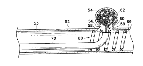
As is shown in Figures 5-7, the retaining device in the variations of this
embodiment may be a helically wound member, wherein the semi-penetrable
space for occlusion device insertion is provided by the space between adjacent
windings of the helix. In a preferred mode, the helically wound member that
forms the retaining device is a metal wire wound into a primary helix which is
further wound into a secondary helix. In this mode, windings of the secondal-y
helix form the semi-penetrabl; space for occlusion device insertion.
In the variation shown in Figure 5, the pre-determined, semi-penetrable
space (60) of retaining device (69) is defined by the space between adjacent
helical windings (58) and (59). This semi-penetrable space (60) is equal to or
greater in diameter than occlusion device (62) when it is being introduced
into
aneurysm (54). However, semi-penetrable space (60) is less than the diameter
of
occlusion device (62) after it is in the aneurysm. Thus, in this embodiment,
the
spacing provided by the retaining device allows the introduction of occlusion
devices into the aneurysm but does not allow significant migration of
occlusion
devices. once implanted, back into the adjacent vessel lumen (~3).
CA 02211512 1997-07-25
More particularly, in Figure 5 the occlusion device (62) is radially
constrained to a first shape having a first outer diameter when within the
delivery
lumen of delivery catheter (70). The delivery catheter distal end is abutting
the
inner surface of the retaining device (69). The occlusion device (62) is then
S advanced out the distal end of the delivery catheter (70) and through the
space in
the retaining device (69), where it is then radially artificially
unconstrained. Once
released into the aneurysm sac through the retaining device semi-penetrable
space,
the occlusion device (62) takes on a second shape having a second outer
diameter
that prevents it from migrating back through the semi-penetrable space and
into
the body lumen (53).
In a more particular embodiment of the artificial occlusion kit shov~m in
Figure S, the semi-penetrable spaces of the retaining device are distensible.
This
distensibility enhances the semi-penetrability of the spaces. More
specifically,
occlusion devices may be introduced through such spaces when an applied force
distends open the spaces. Once the occlusion devices are implanted into the
occlusion site, however, passive migration of the devices back through the
spaces
does not provide the requisite force to distend open these spaces--the passive
migration is thus prevented.
In one aspect of this variation, a particular occlusion device may be used in
conjunction with a retaining device, and be of such construction and dimension
that it may be advanced unaided through the spaces provided in the retaining
device. For example, detachable occlusion devices such as those described in
US
5.122,136 or US 5,354,295 may be constructed with sufficient pushability to be
advanced between adjacent coil winds of the retaining device and into the
aneurysm sac. They may thereafter be detached within the aneurysm for
occlusion.
Another aspect of this variation is shown in Figure 6. Here. retaining
device (119) is shown implanted into vessel (102) such that it radially
engages the
vessel wall adjacent to entrance zone (106) to wide-neck aneun~sm (104) and
brides across entrance zone (106). The helical shape of retaining device (119)
is
21
CA 02211512 1997-07-25
show to have a pre-determined spacing which may be spread when adjacent
helical windings are forced apart. In Figure 6, delivery catheter (110) is
advanced
through the retaining device and into the entrance zone ( 106) of the aneurysm
( 104).
In the particular variation of Figure 6, delivery catheter (110) has a tip
( 1 I 1 ) which is tapered and dimensioned such that adjacent helical windings
of
retaining device (119) are forced apart when delivery catheter (110) is forced
radiallv against the retaining device ( 119) from its inner lumen ( 130) and
toward
the entrance zone ( 106). To achieve this interaction, delivery catheter ( 1 I
0) ma~~,
for example, have a pre-shaped bend in the distal delivery catheter region
ending
in tip ( 111 ). This shape may aid in the advancement of the delivery catheter
through the branching vasculature, or may also be sufficiently straightened
coaxially over a guidewire to avoid proximal vessel trauma while tracking to
the
site.
1 S In a further variation shov~m in Figure 7, an introduces wire (140) may be
forced through spaces provided in the retaining device, such as between
adjacent
winds of a helically shaped retaining device as shown in Figure 7. Once the
introduces wire (140) is advanced through the retaining device and into the
aneurysm, a delivery catheter such as delivery catheter (110) (shown in Figure
6)
may thereafter be advanced coaxially over the introduces wire ( 140) and into
the
entrance zone of the wide-neck aneurysm. An advantage to using an introduces
wire in this technique as compared to that previously described in reference
to
Figure 6 is that the delivery catheter may be introduced into the aneurysm
over the
introduces without the need for preshaping the delivery catheter.
2~ In the particular variation of Figure 7, however, delivery catheter (120)
is
not shown to be advanced into entrance zone ( 106) or aneurysm ( 104), but
rather
is advanced merely to abut the inner diameter of the helical windings forming
retaining device (I 19). Vaso-occlusion coil (108) is shown being advanced
coaxially over introduces wire (140) while advancing through delivery catheter
(120). through adjacent windings of helical retaining device (119). and
ultimately
CA 02211512 1997-07-25
off the distal end of introducer wire (140) and into the sac of aneurysm
(104).
Such coaxial advancement of vaso-occlusion coil ( 108) may occur, for example,
by coaxially advancing a pusher member, located proximally of vaso-occlusion
coil (108), in the distal direction against a proximal end of vaso-occlusion
coil.
S The critical performance of the introducer wire is that it must be
sufficiently stiff and of such diameter and geometry to allow it to pass
through the
spacing provided in the retaining device ( 119). However, it should also not
be too
stiff so as to present risk of trauma or perforation of the thinned aneurysm
wall.
Such introducer wire (140) may also be shapeable such that it is adapted for
tracking to the retaining site adjacent the aneurysm, as well as for advancing
through the spaces in the retaining device barrier at the aneurysm entrance
zone.
Conventional guidewires of the type known in the art may perform sufficiently
as
introducer wire ( 140) in a particular case. Alternatively, the present
invention
further contemplates obvious alterations to known wire designs in order to
function with the individual features of a particular retaining device design,
as
may be apparent to one of ordinary skill. Such particularized retaining device
features that may dictate introducer wire design parameters, for example, may
be
the diameter and degree of distensibility of the semi-permeable space.
"Coil Over Core" Retaining Device
A further embodiment of the present invention is shown in Figure 8. In
this variation, the implantable medical device that functions as a retaining
device
in the novel artificial occlusion kit has a particular construction that
includes a
wire (202) wound into a primary helix over an inner core member (204). The
inner core member (204) and primary wire helix are also wound into a secondary
geometry, and are soldered or welded at both of two ends (210) and (212). The
secured ends (210) and (212) serve to secure the "wire over core" composite
relationship and also provide smooth ends for safety considerations in this
implantable device.
CA 02211512 1997-07-25
Preferably, the inner core member (204) is a metal mandrel, and more
preferably is a superelastic alloy of nickel-titanium. In one particular
variation,
the inner core member (204) is constructed of a nickel-titanium alloy and has
an
outer diameter from 0.003" to 0.006". The heIically wound wire (202) in this
preferred variation may be a radiopaque metal, such as platinum, gold, or
tungsten, and has an outer diameter in the range of 0.001 " to 0.006". The
coil
may have 0-100% spacing. Preferably, wire (202) is wound at a pitch of 0.001"
to 0.008" with 0-100% spacing. For instance, a coil made with 0.003" wire with
0.006'' pitch has 100% spacing; a coil with 0.003'' wire and 0.006" pitch has
0
spacing.
In this variation, wire (202) is secured to the inner core member (204)
using the following process: the coil is secured to the inner core member at
least
two or several locations, preferably at both ends. One method for joining the
components involves resistance welding or a similar such process. Soldering or
brazing is similarly useful in joining the metals.
In the "wire over core" combination structure such as that just described,
the inner core member (204) is chosen such as to provide the requisite shape
memory and stiffness. This inner core member may not by itself provide optimal
radiopacity, since it is not chosen for that purpose. The requisite
radiopacity of
the device may instead be provided by the outer wound coil (202), which might
not provide optimal stiffness or material memory if it were only available
alone in
the device. It is believed that the combined features of this "wire over core"
design may optimally adapt prior known implantable coil technologies to meet
the
particular structural needs of a retaining device in the current invention.
2~ For instance, it is important that the elongate retaining device be
flexible
along its length so that it can be implanted into lumens having bends.
However, it
is believed that too much flexibility may correspond to irregular and random
conformations of the coil when implanted in-vivo, which may produce an
occlusive effect. A primary helical coil wound into a secondary helix, without
~0 more. may be too flexible to effectively engage a vessel wall along the
requisite
~4
CA 02211512 1997-07-25
length to form a barrier against occlusion device migration. However. the
addition of the mandrel in what would otherwise be the primary helix lumen
provides a stiffening structure that still allows for a certain controlled
flexibility of
the secondary helical shape.
Additionally, prior vaso-occlusion coils require substantial space filling for
effective cross-sectional blockage of a body lumen, for example. In these
devices,
only a minimal portion of the device may be required to actually radially
engage a
vessel wall for primarily the purpose of anchoring the device at the occlusion
site.
This means only a small portion of the coil may need to reconfigure from a
first
constrained diameter during delivery to a second diameter at least
approximating
the lumenal wall diameter when delivered. It may be acceptable, even
desirable,
for such occlusion coils to have portions not so significantly altered in
their cross-
sectional diameter when they are delivered at an implantation site, so long as
their
shape presents an occlusion to flow.
1 S In contrast, the present inventive retaining device must take on a shape
at
the retaining site that has sufficient outer diameter along a sufficient
length of the
device to form an effective barrier across the aneurysm entrance zone at the
vessel
wall. Impinging into the lumen's cross-section is generally undesirable. The
reconfiguration to this expanded shape from a first radially constrained shape
during delivery may correspond to a higher degree of requisite material memory
than is possible from a simple fine wire wound into the primary and secondary
helix shapes as previously disclosed. An inner core mandrel, however, may
offer
the structure necessan~ to provide such memory.
In another particular retaining device variation, the wire forming the
?~ primary helical core is wound much tighter than a similar wire might be
wound to
optimally form an occlusion device. It is believed that coil stiffness may be
controlled by adjusting the outer diameter of the primary coil helix (e.g.
tightness
of winding) to which a given wire is wound. It is believed that, by providing
one
preferred retaining device may comprise a wire wound very tightly into a
primary
helix that has also a secondary shape.
CA 02211512 1997-07-25
One preferred application of this "tightly wound" variation comprises a
platinum wire of 0.005" outer diameter wound over a .009" mandrel. In
contrast,
common known occlusion coils for occluding aneurysms is constructed a .005"
wire wound over a .011" mandrel. Similarly, when a smaller diameter primary
S helix i s desired, a wire having an outer diameter of .003" may be wound
over a
mandrel having an outer diameter of 0.007". In any case, the wire is
thereafter
annealed in the wound shape to form a primary coil of pre-determined
dimensions. A secondan~ shape may then be imparted to the primary coil, which
secondary shape ma5- also be a helical coil.
It should be apparent to those skilled in this art that the coil placed within
an aneurysm need not be the random shape described and shown above. indeed,
many shapes would be suitable for use with this invention. We have found that
anatomically shaped oval (222) or semi-oval (224) coils are suitable for this
invention. It should be apparent that both these are regularly wound and are
provided with the shape of the anatomical cavity into which they are placed.
Coils (222) and (224) provide a similar amount a rate of occlusion within the
aneurysm structure and yet do so with a significantly smaller mass of coil
than the
random shapes :-.hown above in many of the drawings.
Other Clinical Applications and Design Embodiments
The ultimate goal of the particular artificial occlusion kits, novel
components thereof, and related methods described above is to occlude
aneurysms
having entrance zones or necks that are of such width and geometry that
conventional techniques would result in unwanted migration of occlusion
devices
from the aneurysm and into the adjacent vessel. However, the assemblies,
components, and methods of the present invention that were conceived of in
order
to meet this need may provide additional benefits in other medical treatments.
Additionally. the invention contemplates retaining device designs that meet
the
general requirements of the novel artificial occlusion kit but vary from the
specific
variations just described.
26
CA 02211512 1997-07-25
In one aspect of the invention, for example, the artificial occlusion kit
embodiments and variations have been described specifically as applied to
aneurysms in vessel walls. However, other occlusion sites adjacent to and in
fluid
communication with body lumens may present similar concerns as to migration of
occlusion devices from an occlusion site and into an adjacent lumen. For
instance, a vessel that branches off of a feeding vessel may be a body space
to be
occluded and the feeding vessel at a region adjacent to the branching vessel
may
be a desired retaining site. The present invention contemplates use of the
apparatus embodiments described in such body spaces and lumens in addition to
aneurysm sites in vessels.
The invention also broadly contemplates a retaining device structure that is
expandable at a retaining site of a body lumen to form a barrier against
migration
of at least one occlusion device through an entrance zone between an occlusion
site and an adjacent lumen, and that also provides a lumen for flow through
the
body lumen at the retaining site. Examples have been provided in the form of
shape memory coils delivered through radially confining delivery sheaths or
over
delivery wires, in addition to an alternative balloon expandable retaining
device
embodiment. Various specific retaining device designs that meet the broad
requirements provided, beyond the particular variations provided, are within
the
scope of this invention.
Also, while various retaining device designs may meet the requirements
of the novel artificial occlusion kit described, at least one novel
electrolytically
detachable implantable medical device has been conceived of for use as a
retaining device in the artificial occlusion kit. This novel implantable
medical
device may have useful medical applications in addition to retaining
artificial
occlusion devices. The scope of this aspect of the invention, while intimately
pertaining to an artificial occlusion kit, should not be limited to the kit
embodiments described for artificial occlusion.
?7
CA 02211512 1997-07-25
Modification of the above-described variations for carrying out the
invention that would be apparent to those of skill in the fields of medical
device
design are intended to be within the scope of the following claims.