Note: Descriptions are shown in the official language in which they were submitted.
CA 02211604 1997-07-28
DFSCRIPTTON
APPARATUS AND METHOD FOR DIAGNOSING OSTEOPOROSIS
T~CHNICAT FIFTD
This invention relates to an ultrasonic reflection
type of apparatus and method for diagnosing osteoporosis
by emitting ultrasonic pulses toward predetermined
cortical bone in a subject so as to measure the echo
levels from the surface of the cortical bone.
BACKGROUND ART
With the advent of an ageing society in recent
years, the bone disease referred to as osteoporosis has
become a problem. This is a disease in which the loss
of bone calcium results in brittleness and susceptibility
to fractures with minimal trauma, and can cause the
elderly to become bedridden. The physical diagnosis of
osteoporosis is managed by the precise measurement of
bone density using a diagnostic apparatus featuring the
use of X-rays such as DXA, but problems involved in
physical diagnosis with X-rays are that the equipment is
large and expensive, and its use is limited in many ways
in the interests of protecting against harm caused by
radiation exposure.
CA 02211604 1997-07-28
Diagnostic apparatuses featuring the use of
transmitted ultrasonic waves or reflected ultrasonic
waves have begun to enjoy more popularity as simple
devices which do not suffer from such drawbacks.
The diagnostic apparatuses noted in Japanese Laid-
Open Patent Application 2-104337 and US Patent
Application 193,295 are known as ultrasonic transmitting
types of diagnostic devices. In these diagnostic
apparatuses, the acoustic velocity in bone is measured by
setting up two ultrasonic transducers facing each other
on either side of a part of a subject's body, so that
ultrasonic pulses are emitted from one ultrasonic
transducer at the osseous tissue, and the ultrasonic
pulses passing through the osseous tissue are received by
the other ultrasonic transducer. The extent of
osteoporosis is diagnosed on the assumption that a slower
acoustic velocity in osseous tissue indicates lower bone
density due to loss of bone calcium.
The theoretical basis linking bone density and
acoustic velocity is uncertain, however. Strictly
speaking, the acoustic velocity in osseous tissue is not
proportional to bone density, but is given by the square
root of [the elastic modulus of bone/bone density].
Furthermore, because the elastic modulus of bone and bone
density play mutually cancelling roles in acoustic
CA 02211604 1997-07-28
velocity, where increases in the bone density
(denominator) are met by increases in the elastic modulus
of bone (numerator), the acoustic velocity in osseous
tissue is not capable of sensitive response to increases
in bone density. As such, there is not that high a
correlation between the acoustic velocity in osseous
tissue and bone density. Reliability is accordingly a
problem in conventional ultrasonic transmission types of
diagnostic apparatuses in which bone density is estimated
on the basis of the acoustic velocity in osseous tissue.
Ultrasonic reflection types of diagnostic
apparatuses have meanwhile been proposed by the applicant
in Japanese Patent Applications 6-310445, 7-140730,
7-140731, 7-140732, 7-140733, and 7-140734, and
International Laid-Open Patent Application WO 96/18342.
In these diagnostic apparatuses, a single ultrasonic
transducer capable of both transmission and reception is
used to emit ultrasonic pulses toward cortical bone in a
subject, echoes reflected on the surface of the cortical
bone are received, and the acoustic impedance of the
subject's cortical bone is calculated on the basis of the
resulting echo data. The progress of osteoporosis is
then diagnosed based on the level of the acoustic
impedance thus calculated.
CA 02211604 1997-07-28
The acoustic impedance of bone is given by the
square root of [the elastic modulus x density] of bone,
and since, as described above, the elastic modulus of
bone increases (or decreases) as bone density increases
(or decreases), the elastic modulus of bone and bone
density play a synergistic role in acoustic impedance.
Thus, the latter ultrasonic reflection type of apparatus
in which acoustic impedance is used as an index can be
considered more reliable because it is capable of more
sensitive response to the extent of the progress of
osteoporosis than is the former ultrasonic transmission
type of apparatus in which acoustic velocity is used as
an index.
Although acoustic impedance can be considered a
sensitive indicator of the progress of osteoporosis, in
the final analysis it is only an index of bone density,
which does not mean that the bone density itself is
determined. Furthermore, when the acoustic impedance of
cortical bone is lower than that of soft tissue, or when
the cortical bone is thinner than the ultrasonic
wavelength, there is a problem in that the acoustic
impedance of cortical bone cannot be measured or that
such measurement is uncertain.
In view of the foregoing, a first object of the
present invention is to provide an ultrasonic reflection
CA 02211604 1997-07-28
type of apparatus and method for diagnosing osteoporosis,
which is simple, with no danger of exposure to radiation,
yet is capable of determining bone density. A second
object of the present invention is to provide an
ultrasonic reflection type of apparatus and method for
diagnosing osteoporosis, which is capable of highly
reliable diagnosis, even when the acoustic impedance of
the cortical bone is lower than that of the soft tissue
and when the cortical bone is thinner than the ultrasonic
wavelength.
SUMMARY OF T~F INVFNTION
In the apparatus (and method) for diagnosing
osteoporosis in the present invention, ultrasonic pulses
are repeatedly emitted toward cortical bone in a subject,
the echoes reflected on the surface of the cortical bone
at that time are received, and osteoporosis is diagnosed
based on the resulting echo data.
As such, a first aspect of the present invention is
to provide an apparatus for diagnosing osteoporosis,
comprising: an echo level detecting means for detecting
the echo level of the echoes reflected on the surface of
the cortical bone when the ultrasonic pulses are emitted;
a maximum echo level extracting means for extracting the
maximum echo level from among the echo levels thus
CA 02211604 1997-07-28
detected; a reflection coefficient calculating means for
calculating the ultrasonic reflection coefficient at the
interface between the soft tissue and cortical bone of
the subject based on said extracted maximum echo level;
and a bone density calculating means for calculating the
density of the subject's cortical bone using a
predetermined recurrence formula for the cortical bone
density relative to the ultrasonic reflection
coefficient.
In a preferred embodiment of the bone density
calculating means, the recurrence formula for the
cortical bone density relative to the ultrasonic
reflection coefficient is given in the form of Formula
(1) or (2)
p = a' R+~' (1)
p: density of cortical bone [kg/m3]
R: ultrasonic reflection coefficient at interface
between soft tissue and cortical bone of subject
a': regression coefficient [kg/m3]
~': section [kg/m3]
The regression coefficient a' should be established
within the range of 588 to 1100, and the section ~'
should be established within the range of 953 to 1060.
CA 02211604 1997-07-28
p = B~ RA~ (2)
A': regression coefficient
B': constant [sec/m]
A second aspect of the present invention is to
provide an apparatus, comprising: an echo level detecting
means for detecting the echo level of the echoes
reflected on the surface of the cortical bone when the
ultrasonic pulses are emitted; a maximum echo level
extracting means for extracting the maximum echo level
. from among the echo levels thus detected; an acoustic
impedance calculating means for calculating the acoustic
impedance of the subject's cortical bone based on said
extracted maximum echo level; and a bone density
calculating means for calculating the density of the
subject's cortical bone using a predetermined recurrence
formula for the cortical bone density relative to the
acoustic impedance.
In a preferred embodiment of the bone density
calculating means, the recurrence formula for cortical
bone density relative to acoustic impedance is given by
Formula (3) or (4).
p = aZb + ~ (3)
CA 022ll604 l997-07-28
p: density of cortical bone [kg/m3]
Zb: acoustic impedance of cortical bone in subject
[ kg/m2sec]
a: regression coefficient [sec/m]
~: section [kg/m3]
The regression coefficient a should be established
within the range of 1.27 x 10⁻⁴ to 2.34 x 10⁻⁴, and the
section ~ should be established within the range of 646
to 887.
p = BZbA (4)
A: regression coefficient
B: constant [sec/m]
The regression coefficient A should be established
with the range of 0.239 to 0.445, and the constant B
should be established within the range of 10~239 to 101-55.
A third aspect of the present invention is to
provide an apparatus for diagnosing osteoporosis,
comprising: an echo waveform detecting means for
detecting the reception waveform of the echoes reflected
on the surface of the cortical bone when the ultrasonic
pulses are emitted; a maximum echo waveform extracting
CA 02211604 1997-07-28
means for extracting the maximum echo reception waveform
by comparing the plurality of echo reception waveforms
that have been detected; a Fourier transform treatment
means for finding the maximum echo spectrum by the
Fourier transform treatment of the maximum echo reception
waveform; and a complex reflection coefficient
calculating means for calculating the ultrasonic complex
reflection coefficient (complex acoustic characteristics
data) of cortical bone relative to the soft tissue of the
subject based on the maximum echo spectrum thus
determined, wherein osteoporosis is diagnosed on the
basis of the ultrasonic complex reflection coefficient
thus calculated.
A preferred embodiment of the third aspect further
comprises a diagnostic means for obtaining amplitude data
and phase data from the ultrasonic complex reflection
coefficient thus calculated, and for diagnosing
osteoporosis based on the resulting amplitude and phase
data.
A fourth aspect of the present invention is to
provide an apparatus for diagnosing osteoporosis,
comprising: an echo waveform detecting means for
detecting the reception waveform of the echoes reflected
on the surface of the cortical bone when the ultrasonic
pulses are emitted; a maximum echo waveform extracting
CA 02211604 1997-07-28
means for extracting the maximum echo reception waveform
by comparing the plurality of echo reception waveforms
that have been detected; a Fourier transform treatment
means for finding the maximum echo spectrum by the
Fourier transform treatment of the maximum echo reception
waveform; and a complex acoustic impedance calculating
means for calculating the complex acoustic impedance
(complex acoustic characteristics data) of the subject's
cortical bone based on the maximum echo spectrum thus
determined, wherein osteoporosis is diagnosed on the
basis of the complex acoustic impedance thus calculated.
A preferred embodiment of the fourth aspect further
comprises a diagnostic means for obtaining amplitude data
and phase data from the complex acoustic impedance thus
calculated, and for diagnosing osteoporosis based on the
resulting amplitude and phase data.
A fifth aspect of the present invention is to
provide a method for diagnosing osteoporosis, wherein an
ultrasonic transducer is placed on a predetermined area
on the surface of a subject's skin, ultrasonic pulses are
repeatedly emitted toward cortical bone below the skin,
the echoes reflected on the surface of the cortical bone
at that time are received so as to detect the echo level,
the maximum echo level is extracted from the echo levels
thus detected, the ultrasonic reflection coefficient at
CA 02211604 1997-07-28
the interface between the soft tissue and the cortical
bone of the subject is calculated based on said extracted
maximum echo level, and the density of the subject's
cortical bone is then calculated using a predetermined
recurrence formula for the cortical bone density relative
to the ultrasonic reflection coefficient.
A sixth aspect of the present invention is to
provide a method for diagnosing osteoporosis, wherein an
ultrasonic transducer is placed on a predetermined area
on the surface of a subject's skin, ultrasonic pulses are
repeatedly emitted toward cortical bone below the skin,
the echoes reflected on the surface of the cortical bone
at that time are received so as to detect the echo level,
the maximum echo level is extracted from the echo levels
thus detected, the acoustic impedance of the cortical
bone of the subject is calculated based on said extracted
maximum echo level, and the density of the subject's
cortical bone is then calculated using a predetermined
recurrence formula for the cortical bone density relative
to the acoustic impedance.
A seventh aspect of the present invention is to
provide a method for diagnosing osteoporosis, wherein an
ultrasonic transducer is placed on a predetermined area
on the surface of a subject's skin, ultrasonic pulses are
repeatedly emitted toward cortical bone below the skin,
CA 02211604 1997-07-28
the reception waveforms of the echoes reflected on the
surface of the cortical bone at that time are received so
as to detect the echo reception waveforms, the maximum
echo is extracted from the echo reception waveforms thus
detected, the maximum echo spectrum is determined by the
Fourier transform treatment of the maximum echo reception
waveform, the ultrasonic complex reflection coefficient
of the cortical bone relative to the soft tissue of the
subject is calculated based on the maximum echo spectrum
that has been determined, and osteoporosis is diagnosed
based on the amplitude data and phase data obtained from
the ultrasonic complex reflection coefficient thus
calculated.
An eighth aspect of the present invention is to
provide a method for diagnosing osteoporosis, wherein an
ultrasonic transducer is placed on a predetermined area
on the surface of a subject's skin, ultrasonic pulses are
repeatedly emitted toward cortical bone below the skin,
the reception waveforms of the echoes reflected on the
surface of the cortical bone at that time are received so
as to detect the echo reception waveforms, the maximum
echo is extracted from the echo reception waveforms thus
detected, the maximum echo spectrum is determined by the
Fourier transform treatment of the maximum echo reception
waveform, the complex acoustic impedance of the cortical
bone of the subject is calculated based on the maximum
CA 02211604 1997-07-28
echo spectrum that has been determined, and osteoporosis
is diagnosed based on the amplitude data and phase data
obtained from the complex acoustic impedance thus
calculated.
CA 02211604 1997-07-28
BRI~F DFSCRIPTION OF THF DRAWINGS
Fig. l is a block diagram depicting the electrical
structure of the apparatus for diagnosing osteoporosis in
a first embodiment of the invention;
Fig. 2 is a schematic outer view of the same
apparatus;
Figs. 3 through 6 are illustrations used to describe
the operation of the apparatus;
Fig. 7 is a flow chart of the operation of the same
apparatus;
Fig. 8 is a graph of the regression line for
cortical bone density p relative to the acoustic
impedance Zb, and is used to describe the contents of the
bone density calculating subprogram constituting the same
apparatus;
Fig. 9 is a graph of the regression line for
cortical bone density p relative to the acoustic
impedance Zb, and is used to describe the contents of the
bone density calculating subprogram in a fourth
embodiment;
14
CA 02211604 1997-07-28
Fig. 10 is a graph of the regression line for
cortical bone density p relative to the acoustic
impedance Zb, and is used to describe the contents of the
bone density calculating subprogram in a sixth
embodiment;
Fig. 11 is a block diagram of the electrical
structure of the apparatus for diagnosing osteoporosis in
an eighth embodiment of the present invention;
Fig. 12 is a flow chart of the operation and
processing procedures of the same apparatus;
Fig. 13 schematically depicts the apparatus for
diagnosing osteoporosis while in use in a ninth
embodiment of the present invention;
Fig. 14 is a flow chart of the operation and
processing procedures of the same apparatus; and
Fig. 15 is a flow chart of the operation and
processing procedures of the apparatus for diagnosing
osteoporosis in a tenth embodiment of the present
invention.
B~ST MODF FOR CARRYING OUT THF INVFNTION
CA 02211604 1997-07-28
The best modes for carrying out the invention are
described below with reference to the drawings. The
invention is described in detail using embodiments.
Embodiment 1
Fig. 1 is a block diagram depicting the electrical
structure of the apparatus for diagnosing osteoporosis in
a first embodiment of the invention; Fig. 2 is a
schematic outer view of the same apparatus; Figs. 3
through 6 are illustrations used to describe the
operation of the apparatus; Fig. 7 is a flow chart of the
operation of the same apparatus; Fig. 8 is a graph of the
regression line for cortical bone density p relative to
the acoustic impedance Zb, and is used to describe the
bone density calculating subprogram constituting the same
apparatus.
As shown in Figs. 1 and 2, the apparatus for
diagnosing osteoporosis in this example comprises: an
ultrasonic transducer 1 (hereinafter simply referred to
as transducer), which emits ultrasonic pulses toward
predetermined cortical bone in a subject at a measuring
location in response to electrical pulse signals that are
input at a predetermined period, and receives echoes
(reflected waves) from the surface of the cortical bone
and converts them to a reception signal (electrical
16
CA 02211604 1997-07-28
signal); an apparatus main unit 2, which carries out the
diagnosis of osteoporosis by supplying electrical puIse
signals to the transducer 1 and processing the reception
signals output from the transducer 1 so as to extract the
echo level (reflection wave amplitude) from the cortical
bone; and a cable 3 connecting the transducer 1 and
apparatus main unit 2.
The aforementioned transducer 1 consists primarily
of an ultrasonic oscillator la having electrode layers on
either side of a disk-shaped thickness oscillation type
of piezo-electric element of lead zirconate titanate
(PZT) or the like. An ultrasonic delay spacer lb of a
polyethylene bulk or the like is fixed to one of the
electrode surfaces (ultrasonic pulse transducer surface)
of the ultrasonic oscillator la in order to eliminate the
effects of transmission reverberation. Here, cortical
bone can be irradiated with nearly flat ultrasonic pulses
from the transducer surface of the transducer 1 to carry
out highly accurate measurements, and nearly flat echoes
should be reflected from the cortical bone to the
transducer surface, so the transducer 1 is ideally
constructed of a piezo-electric element with a relatively
large disk radius to make the transducer surface as wide
as possible (in this example, the diameter D of the
transducer surface is 15 mm). From a similar
perspective, the measuring site that is used should be
CA 02211604 1997-07-28
the cortical bone of the heel, top of the patella, tibia,
scapula, cranial bone, o~ the like, which can be regarded
as being flat, with a large curvature radius, and which
is close to the surface of the skin. As a result of
detailed measurements at various locations of cortical
bone in humans, the inventors of the present application
found that an ideal measuring location was the cortical
bone of the lower tibia, especially within a range K of
40 mm to 100 mm over the ankle Mc, as shown in Fig. 3,
because virtually noise-free echoes alone were extracted
independently from the lower tibia Mb far more frequently
than with the cortical bone in other locations. When the
influence of transmission reverberation is negligible,
the ultrasonic delay spacer lb can be omitted.
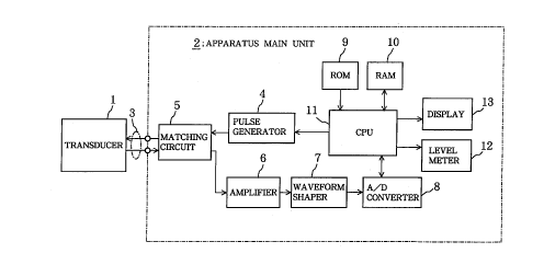
The aforementioned apparatus main unit 2 comprises a
pulse generator 4, matching circuit 5, amplifier 6,
waveform shaper 7, A/D convertor 8, ROM 9, RAM 10, CPU
(central processing unit) 11, level meter 12, and display
13.
The pulse generator 4 is connected via a cable 3 to
the transducer 1, and repeatedly produces an electrical
pulse signal with a central frequency of, for example, 1
MHz or 2.5 MHz at a predetermined period (100 msec, for
example), which is sent to the transducer 1. The
matching circuit 5 matches impedance, to allow the
18
CA 02211604 1997-07-28
signals to be transmitted and received at optimal energy
efficiency between the transducer 1 and apparatus main
unit 2 which are connected by the cable 3. Thus, when
the ultrasonic oscillator la of the transducer 1 receives
echoes from cortical bone, the reception signal is output
from the transducer 1 and is input to the amplifier 6 via
the matching circuit 5 with no loss of energy. The
amplifier 6 amplifies the reception signal input through
the matching circuit to a predetermined amplification
level and then inputs it to the waveform shaper 7. The
waveform shaper 7 consists of a band pass filter having
an LC structure, and filters the reception signal that
has been amplified by the amplifier 6 to shape the
waveform to a linear form in order to eliminate noise,
and the signal is then input to the A/D convertor. The
A/D convertor 8 is equipped with a sample holder circuit
not shown in the figure, sampling memory (SRAM), and the
like, and samples the output signal from the waveform
shaper 7 (waveform shaped analog reception signal) at a
predetermined frequency (such as 12 MHz) when the CPU 11
sends a command to start sampling, so as to sequentially
convert the signals to digital signals, and the resulting
digital signals are temporarily stored in the sampling
memory itself and then sent to the CPU 11.
The ROM 9 stores the operating system (OS) as well
as the various processing programs of the CPU 11,
19
CA 02211604 1997-07-28
specifically, the maximum echo level extracting
subprogram, reflection coefficient calculating
subprogram, acoustic impedance calculating subprogram,
and bone density calculating subprogram.
A procedure for taking in the digital signal from
the sampling memory of the A/D convertor 8 for each pulse
and echo to detect the echo level for each echo, and a
processing procedure for extracting the maximum echo
level from the echo levels that are detected for each
echo, are written to the maximum echo level extraction
subprogram. A processing procedure for calculating the
ultrasonic reflection coefficient R during roughly
perpendicular reflection at the interface between the
soft tissue and cortical bone (measuring location~ of the
patient based on the maximum echo level value given by
the maximum echo level extraction subprogram is written
to the reflection coefficient calculating subprogram. A
procedure for calculating the acoustic impedance Zb using
Formula (5) based on the values calculated for the
ultrasonic reflection coefficient R given by the
reflection coefficient calculating subprogram is written
to the acoustic impedance calculating subprogram.
Zb = Za (R + 1)/(1 - R) (5)
Za: acoustic impedance of soft tissue
CA 02211604 1997-07-28
Formula (5) is derived from Formula (6). As shown
in Fig. 6(a), the surface Y of cortical bone Mb can be
regarded as being flat, and the ultrasonic pulse Ai
generated from the transducer 1 can also be regarded as
being flat, so when the wavefront is parallel to the
surface Y of the cortical bone Mb (lands roughly
perpendicular), the ultrasonic reflection coefficient is
expressed by Formula (6). As will be described below,
the echo level is greatest when the wavefront of a flat
wave and the surface Y of cortical bone Mb are parallel.
Accordingly, the ultrasonic reflection coefficient given
by Formula (6) is the ultrasonic reflection coefficient
when the maximum echo level is obtained. Formula (5) is
thus obtained by transforming Formula (6).
R = (Zb - Za)/(Zb + Za) (6)
The bone density calculating subprogram contains a
processing procedure for the bone density (cortical bone
density) p of a patient using Formula (7) based on the
value calculated for the acoustic impedance Zb given by
the acoustic impedance calculating subprogram.
Here, Formula (7) is the recurrence formula of the
bone density p relative to the acoustic impedance Zb,
CA 02211604 1997-07-28
which is obtained by prior examination of a specimen, as
shown in Fig. 8.
p = aZb + ~ (7)
= 1.80 x 10⁻⁴ZB + 766
p: cortical bone density [kg/m3]
Zb: acoustic impedance of cortical bone [kg/m2sec]
a: regression coefficient of bone density relative
10 to acoustic impedance [sec/m]
~: section [kg/m3]
In the examination of the specimen described above,
the acoustic impedance Zb was measured for the cortical
bone of the tibia using ultrasonic reflection, and the
density p of the cortical bone Mb was measured by X-ray
(QCT) of the radius (arm bone). Examination of the
specimen revealed a high correlation (r = 0.67) between
the acoustic impedance Zb and the bone density p measured
by X-ray (QCT). Statistical hypothesis testing resulted
in a 95% probability (reliability) that a patient's bone
density p would fall within the pmin to pmax range when
the value for the acoustic impedance Zb of the cortical
bone of any patient is Zb. The significance level is
thus 5%.
CA 022ll604 l997-07-28
Here, pmin is given by Formula (8), and pmax is
given by Formula (9).
pmin = (1.80 x 10⁻⁴ -- 30%) Zb + (766 -- 16%) (8)
pmax = (1.80 x 10⁻⁴ + 30%) Zb + (766 + 16%) (9)
RAM 10 has a working area in which the working area
for the CPU 11 is established, and a data area in which
various data are temporarily stored. The data area
contains an echo level memory area for storing the most
recently detected echo level (hereinafter referred to as
current echo level) or maximum echo level, an echo
waveform memory area for storing the most recently
detected echo waveform (current echo waveform) or maximum
echo waveform, and a measurement continue flag or the
like for storing data on whether or not to continue
measurement.
The CPU 11 executes the various processing programs
stored in the ROM 9 using RAM 10 to start the pulse
generator 4 or A~D convertor 8, controls the various
components of the apparatus to detect echo levels for
each pulse and echo, extracts the maximum echo level, and
calculates the bone density p of the patient based on the
maximum echo level value detected, so as to diagnose
osteoporosis.
CA 02211604 1997-07-28
The level meter 12 is controlled by the CPU 11 and
displays the current echo level stored in RAM 10 by the
deflection of the liquid crystal needle pattern 12a
indicated by the broken line in Fig. 2 as well as the
maximum echo level, which is the greatest echo level
among those thus far detected, by the deflection of the
liquid crystal needle patten 12b indicated by the solid
line in the figure. The display 13 consists of a CRT
display or liquid crystal display. The measured values
of the echo levels and the like, the ultrasonic
reflection coefficient R, the acoustic impedance Zb, the
calculated values of the bone density p, and the echo
waveforms are displayed on screen under the control of
the CPU 11.
The operation of this example (course of CPU 11
processing during diagnosis of osteoporosis) is described
below with reference to Figs. 4 through 7.
First, the cortical bone of the lower tibia,
particularly within a range K of 40 mm to 100 mm above
the ankle Mc, is selected. Of course, the cortical bone
of other desirable locations such as the heel, top of the
patella, scapula, and cranial bone may also be selected
as needed. When the power source is turned on in the
apparatus, the CPU 11 presets the various components of
24
CA 02211604 1997-07-28
the apparatus and initializes the counter, the various
registers, and the various flags, and waits for the
measurement begin switch to be pressed (step SP10 (Fig.
7)). Here, as shown in Fig. 4, the operator applies
ultrasonic gel 14 over the surface of the soft tissue Ma
(skin surface X) on the cortical bone Mb at the patient
measuring location, presses the transducer 1 against the
skin surface X via the ultrasonic gel 14, and turns the
measurement begin switch on, with the transducer surface
facing the cortical bone Mb. When the measurement begin
switch is turned on (step SP11), the CPU 11 writes "1" to
the measurement continue flag to raise the measurement
continue flag, and the diagnostic operations are then
started according to the processing procedure (primarily
the procedure in the maximum echo level extraction
subprogram) shown in Fig. 7. The CPU 11 first issues a 1
pulse generating command to the pulse generator 4 (step
SP12). When the pulse generator 4 receives the 1 pulse
generating command from the CPU 11, it sends an
electrical pulse signal to the transducer 1. When the
transducer 1 receives the electrical pulse signal from
the pulse generator 4, it emits a nearly flat ultrasonic
pulse Ai toward the patient's cortical bone Mb. As shown
in Fig. 5, the ultrasonic pulse Ai thus emitted is
introduced from the skin surface X into the soft tissue
Ma and is propagated toward the cortical bone Mb. A
portion is reflected at the surface Y of the cortical
CA 02211604 1997-07-28
bone Mb, resulting in echo Ae, and a portion is absorbed
by the cortical bone Mb, but the remainder passes through
the cortical bone Mb. The echo Ae follows a path
opposite that of the incident ultrasonic pulse Ai and is
received back at the ultrasonic oscillator la of the
transducer 1. When the ultrasonic pulse Ai is emitted
from the transducer 1 toward the cortical bone Mb, as
shown in the figure, first the transmission resonance
An1, then the echo An2 from the skin surface X, and a
little later the echo Ae from the cortical bone Mb are
received by the ultrasonic oscillator la and are
converted to a reception signal (electrical signal)
corresponding to the ultrasonic waveform and amplitude.
The resulting reception signal is input via the cable 3
to the apparatus main unit 2 (matching circuit 5),
amplified to a predetermined amplification level by the
amplifier 6, shaped into a linear waveform by the
waveform shaper 7, and then input to the A/D convertor 8.
After the CPU 11 has sent a 1 pulse generating
command to the pulse generator 4 (step SP12), it issues a
sampling start command (step SP13) to the A/D convertor 8
upon measuring the time in which the transmission
resonance An1 is received by the ultrasonic oscillator la
of the transducer 1, the echo An2 from the skin surface X
is then received, and the echo Ae from the cortical bone
26
CA 02211604 1997-07-28
Mb returns to the transducer surface of the oscillator la
of the transducer 1.
When the A/D convertor 8 receives the sampling start
command from the CPU 11, it samples the reception signal
for one echo from the cortical bone Mb, which has been
input after undergoing waveform shaping from the waveform
shaper 7, at a predetermined frequency (such as 12 MHZ )
to convert it to a digital signal, and the resulting N
sample value (digital signal for 1 echo) is temporarily
stored in the sample memory itself. Subsequently, when
there is a transmission command from the CPU 11, the N
sample values stored in the sampling memory are
sequentially transmitted to the CPU 11. The CPU 11
sequentially takes in the N sample values from the A/D
convertor 8 and stores the current echo waveform in the
echo waveform memory area of RAM 10, the maximum value
among the N sample values is extracted so as to detect
the current echo level, and the detected results are
stored in the echo level memory area of RAM 10 ( step
SP14). The current echo level stored in the echo level
memory area of RAM 10, as shown by the broken line in
Fig. 4, is displayed by the deflection of the liquid
crystal needle pattern 12a in the level meter 12 (step
SP15).
CA 02211604 1997-07-28
The CPU 11 then reads out the current echo level and
the maximum echo level from the echo level memory area of
RAM 10 to determine whether or not the current echo level
value is greater than the maximum echo level value (step
SP16). This is the first determination, and since the
maximum echo level value is the initialized value "0,"
the CPU 11 determines that the current echo level value
is greater than the maximum echo level value, the maximum
echo level value stored in the echo level memory area of
RAM 10 is replaced by the current echo level value, and
the maximum echo waveform stored in the echo waveform
memory area of RAM 10 is also replaced by the current
echo waveform (step SP17). The new maximum echo waveform
is displayed on the screen of the display 13, and the new
maximum echo level is displayed by the deflection of the
liquid crystal needle pattern 12b on the level meter 12,
as shown by the solid line in Fig. 4 (step SP18). Then,
when the CPU 11 looks for the measurement continue flag
in RAM 10 (step SP19) and raises the measurement continue
flag (when the contents of the measurement flag are "1"),
the CPU 11 determines that measurement is to continue,
repeats the 1 pulse emission and 1 echo reception
described above (steps SP12 through SP15), and then again
reads out the current echo level and maximum echo level
from the echo level memory area in RAM 10 in step SP16 to
determine whether or not the current echo level value is
greater than the value of the maximum echo level. When
28
CA 02211604 1997-07-28
it is determined that the current echo level is not
greater than the maximum echo level, the system jumps
directly to step SP19 without modifying the values, and
looks for the measurement continue flag. As long as the
operator does not press the measurement end switch, the
contents of the measurement continue flag are "1," and
the CPU 11 repeats the 1 pulse emission 1 echo
transmission described above (steps SP12 through SP15)
and the maximum echo level extraction (steps SP16 through
SP19).
While the CPU 11 is repeating the process described
above (steps SP12 through SP19), the operator aims the
transducer 1 at the skin surface X, as indicated by the
arrow W in Fig. 4, and changes the direction of the
transducer 1 by sometimes describing a circle in the
manner of the precession of a top and sometimes
oscillating it in any direction in the manner of a seesaw
on the cortical bone Mb at the measuring site while
checking the direction in which the liquid crystal needle
patterns 12a and 12b of the level meter 12 oscillate the
greatest, that is, the direction in which the maximum
echo level is detected. As shown in Fig. 6(a), the
maximum oscillation of the liquid crystal needle patterns
12a and 12b of the level meter 12 is where the normal of
the cortical bone Mb and the normal of the transducer
surface of the transducer 1 are aligned and thus when the
29
CA 02211604 1997-07-28
wavefront of the flat ultrasonic pulse Ai is roughly
parallel to the surface Y of the cortical bone Mb (when
the flat ultrasonic pulse Ai lands roughly perpendicular
on the surface Y of the cortical bone Mb).
That is because, when both normals are aligned, as
shown in Fig. 6(a), the echo Ae reflected perpendicular
on the surface Y of the cortical bone Mb returns
perpendicular to the transducer surface of the transducer
1, so the wavefront of the echo Ae is also roughly
parallel to the transducer surface. There is thus
minimal deviation of the echo Ae phase due to differences
in the reception position on the transducer surface, so
the crests and troughs of the reception signal do not
cancel each other out very much, allowing echoes Ae to be
received at maximum echo levels. In contrast, when both
normals are not aligned, as shown in Fig. 6(b), the
wavefront of the echo Ae does not line up with the
transducer surface, so the reception signal is lower
because the crests and troughs cancel each other out.
Diagnostic accuracy is increased in the diagnostic
apparatus in this embodiment, based on the extraction of
the perpendicularly reflected echo Ae. That is because
Formula (5) for deriving the acoustic impedance Zb from
the ultrasonic reflection coefficient R during roughly
perpendicular reflection in the acoustic impedance
CA 02211604 1997-07-28
calculating subprogram described above is established
when the echo Ae is reflected roughly perpendicularly
from the cortical~bone Mb, as described above. Hence,
when the echo level peaks out as the operator varies the
angle of the transducer 1 around the normal of the
cortical bone Mb, it can be concluded that echoes Ae are
reflected roughly perpendicularly on the surface Y of the
cortical bone Mb back to the transducer surface of the
transducer 1.
The liquid crystal patterns 12a and 12b of the level
meter 12 change in a sensitive manner (oscillate
vigorously) in the event of pronounced nonalignment
between the normal of the cortical bone Mb and the normal
of the transducer surface, but since such changes are
blunted (the oscillation abates) when the normals are
roughly aligned, it is relatively easy to find a
perpendicularly reflected echo Ae.
When the operator looks at the extent of oscillation
in the liquid crystal needle patterns 12a and 12b of the
level meter and determines that the maximum echo level
can be extracted, the measurement end switch is pressed.
When the measurement end switch is pressed, the CPU 11
rewrites the contents of the measurement continue flag as
"0" by an interrupt process so as to lower the
measurement continue flag. When the measurement continue
CA 02211604 1997-07-28
flag is lowered, the CPU 11 stops any subsequent 1 pulse
emissions (step SP19). The maximum echo level stored in
the echo level memory area of RAM 10 is read out and
displayed on the screen of the display 13 (step SP20).
The CPU 11 then executes the reflection coefficient
calculating subprogram to calculate the ultrasonic
reflection coefficient R at the interface between the
soft tissue Ma and cortical bone Mb of the patient based
on the maximum echo level V1 stored in the echo level
memory area of RAM 10 and the complete echo level V0
previously written to the reflection coefficient
calculating subprogram (step SP21), and the calculated
value is displayed on the screen of the display 13 (step
SP22).
Here, the ultrasonic reflection coefficient R is
derived from the ratio [R=V1/V0] between the complete
echo level V0 during completely perpendicular reflection
and the maximum echo level V1. The complete echo level
V0 can be calculated theoretically, but it can also be
determined by preparing a dummy block made of plastic or
the like to measure the echo levels.
The CPU 11 then substitutes the value for the
ultrasonic reflection coefficient R given by the
reflection coefficient calculating subprogram into
CA 02211604 1997-07-28
Formula (5) to calculate the acoustic impedance Zb
[kg/m2sec] of the cortical bone Mb in accordance with the
acoustic impedance calculating subprogram (step SP23),
and the results of the calculation are displayed on the
screen of the display 13 (step SP24). The CPU 11 then
substitutes the value for the acoustic impedance Zb of
the cortical bone Mb given by the acoustic impedance
calculating subprogram into Formula (7) to calculate the
bone density in accordance with the bone density
calculating subprogram (step SP25), and the results of
the calculation are displayed on the screen of the
display 13 (step SP26).
Thus, in the structure described above, the maximum
echo level is easily extracted, with good extraction
reproducibility, because of the use of perpendicularly
reflected echoes Ae in which the changes in echo levels
from the cortical bone due to displacement (oscillation
of the transducer 1) are blunted. Because the cortical
bone of the lower tibia is used as a measuring location,
there is less contamination by noise of unknown origin,
thus ensuring reliable detection of echoes from the
cortical bone. In addition, the current echo levels are
displayed moment by moment by the liquid crystal needle
pattern 12a of the level meter 12, and the maximum echo
level is also constantly displayed by the liquid crystal
needle pattern 12b, so the maximum echo level is easy to
CA 02211604 1997-07-28
find. The acoustic impedance Zb of the cortical bone Mb
can thus also be accurately determined.
The acoustic impedance Zb of the cortical bone Mb is
expressed by the square root of the [elastic modulus x
density] of cortical bone Mb, and thus increases with
extreme sensitivity in response to increases in the
cortical bone density as a result of the synergistic
effects in which the elastic modulus of cortical bone
increases as the cortical bone density increases.
Similarly, the elastic modulus of cortical bone decreases
with decreases in cortical bone density, so the acoustic
impedance Zb decreases with extreme sensitivity in
response to decreases in cortical bone density. The
acoustic impedance Zb of cortical bone Mb is thus a good
index for determining bone density.
Furthermore, a recurrence formula for bone density p
relative to acoustic impedance Zb has also been prepared,
allowing the bone density (cortical bone density) p of a
patient to be calculated with a 95% reliability based on
the acoustic impedance Zb. The extent of osteoporosis
can thus be directly ascertained.
Embodiment 2
34
CA 02211604 1997-07-28
A second embodiment of the present invention is
described below.
The reflection coefficient calculating subprogram
(algorithm) used in the second embodiment is different
from that in the first embodiment described above. Other
than this, the embodiment is roughly the same in
structure as the first embodiment. That is, in the
reflection coefficient calculating subprogram in the
second embodiment, the ultrasonic reflection coefficient
R for when the ultrasonic pulse Ai is roughly
perpendicularly reflected at the interface between the
soft tissue Ma and cortical bone Mb can be determined
using Formula (10), assuming that the ultrasonic pulse Ai
and echo Ae are regarded as being sufficiently flat and
that the attenuation of the ultrasonic waves in the soft
tissue Ma can be disregarded.
R = Ve/P-Q-B-Vi (10)
R: ultrasonic reflection coefficient for when
ultrasonic pulse Ai is roughly perpendicularly
reflected at the interface between soft tissue Ma
and cortical bone Mb
P: sound pressure of ultrasonic pulse Ai output from
transducer 1 in roughly perpendicular direction
When unit electrical signal (voltage, current,
CA 02211604 1997-07-28
scattering parameter, or the like) is applied to
transducer 1
Q: amplitude of reception signal (electrical signal)
output from transducer 1 when unit sound pressure
of echo Ae lands roughly perpendicular on
transducer surface of transducer 1
B: product of amplification level amplifier 6 and
amplification level of waveform shaper 7
Vi: amplitude of electrical signal applied from
pulse generator 4 to transducer 1
Ve: maximum echo level
P, Q, B, and Vi are all functions of frequency.
Components at a central frequency (such as 2.5 MHz) are
used here. The measured and set values for P, Q, B, and
Vi are previously written to ROM 9 (the reflection
coefficient calculating subprogram in this example).
Formula (10) is derived as follows. First, when an
electrical signal of amplitude Vi is applied from the
pulse generator 4 to the transducer 1, an ultrasonic
pulse Ai of sound pressure PVi is output from the
transducer surface of the transducer 1 toward the
cortical bone Mb. As a result, a bone echo Ae of sound
pressure RPVi is returned perpendicularly to the
transducer surface of the transducer 1. The maximum echo
level Ve is accordingly given by Formula (11).
36
CA 02211604 1997-07-28
Ve = Q-RP-B-Vi (11)
Formula (10) is derived from Formula (11).
Roughly the same effects as those in the first
embodiment can thus also be obtained in the second
embodiment because the acoustic impedance Zb of cortical
bone Mb is calculated by the CPU 11 from the ultrasonic
reflection coefficient R at the interface between soft
tissue Ma and cortical bone Mb.
Embodiment 3
A third embodiment of the present invention is
described below.
The third embodiment differs from the first
embodiment in that pmin to pmax is calculated and output
with a 95% reliability or 5% significance level when the
bone density calculating subprogram is executed to carry
out the regression of the bone density p. Other than
this, the embodiment is roughly the same in structure as
the first embodiment. That is, Formula (12) is used as a
recurrence formula giving the bone density p in the bone
density calculating subprogram in this example.
CA 022ll604 l997-07-28
p = aZb + ~ (12)
p: cortical bone density [kg/m3]
Zb: acoustic impedance of cortical bone (kgm2sec]
a: regression coefficient [sec/m]
~: section [kg/m3]
Formula (12) differs from Formula (5) in the first
embodiment described above in that the value of the
regression coefficient a has a wider range from 1.27
X 10-4 to 2.34 x 10⁻⁴, and the value for section ,B has
a wider range from 646 to 887.
As such, the bone density pmin to pmax that is
determined is also wider.
pmin = 1.27 x 10⁻⁴ Zb + 646
(1.80 x 10⁻⁴ -- 30%) Zb + (766 -- 16%) (13)
pmax = 2.34 x 10⁻⁴ Zb + 887
= (1.80 x 10⁻⁴ + 30%) Zb + (766 + 169~) (14)
Expressed in percentage, Formulas (13) and (14) are
the same as Formulas (8) and (9) in the first embodiment,
and it may be seen that the recurrence formula in the
first embodiment is the equivalent of the centerline of
the wider range of the recurrence formula in the third
embodiment. As a result, the patient's bone density p
38
CA 02211604 1997-07-28
has a 95% probability (reliability) and 5% significance
level of falling within the pmin to pmax range when the
acoustic impedance Zb value for the cortical bone of any
patient is Zb. The same specimen analysis data as that
used in the first embodiment is used to derive the
recurrence formula in the third embodiment, so the bone
density p calculated by X-ray (QCT) has a high
correlation (r = 0.67) with the acoustic impedance Zb in
the third embodiment as well.
Thus, roughly the same effects as in the first
embodiment above can be obtained with the structure of
this example, allowing the estimated value of the bone
density p to be assessed in terms of probability
(statistics).
Embodiment 4
A fourth embodiment of the present invention is
described below.
The structure of the fourth embodiment differs
substantially from the structure of the first through
third embodiments in that a linear recurrence formula (p
= aZb + ~) was used in the bone density calculating
subprograms of the first through third embodiments,
whereas a nonlinear recurrence formula, as indicated in
39
CA 02211604 1997-07-28
Formula (15), is used in the fourth embodiment. Here,
Formula (15) is a recurrence formula for bone density p
relative to the acoustic impedance Zb, and, as shown in
Fig. 9, is obtained by the statistical treatment of data
from the specimen examination.
p = BZbA = 10~394 Zb0342 (15)
p: cortical bone density [kg/m3]
Zb: acoustic impedance of cortical bone [kg/m2sec]
A: regression index
B: constant [sec/m]
Statistical hypothesis testing resulted in a 95%
probability (reliability) that a patient's bone density p
would fall within the pmin to pmax range when the value
for the acoustic impedance Zb of the cortical bone of any
patient is Zb. The significance level is thus 5%. Here,
pmin is given by Formula (16), and pmax is given by
Formula (17).
pmin = 1o(0894-73~) zb(0.342-30~ (16)
pmax = 1o(0894+73~) zb(0.342+30~ (17)
The same specimen analysis data as that used in the
first embodiment is used to derive the recurrence formula
CA 02211604 1997-07-28
in the fourth embodiment, so the bone density p
calculated by X-ray (QCT) has a high correlation (r =
0.67) with the acoustic impedance Zb in the fourth
embodiment as well. Thus, roughly the same effects as in
the first embodiment above can be obtained with the
structure of this example.
Embodiment 5
The fifth embodiment differs from the fourth
embodiment in that pmin to pmax is calculated and output
with a 95% reliability or 5% significance level when the
bone density calculating subprogram is executed to carry
out the regression of the bone density p. Other than
this, the embodiment is roughly the same in structure as
the fourth embodiment. That is, Formula (18) is used as
a recurrence formula giving the bone density p in the
bone density calculating subprogram in this example.
p = BZbA = 10~394 Zb0342 (18)
p: cortical bone density [kg/m3]
Zb: acoustic impedance of cortical bone [kg/m2sec]
A: regression index
B: constant [sec/m]
41
CA 02211604 1997-07-28
Formula (18) differs from Formula (15) in the fourth
embodiment in that the value of the regression index A
has a wider range of 0.239 to 0.445, and the value of the
constant B also has a wider range of 10~239 to 10155. As
such, the bone density pmin to pmax that is determined
also has a wider range.
pmin = 1oO239 zb0239
lo~o-894 - 73O Zb(~ 342 - 309~) (19)
~0
pmax = 101 55 Zb~ 445
= 10~~ 594 +730 Zb(~ 342 + 30~ (20)
Expressed in percentage, Formulas (19) and (20) are
the same as Formulas (16) and (17) in the fourth
embodiment, and it may be seen that the recurrence
formula in the fourth embodiment is the equivalent of the
centerline of the wider range of the recurrence formula
in the fifth embodiment. As a result, the patient's bone
density p has a 95% probability (reliability) and 5%
significance level of falling within the pmin to pmax
range when the acoustic impedance Zb value for the
cortical bone of any patient is Zb. The same specimen
analysis data as that used in the first embodiment is
used to derive the recurrence formula in the fifth
embodiment, so the bone density p calculated by X-ray
42
CA 02211604 1997-07-28
(QCT) has a high correlation (r = 0.67) with the acoustic
impedance Zb in the fifth embodiment as well.
Thus, roughly the same effects as in the first
embodiment above can be obtained with the structure of
this example, allowing the estimated value of the bone
density p to be assessed in terms of probability
(statistics).
Embodiment 6
A sixth embodiment of the present invention is
described below.
The structure of the sixth embodiment differs
substantially from the structure of the first through
fifth embodiments in that the patient's bone density is
calculated using a predetermined recurrence formula for
bone density p relative to acoustic impedance Zb in the
bone density calculating subprogram in the first through
fifth embodiments, whereas the patient's bone density p
is calculated using a recurrence formula for bone density
p relative to the ultrasonic reflection coefficient R in
the bone density calculating program in the sixth
embodiment. Here, Formula (21) is a recurrence formula
for bone density p relative to the ultrasonic reflection
coefficient R, and, as shown in Fig. 10, is obtained by
43
CA 02211604 1997-07-28
the statistical treatment of data from the specimen
examination.
p = a' R + ~' = 843R + 1000 (21)
p: cortical bone density [kg/m3]
R: ultrasonic reflection coefficient at interface
between soft tissue and cortical bone
a': regression coefficient [kg/m3]
~': section [kg/m3]
In the specimen examination described above, the
ultrasonic reflection coefficient R was calculated for
the cortical bone of the tibia using ultrasonic
reflection, and the density p of the cortical bone Mb was
determined by X-ray (QCT) of the radius (arm bone).
Specimen analysis revealed that the bone density measured
by X-ray (QCT) had a high correlation (r = 0.67) with the
ultrasonic reflection coefficient R.
Statistical hypothesis testing resulted in a 95%
probability (reliability) that a patient's bone density p
would fall within the pmin to pmax range when the value
for the ultrasonic reflection coefficient R of the
cortical bone of any patient is R. The significance
level is thus 5%.
CA 02211604 1997-07-28
Here, pmin and pmax are given by Formulas (22) and
(23), respectively.
pmin = (843 - 30%) R + (1000 - 6%) (22)
pmin = (843 + 30%) R + (1000 + 6%) (23)
The same specimen analysis data as that used in the
first embodiment is used to derive the recurrence formula
in the sixth embodiment, so the bone density p calculated
by X-ray (QCT) has a high correlation (r = 0.67) with the
acoustic impedance Zb in the sixth embodiment as well.
Thus, roughly the same effects as in the first embodiment
above can be obtained using the ultrasonic reflection
coefficient R at the interface between the soft tissue Ma
and cortical bone Mb, which is a monotone increasing
function of the acoustic impedance Zb of cortical bone
Mb, as an index of bone density instead of using the
acoustic impedance Zb of cortical bone MB as an index of
bone density p.
Embodiment 7
The seventh embodiment differs from the sixth
embodiment in that pmin to pmax is calculated and output
with a 95% reliability or 5% significance level when the
bone density calculating subprogram is executed to carry
CA 02211604 1997-07-28
out the regression of the bone density p. Other than
this, the embodiment is roughly the same in structure as
the sixth embodiment. That is, Formula (24) is used as a
recurrence formula giving the bone density p in the bone
density calculating subprogram in this example.
p = a' R + ~' (24)
p: cortical bone density [kg/m3]
R: ultrasonic reflection coefficient at interface
between soft tissue and cortical bone
a': regression coefficient [kg/m3]
~': section [kg/m3]
Formula (24) differs from Formula (21) in the sixth
embodiment in that the value of the regression
coefficient a' has a wider range from 588 to 1100, and
the value for section ~' has a wider range from 953 to
1060. As such, the bone density pmin to pmax that is
determined is also wider.
pmin = 588 Zb + 953
= (843 - 30%) R + (100 - 6%) (25)
pmax = 1100 Zb + 1060
= (843 + 30%) R + (1000 + 6%) (26)
46
CA 02211604 1997-07-28
Expressed in percentage, Formulas (25) and (26) are
the same as Formulas (22) and (23) in the sixth
embodiment, and it may be seen that the recurrence
formula in the sixth embodiment is the equivalent of the
centerline of the wider range of the recurrence formula
in the seventh embodiment. As a result, the patient's
bone density p has a 95% probability (reliability) and 5%
significance level of falling within the pmin to pmax
range when the acoustic impedance Zb value for the
cortical bone of any patient is Zb. The same specimen
analysis data as that used in the first embodiment is
used to derive the recurrence formula in the seventh
embodiment, so the bone density p calculated by X-ray
(QCT) has a high correlation (r = 0.67) with the acoustic
impedance Zb in the seventh embodiment as well. Thus,
roughly the same effects as in the first embodiment above
can be obtained with the structure of this example,
allowing the estimated value of the bone density p to be
assessed in terms of probability (statistics).
Embodiment 8
Fig. 11 is a block diagram of the electrical
structure of the apparatus for diagnosing osteoporosis in
an eighth embodiment of the present invention, and Fig.
12 is a flow chart of the operation and processing
procedures of the same apparatus.
47
CA 02211604 1997-07-28
The structure of the apparatus for diagnosing
osteoporosis in the eighth embodiment differs
substantially from the structure of the first through
seventh embodiments described above by having a Fourier
transform function, allowing the complex acoustic
impedance to be calculated.
As shown in Fig. 11, the apparatus main unit 2A in
this example has a new timing circuit 15 in addition to
the structure of the first embodiment.
The pulse generator 4 repeatedly produces an
electrical pulse signal of 1 MHz or 2.5 MHz, for example,
at a predetermined period (100 msec, for example) and
transmits these pulses to the transducer 1, and a timing
start signal Tp is supplied to the timing circuit 15 with
the same timing as that in the transmission of the
electrical pulse signal. The ultrasonic pulse period is
set sufficiently longer than the echo arrival time
described below.
The following processing program is stored in the
ROM 9 in this example to allow the CPU 11 to calculate
the complex acoustic impedance in order to diagnose
osteoporosis. That is, the processing program in this
example includes: a procedure in which the echo waveform
48
CA 02211604 1997-07-28
(echo signal) is taken from the A/D converter 8 for each
pulse and echo so as to check the echo level; a procedure
in which the maximum echo level is extracted from the
many echo levels thus detected; a processing procedure in
which the high speed Fourier transform means is actuated
to rapidly determine the maximum echo waveform spectrum
on the basis of the maximum echo waveform during the
extraction of the maximum echo level; a procedure in
which the ultrasonic complex reflection coefficient R(~)
of the cortical bone Mb relative to the soft tissue Ma of
the patient at an angular frequency ~ is calculated
based on the spectrum; and a procedure in which the
complex acoustic impedance Zb(~) of the patient's
cortical bone Mb at an angular frequency ~ is calculated
based on the ultrasonic complex reflection
coefficient R(0) thus calculated.
In this processing program, the complex acoustic
impedance Zb(~) for the patient's cortical bone Mb is
given by Formula (27).
Zb(~) = Za(~){R)~) + 1}/{1 - R(~)} (27)
Za(~): complex acoustic impedance (known) of soft
tissue Ma at angular frequency ~
R(~): ultrasonic complex reflection coefficient of
cortical bone Mb relative to soft tissue Ma of
49
CA 02211604 1997-07-28
patient at angular frequency ~
Formula (27) is derived from Formula (28). That is,
as shown in Fig. 6(a), the ultrasonic complex reflection
coefficient R(~) of the cortical bone Mb relative to the
soft tissue Ma of the patient is expressed by Formula
(28) when the surface Y of the cortical bone Mb is
roughly flat, the ultrasonic pulse Ai emitted from the
transducer 1 is also flat, and the wavefront is roughly
parallel to the surface Y of the cortical bone Mb (in
other words, when the ultrasonic pulse Ai lands roughly
perpendicular on the surface Y of the cortical bone Mb).
Meanwhile, the echo level is greatest when the ultrasonic
pulse Ai lands roughly perpendicular on the surface Y of
the cortical bone Mb. As such, the ultrasonic complex
reflection coefficient R(~) given by Formula (28) is the
ultrasonic complex reflection coefficient R(~) when the
maximum echo level is obtained. Formula (27) is thus
obtained by the rearrangement of Formula (28).
R(~) = {Zb(~) - Za(~)}/{Zb(~) + Za(~)} (28)
The CPU 11 uses RAM 10 to execute the processing
program described above stored in ROM 9 so as to start
the pulse generator 4 or A/D convertor 8, controls each
component of the apparatus to take in an echo signal from
the A/D convertor 8 for each pulse and echo to detect the
CA 02211604 1997-07-28
echo level, extracts the maximum echo level, determines
the maximum echo waveform spectrum based on the maximum
echo waveform, calculates the ultrasonic complex
reflection coefficient R(~) of the cortical bone Mb
relative to the soft tissue Ma of the patient at an
angular frequency ~ on the basis of the spectrum,
calculates the complex acoustic impedance Zb(~) of the
patients' cortical bone Mb at the angular frequency ~
based on the ultrasonic complex reflection coefficient
R(w) thus calculated, and produces a diagnosis of
osteoporosis on the basis of the phase data and amplitude
data obtained form the complex acoustic impedance thus
calculated. The measured values of the echo levels and
the like, the ultrasonic complex reflection coefficient
R(~), the complex acoustic impedance Zb(~), the
calculated value for bone density p, the echo waveform,
and the like are displayed on the screen of the display
13 under the control of the CPU 11. The timing
circuit 15 measures the echo arrival time, which is the
time elapsed after the ultrasonic pulse Ai is emitted
from the transmitting surface of the transducer 1 until
the echo Ae is reflected on the surface Y of the cortical
bone Mb back to the receiving surface. The timing
circuit 15 comprises a clock generator and counter
circuit which are not shown in the figure, wherein the
timing is started whenever a timing start signal Tp is
received from the pulse generator 4, and the timing is
CA 02211604 1997-07-28
concluded when a stop signal is sent from the A/D
convertor 8. Here, the transmission of the stop signal
from the A/D convertor 8 is the timing by which the A/D
convertor 8 detects the reception of the echo Ae. The
timing value is thus kept until it is reset, and the
timing value that is kept is given to the CPU 11 as the
echo arrival time as needed.
The operation of this example (primarily the CPU 11
processing during the diagnosis of osteoporosis) is
described below with reference to Fig. 12.
First, the cortical bone Mb of the tibia, for
example, which has a substantial curvature radius, which
is close to the surface of the skin, and which is
relatively thick, is selected as the measuring site.
When the power source is turned on in the apparatus,
the CPU 11 presets the various components of the
apparatus and initializes the counter, the various
registers, and the various flags (step SQ10), and then
waits for the measurement begin switch to be pressed
(step SQ11). Here, as shown in Fig. 4, the operator
applies ultrasonic gel 14 over the surface of the soft
tissue Ma (skin surface X) on the cortical bone Mb at the
patient measuring location, presses the transducer 1
against the skin surface X via the ultrasonic gel 14, and
CA 02211604 1997-07-28
turns the measurement begin switch on, with the
transducer surface facing the cortical bone Mb. When the
measurement begin switch is turned on (step SQ11), the
CPU 11 writes "1" to the measurement continue flag to
raise the measurement continue flag, and the diagnostic
operations are then started according to the processing
procedure given in Fig. 12.
The CPU 11 first issues a 1 pulse generating command
to the pulse generator 4 (step SQ12). When the pulse
generator 4 receives the 1 pulse generating command from
the CPU 11, it sends an electrical pulse signal to the
transducer 1, and a timing start signal Tp is supplied to
the timing circuit 15 with the same timing as the
transmission of the ultrasonic pulse.
When the transducer 1 receives the electrical pulse
signal from the pulse generator 4, it emits an ultrasonic
pulse Ai (which may be regarded as being flat during the
short period of treatment) toward the patient's cortical
bone Mb. Meanwhile, the timing circuit 15 begins timing
at the same time that the timing start signal Tp is
received from the generator 4. As shown in Fig. 5, a
portion of the ultrasonic pulse Ai thus emitted from the
transducer 1 is reflected at the surface X of the skin,
and the remainder is introduced from the surface X of the
skin into the soft tissue Ma and is propagated toward the
CA 02211604 1997-07-28
cortical bone Mb. A portion is reflected at the surface
Y of the cortical bone Mb, resulting in echo Ae, and a
portion is absorbed by the cortical bone Mb, while the
remainder passes through the cortical bone Mb. The echo
Ae from the cortical bone Mb follows a path opposite that
of the incident ultrasonic pulse Ai and is received back
at the ultrasonic oscillator la of the transducer 1.
After the emission of the ultrasonic pulse Ai by the
transducer 1, first the transmission resonance An1, then
the echo An2 from the skin surface X, and a little later
the echo Ae from the cortical bone Mb are received by the
ultrasonic oscillator la and are converted to a reception
signal corresponding to the ultrasonic waveform and
amplitude. The resulting reception signal is input via
the cable 3 to the apparatus main unit 2 (matching
circuit 5), amplified to a predetermined amplification
level by the amplifier 6, shaped into a linear waveform
by the waveform shaper 7, and then input to the A/D
convertor 8.
After the CPU 11 has sent a 1 pulse generating
command to the pulse generator 4 (step SQ12), it issues a
sampling start command (step SQ13) to the A/D convertor 8
upon measuring the time in which the transmission
resonance An1 is received by the ultrasonic oscillator la
of the transducer 1, the echo An2 from the skin surface
- is then received, and the echo Ae from the cortical bone
54
CA 02211604 1997-07-28
Mb returns to the transducer surface of the ultrasonic
oscillator la of the transducer 1. When the A/D
convertor 8 receives the sampling start command from the
CPU 11, it samples the reception signal for one echo from
the cortical bone Mb, which has been input after
undergoing waveform shaping from the waveform shaper 7,
at a predetermined frequency (such as 12 MHz) to convert
it to a digital signal, and the resulting N sample value
(digital signal for 1 echo) is temporarily stored in the
sample memory itself. A stop signal is meanwhile sent to
the timing circuit 15, and the timing is stopped.
Subsequently, when there is a transmission command from
the CPU 11, the N sample values stored in the sampling
memory are sequentially transmitted to the CPU 11. The
CPU 11 sequentially takes in the N sample values from the
A/D convertor 8 and stores the current echo waveform in
the waveform memory area of RAM 10, the maximum value
among the N sample values is extracted to detect the
current echo level (current echo amplitude), and the
detected results are stored in the echo data memory area
of RAM 10 (step SQ14). Meanwhile, the echo arrival time
is read from the timing circuit 15 when the echo signal
is read in, and the current echo arrival time thus read
in is stored in the data memory area of the RAM 10. The
current echo level stored in RAM 10, as shown by the
broken line in Fig. 4, is displayed by the deflection of
CA 02211604 1997-07-28
the liquid crystal needle pattern 12a in the level meter
12 (step SQ15).
The CPU 11 then reads out the current echo level and
the maximum echo level from the echo data memory area of
RAM 10 to determine whether or not the current echo level
value is greater than the maximum echo level value (step
SQ16). This is the first determination, and since the
maximum echo level value is the initialized value "0,"
the CPU 11 determines that the current echo level value
is greater than the maximum echo level value, the maximum
echo level value stored in the echo data memory area of
RAM 10 is replaced by the current echo level value, the
maximum echo arrival time corresponding to the maximum
echo level is replaced by the current echo arrival time,
and the maximum echo waveform stored in the waveform
memory area of RAM 10 is also replaced by the current
echo waveform (step SQ17).
The new maximum echo waveform is displayed on the
screen of the display 13, and the new maximum echo level
is displayed by the deflection of the liquid crystal
needle pattern 12b on the level meter 12, as shown by the
solid line in Fig. 4 (step SQ18). Then, when the CPU 11
looks for the measurement continue flag in RAM 10 (step
SQ19) and raises the measurement continue flag (when the
contents of the measurement flag are "1"), the CPU 11
determines that measurement is to continue, repeats the 1
56
CA 02211604 1997-07-28
pulse emission and 1 echo reception described above
(steps SQ12 through SQ15), and then again reads out the
current echo level and maximum echo level from the echo
data memory area in RAM 10 in step SQ16 to determine
whether or not the current echo level value is greater
than the value of the maximum echo level. When it is
determined that the current echo level is not greater
than the maximum echo level, the system jumps directly to
step SQ19 without modifying the values, and Iooks for the
measurement continue flag.
As long as the operator does not press the
measurement end switch, the contents of the measurement
continue flag are "1," and the CPU 11 repeats the 1 pulse
emission 1 echo transmission described above (steps SQ12
through QP15) and the maximum echo level extraction
(steps SQ16 through SQl9). While the CPU 11 is repeating
the process described above (steps SQ12 through SQ19),
the operator aims the transducer 1 at the skin surface X,
as indicated by the arrow W in Fig. 4, and changes the
direction and angle of the transducer 1 by sometimes
describing a circle or spiral in the manner of the
precession of a top and sometimes oscillating it in any
direction in the manner of a seesaw on the cortical bone
Mb at the measuring site while checking the direction in
which the liquid crystal needle patterns 12a and 12b of
the level meter 12 oscillate the greatest, that is, the
CA 02211604 1997-07-28
direction in which the maximum echo level is detected.
As described in the first embodiment, echoes Ae reflected
roughly perpendicularly at the surface Y of the cortical
bone Mb can be considered to have returned to the
transducer surface of the transducer 1 when the echo
level is greatest. Thus, the echo arrival time Ta during
the maximum level measured at this time is the time the
echo An2 perpendicularly reflected at the surface Y of
the cortical bone Mb takes to return to the transducer
surface of the transducer 1 after the ultrasonic pulse Ai
has been emitted. The liquid crystal patterns 12a and
12b of the level meter 12 change in a sensitive manner
(oscillate vigorously) in the event of pronounced
nonalignment between the normal of the cortical bone Mb
and the normal of the transducer surface, but since such
changes are blunted (the oscillation abates) when the
normals are roughly aligned, it is relatively easy to
find a perpendicularly reflected echo Ae.
When the operator looks at the extent of oscillation
in the liquid crystal needle patterns 12a and 12b of the
level meter and determines that the maximum echo level
can be extracted, the measurement end switch is pressed.
When the measurement end switch is pressed, the CPU 11
rewrites the contents of the measurement continue flag as
"0" by an interrupt process so as to lower the
measurement continue flag. When the measurement continue
58
CA 02211604 1997-07-28
flag is lowered, the CPU 11 stops any subsequent 1 pulse
emissions (step SQ19). The maximum echo level stored in
the echo data memory area of RAM 10 is read out and
displayed on the screen of the display 13 (step SQ20).
The CPU 11 then moves to a high speed Fourier
transform routine, reads out the maximum echo waveform
ve(t) from the waveform memory area in RAM 10 for Fourier
transformation, and determines the maximum echo waveform
spectrum (hereinafter referred to as maximum echo
spectrum). The maximum echo spectrum Ve(~) is converted
to a frequency f, for example, to determine the frequency
components within a range from about 300 kHz to 2.5 MHz.
The complex reflection coefficient calculating routine is
then executed so as to calculate the ultrasonic complex
reflection coefficient R(~) (step SQ21) at the interface
between the soft tissue Ma and cortical bone Mb of the
patient at an angular frequency ~ based on the maximum
echo spectrum Ve(~) thus calculated, and the calculated
value is displayed on the screen of the display 13 (step
SQ22).
In step SQ21, the ultrasonic complex reflection
coefficient R(~) is given by Formula (29).
59
CA 02211604 1997-07-28
R( ) Ru(~)Ve(~) e ( 2 9 )
j: imaginary unit
Ve(~): maximum echo spectrum for echo Ae
perpendicularly reflected at surface Y of
cortical bone Mb
Ta: echo arrival time during maximum level of echo
Ae perpendicularly reflected at surface Y of
Cortical bone Mb
Vu(~): maximum echo spectrum (known) of echo
perpendicularly reflected at pseudo-cortical
bone
Tu: echo arrival time (known) during maximum level
of echo perpendicularly reflected at pseudo-
cortical bone
Ru(~): ultrasonic complex reflection coefficient
(known) of pseudo-cortical bone relative to
pseudo-soft tissue Ma at angular frequency
Here, exp {- j~(Ta - Tu)} is a factor expressing the
phase difference between an echo Ae from the surface Y of
the cortical bone Mb and an echo Au from the surface of
the pseudo-cortical bone, which are each received at the
transducer surface of the transducer 1, and is intended
to compensate for the difference between the thickness of
the soft tissue Ma of the patient and the standard
CA 02211604 1997-07-28
thickness of pseudo-soft tissue during the measurement of
the pseudo-cortical bone.
A substance having acoustic properties similar to
those of cortical bone Mb (in this case, an acrylic
- resin) may be used as the pseudo-cortical bone. A
substance having acoustic properties similar to those of
soft tissue Ma (in this case, water) may be used as the
pseudo-soft tissue placed directly in front of the
pseudo-cortical bone. The values for the maximum echo
spectrum Vu(~) for pseudo-cortical bone and echo arrival
time Tu during the maximum level are obtained by
previously introducing an acrylic resin block (pseudo-
cortical bone) having a known ultrasonic complex
reflection coefficient Ru(~) in a water tank (pseudo-
soft tissue), arranging the transducer 1 at a distance
corresponding to the standard thickness of the soft
tissue Ma with respect to the block, emitting ultrasonic
pulses Ai at the pseudo-cortical bone, and effecting the
Fourier transform process described above or the like on
the echo data thus obtained. The resulting values for
the maximum echo spectrum Vu(~) for pseudo-cortical bone
and echo arrival time Tu during the maximum level are
stored in ROM 9 along with the known ultrasonic complex
reflection coefficient Ru(~).
61
CA 02211604 1997-07-28
The CPU 11 then executes the complex acoustic
impedance calculating routine so as to calculate the
complex acoustic impedance Zb(~) for cortical bone Mb by
substituting the ultrasonic complex reflection
coefficient R(~) given by the complex reflection
coefficient calculating routine into Formula (27) (step
SQ23).
When the patient's osteoporosis is advanced,
resulting in [IZa(~)l>lZb(~)l], the real part of R(~),
from Formula (28), is negative. This means that the
phase of echo Ae is inverted at the surface Y of the
cortical bone Mb. The CPU 11 displays the calculated
results of the complex acoustic impedance Zb(~) for
cortical bone Mb on the screen of the display 13 (step SQ
24).
The acoustic impedance of bone is given by the
square root of [elastic modulus x density] of bone, and
the elastic modulus of bone increases (or decreases) with
increases (or decreases) in bone density, so the elastic
modulus of bone and bone density play a synergistic role
in acoustic impedance. Thus, because the acoustic
impedance serves as an index of osteoporosis in the
structure of this embodiment, it is capable of sensitive
response to the extent to which osteoporosis has
progressed. For example, when the acoustic impedance of
62
CA 02211604 1997-07-28
cortical bone is far lower than the mean value for a
given age level, the osteoporosis of the cortical bone
can be considered to have deteriorated.
Furthermore, from Formula (29), phase data can be
determined along with the magnitude of the ultrasonic
complex reflection coefficient R(~), so the diagnosis
will not be erroneous even when the acoustic impedance of
cortical bone is lower than the acoustic impedance for
soft tissue. In contrast, in methods where the
reflection coefficient R is not given in the form of
complex numbers, the diagnosis is sometimes erroneous
because in such cases the CPU 11 takes an absolute value
IRI for the ultrasonic reflection coefficient R in
calculations using [Zb = Za (1 + IRI)/(1 - IRI) = Za (1 -
R)/(1 + R)] (Formula (5)).
Embodiment 9
Fig. 13 schematically depicts the apparatus for
diagnosing osteoporosis while in use in a ninth
embodiment of the present invention, and Fig. 14 is a
flow chart of the operation and processing procedures of
the same apparatus.
The complex acoustic impedance could not be
calculated unless the cortical bone was of a certain
63
CA 02211604 1997-07-28
thickness in the apparatus for diagnosing osteoporosis in
the eighth embodiment, whereas a feature of the apparatus
for diagnosing osteoporosis in this example is that even
thin cortical bone (such as the thin heel bone, which is
immediately adjacent to cancellous bone Mc on the side
opposite the soft tissue Ma) can be selected as a
measuring site.
Echoes produced in cortical bone that is not thick
are different from those produced in cortical bone of a
certain thickness. In cortical bone that is not thick,
as shown in Fig. 13, a portion of the ultrasonic pulse Ai
emitted at the cortical bone Mb is reflected at a
reflection coefficient Sb at the surface Y, resulting in
echo AeO, and a portion passes through at a transmission
coefficient Tb in the form of a transmission ultrasonic
wave AtO, penetrating the cortical bone Mb and arriving
at the interface Q with cancellous bone Mc. At the
interface Q with the cancellous bone Mc, a portion of the
transmission ultrasonic wave AtO is reflected at a
reflection coefficient Sc, resulting in a reflected
ultrasonic wave Arl, and returns through the cortical
bone Mb. A portion of the reflected ultrasonic wave Arl
passes through the interface Y with soft tissue Ma at a
transmission coefficient Tb, resulting in an echo Ael
toward the transducer 1, and a portion is reflected at
the interface Y with the soft tissue Ma, resulting in a
64
CA 02211604 1997-07-28
reflected ultrasonic wave Ar2, and arrives back at the
interface Q with the cancellous bone Mc. A portion of
the reflected ultrasonic wave Ar2 is reflected here
again, resulting in a reflected ultrasonic wave Ar3, and
returns through the cortical bone Mb, and a portion
passes through the interface Y with the soft tissue Ma at
a transmission coefficient Tb, resulting in an echo Ael
toward the transducer 1. Accordingly, the echo Ae
returning from the cortical bone Mb involves an
overlapping of echoes AeO, Ael, Ae2, etc. which are
obtained in the course of the multiple reflections
described above. The ultrasonic complex reflection
coefficient R(~) of the patient's bone is thus given by
Formula (30).
R(~)=Sb(~)+ Tb(~)Sc(~) e
1 + Sb (~I)) SC (CL~) e- 2 ~ ~ r
T: time for ultrasonic wave to propagate through
cortical bone Mb of a thickness L
In the case of perpendicular incidence, Formulas
(31) and (32) are used for the interface Y between
cortical bone Mb and soft tissue Ma, and Formula (33) is
used for the interface Q between the cancellous bone Mc
and cortical bone Mb.
Sb(~) = {Zb(~) - Za(~)}/{Zb(~) + Za(~)} (31)
CA 02211604 1997-07-28
Tb(~) = 2 {Zb(~)Za(~)}1/2/{Zb(~) + Za(~)} (32)
Sc(~) = {Zc(~) - Zb(~)}/{Zc(~) + Zb(~)} (33)
Zc: complex acoustic impedance for
- cancellous bone Mc
Formulas (31), (32), and (33) are each substituted
into Formula (30), and are arranged when the thickness L
of the cortical bone Mb is sufficiently smaller than the
ultrasonic wavelength to obtain Formula (34) giving the
complex acoustic impedance Z(~) which takes into account
the multiple echoes AeO, Ael, Ae2, etc.
Z(~)=Zc(~)+ i~rtzb(~)-zc(~)]
- As shown in Formula (35), Formula (34) is simplified
when taking into account Zb(~)>>Zc(~).
Z(~) = Zc(~) + j~Zb(~) = Zc(~) + j~pL (35)
p: bone density of cortical bone Mb
Here, pL is the mass per unit surface area of
cortical bone Mb, that is, the area density ~.
66
CA 02211604 1997-07-28
Although the complex acoustic impedance Z(~) of bone
can be calculated on the basis of echo data in
conformance with Formula (27), the real and imaginary
parts of Formulas (27) and (35) are equal, so the complex
acoustic impedance Zc(~) of cancellous bone Mc can be
determined from the real parts, and the area density ~ of
the cortical bone Mb can be determined from the imaginary
parts. In this case, the bone density p of the cortical
bone Mb can be determined if the thickness L of the
cortical bone Mb is known. The processing program in
this example includes Formula (35) and the like, and
takes into account the multiple echoes AeO, Ael, Ae2,
etc.
The operation of this example (primarily the CPU 11
processing during the diagnosis of osteoporosis) is
described below with reference to Fig. 14.
Steps SR10 to SR20 in the processing in this example
are roughly the same as those in the eighth embodiment
(steps SQ10 to SQ20 (Fig. 12)), so the description here
will begin with step SR20 for the sake of convenience.
The CPU 11 displays the maximum echo level on the
screen of the display 13 (step SR20), and then advances
to step SR201 and executes the high speed Fourier
transform routine, so as read out the maximum echo
67
CA 02211604 1997-07-28
waveform ve(t) from the waveform memory area of RAM 10
for Fourier transformation and determine the maximum echo
spectrum Ve(~). The complex reflection coefficient
calculating routine is then executed so as to calculate
the ultrasonic complex reflection coefficient R(~) at
the interface between the patient's soft tissue Ma and
bone at an angular frequency ~ on the basis of the
maximum echo spectrum Ve(~) thus calculated. The
ultrasonic complex reflection coefficient R(~) in this
example is derived by means of Formula (29), in the same
manner as in the eighth embodiment, using the maximum
echo spectrum Ve(~) and echo arrival time Ta during the
maximum level for the patient's bone, as well as the
maximum echo spectrum Vu(~), echo arrival time Tu during
the maximum level, and the ultrasonic complex reflection
coefficient Ru(~) for pseudo-cortical bone.
The maximum echo spectrum Vu(~) and the echo arrival
time Tu during the maximum level for pseudo-cortical bone
were determined for pseudo-cortical bone of known
ultrasonic complex reflection coefficient Ru(~) by
roughly the same procedure as that when the maximum echo
waveform and echo arrival time during the maximum level
were determined for pseudo-cortical bone in the eighth
embodiment, and were stored along with the known
ultrasonic complex reflection coefficient Ru(~) in ROM
9. In this example, however, the maximum echo spectrum
68
CA 02211604 1997-07-28
Vu(~) and echo arrival time Tu during the maximum level
for pseudo-cortical bone were obtained by first immersing
pseudo-cancellous bone consisting of a substance having
acoustic properties similar to those of cancellous bone
Mc in a water tank filled with water or the like, placing
pseudo-cortical bone of a predetermined thickness on the
pseudo-cancellous bone, then arranging the transducer 1
at a distance corresponding to a standard thickness of
soft tissue Ma with respect to the pseudo-cortical bone,
emitting an ultrasonic pulse Ai at the pseudo-cortical
bone, and executing the Fourier transform process or the
like described above on the echo data thus obtained. The
CPU 11 then moves to the complex acoustic impedance
calculating routine, and the ultrasonic complex
reflection coefficient R(~) thus calculated is
substituted into Formula (27) to determine the complex
acoustic impedance Z(~) for the patient's bone (step
SR202). The CPU 11 then determines the complex acoustic
impedance Zc(~) for the patient's cancellous bone and
the area density ~ of the cortical bone Mb from the
resulting complex acoustic impedance Z(~) for bone and
Formula (35)(step SR203), and displays them on the screen
of the display 13 (step SR204).
The structure of the ninth embodiment allows data
such as the area density ~ to be obtained even when the
cortical bone Mb is thinner than the ultrasonic
69
CA 02211604 1997-07-28
wavelength. In this case, the bone density p of the
cortical bone Mb can also be learned when the thickness L
of the patient's cortical bone Mb is known. The complex
acoustic impedance Zc for cancellous bone Mc can also be
learned.
Embodiment 10
Fig. 15 is a flow chart of the operation and
processing procedures of the apparatus for diagnosing
osteoporosis in a tenth embodiment of the present
invention.
The hardware in the tenth embodiment has roughly the
same structure as that in the eighth embodiment, so the
structure in this example will be described with
reference to Fig. 11. In the structure of the
apparatus for diagnosing osteoporosis in this example,
the attenuation A(T) during the reciprocal movement of
the ultrasonic wave through soft tissue Ma is taken into
account, allowing the acoustic impedance Zb for cortical
bone Mb to be measured with even greater accuracy. The
apparatus main unit 2a in this example has a timing
circuit 14 for measuring the echo arrival time T from
after the emission of the ultrasonic impulse Ai from the
transducer surface of the transducer 1 until the echo Ae
returns to the transducer surface. The processing
CA 02211604 1997-07-28
program in this example includes a procedure in which the
ultrasonic reflection coefficient R for cortical bone Mb
relative to the soft tissue Ma of the patient is
calculated on the basis of the maximum echo level and the
echo arrival time T at this time.
The operation of this example (primarily the
processing of the CPU 11 during the diagnosis of
osteoporosis) is described below with reference to Fig.
15.
After the CPU 11 has sent a 1 pulse generating
command to the pulse generator 4 (step ST12), it issues a
sampling start command (step SP13) to the A/D convertor 8
upon measuring the time in which the transmission
resonance An1 is received by the ultrasonic oscillator la
of the transducer 1, the echo An2 from the skin is then
received, and the echo Ae from the cortical bone Mb
returns to the transducer surface of the oscillator la of
the transducer 1. In step ST14, the CPU 11 then reads
the echo waveform (echo signal) from the A/D convertor
8a, reads the echo arrival time T from the timing circuit
14, and stores the current echo waveform (current echo
signal) and echo arrival time T thus read in the echo
data memory area of RAM 10. After the conclusion of the
measurement (steps ST19 and ST20), the CPU 11 first moves
to the ultrasonic attenuation calculating routine to read
CA 02211604 1997-07-28
RAM 10, and substitutes the echo arrival time Tsec into
Formula (36) to calculate the ultrasonic attenuation A(T)
in the patient's soft tissue Ma (step ST201).
A(T)= 10 20-001/l500 (36)
Here, the attenuation A(T) means the level of
attenuation during the reciprocal movement of the
ultrasonic waves in soft tissue Ma, that is, the
attenuation when the ultrasonic waves are propagated from
the surface X of the skin to the surface Y of the of the
cortical bone Mb and are reflected at the surface Y of
the cortical bone Mb back again to the surface X of the
skin (the lower the A(T), the greater the attenuation).
The attenuation A(T) is a function of the echo arrival
time T, the relation of which is determined by experiment
or simulation.
Ultrasonic waves undergo attenuation in the soft
tissue Ma because, first, the ultrasonic waves used in
this example are not completely flat, but also contain
multiple spherical wave components, resulting in the
scattering of acoustic energy (ultrasonic scattering),
and second, because acoustic energy is converted to
thermal energy (ultrasonic absorption) by friction with
the soft tissue Ma. The extent of attenuation caused by
ultrasonic scattering can be determined by measurement or
72
CA 02211604 1997-07-28
experiment from the aperture of the transducer 1,
ultrasonic frequency, acoustic velocity of soft tissue
Ma, and the like.
The extent of attenuation caused by ultrasonic
absorption is lower when the ultrasonic frequency is
lower, and an absorption constant typical of soft tissue
Ma (ultrasonic attenuation rate per unit length) can be
used even when the frequency is not sufficiently low.
Formula (36) giving the ultrasonic attenuation A(T) is an
empirical formula that is obtained when the central
ultrasonic frequency used is set at 2.5 MHz, and the
aperture of the transducer 1 is set at 15 mm.
The CPU 11 then reads the maximum echo level Ve from
the echo data memory area of RAM 10 and substitutes it
along with the calculated attenuation A(T) into Formula
(37) to calculate the ultrasonic reflection coefficient R
at the interface between soft tissue Ma and cortical bone
Mb when the ultrasonic waves land perpendicular on the
cortical bone Mb from the soft tissue Ma (step ST21).
R = Ve/P-Q-B-Vi-A(T) (37)
Here, P, Q, B, and Vi mean the same as in Formula
(10). Formula (37) is derived in the following manner.
First, when an electrical signal of amplitude Vi is
CA 02211604 1997-07-28
applied from the pulse generator 4 to the transducer 1,
an ultrasonic impulse Ai of a sound pressure PVi is
introduced from the transducer surface of the transducer
1 into the soft tissue Ma. As the ultrasonic impulse Ai
thus introduced is attenuated during its propagation in
the soft tissue Ma (assuming it lands perpendicular to
the surface Y of the cortical bone Mb), it is reflected
perpendicularly at the surface Y of the cortical bone Mb,
resulting in echo Ae, and returns perpendicular to the
transducer 1. The sound pressure P(e) of the echo Ae
returning to the transducer surface of the transducer 1
is thus given by Formula (38), taking into account the
attenuation A(T) during the reciprocal movement of the
ultrasonic waves in soft tissue Ma determined from
Formula (36).
P(e) = P-Vi-R-A(T) (38)
When an echo Ae having a sound pressure P(e) is
received at the transducer surface of the transducer 1,
the transducer 1 outputs a reception signal having an
amplitude Q-P(e), and this reception signal is amplified
at an amplitude B by the amplifier 6 (and waveform shaper
7). Following digital conversion by the A/D convertor
8a, the signal is taken in by the CPU 11 and detected in
the form of the maximum echo level Ve (= B-Q-P(e)). The
maximum echo level Ve is thus given by Formula (39).
74
CA 02211604 1997-07-28
Ve = P-Vi-R-A(T)-B-Q (39)
Formula (37) is obtained when Formula (39) is solved
for the ultrasonic reflection coefficient R. To return
to the description of the flow chart in Fig. 15, the CPU
11 calculates the ultrasonic reflection coefficient R at
the interface between soft tissue Ma and cortical bone Mb
using Formula (37) (step ST21), and displays the
calculated results on the screen of the display 13 (step
ST22). The CPU 11 calculates the acoustic impedance Zb
for the patient's cortical bone Mb using Formula (5)
(step ST23), and displays the calculated results on the
screen of the display 13 (step ST24).
Subsequently, the bone density of the patient's
cortical bone Mb (density of cortical bone) p is
calculated (step ST25) on the basis of the calculated
value of the acoustic impedance Zb by the same means as
in the first through seventh embodiments described above,
and the calculated results are displayed on the screen of
the display 13 (step ST26).
In the structure described above, the attenuation
A(T) during the reciprocal movement of the ultrasonic
waves in soft tissue Ma is taken into consideration in
addition to the effects of the first embodiment described
CA 02211604 1997-07-28
above, allowing the acoustic impedance Zb of cortical
bone Mb and the bone density p of cortical bone Mb to be
measured with even greater accuracy.
Embodiments of the present invention were described
in detail above with reference to figures, but the
specific structure is not limited to these embodiments,
and the present invention includes modifications in
design and the like which are within the essential scope
of the present invention. For example, the bone serving
as the measuring site is not limited to cortical bone
such as the tibia, the top of the patella, or the heel,
as long as it can be considered flat. The ultrasonic
oscillator constituting the transducer is not limited to
a thickness oscillation type and may be a flexural
oscillation type.
Since the acoustic impedance of soft tissue Ma is
close to the acoustic impedance of 1.5 x 10⁶ kg/m2sec for
water, the acoustic impedance for water may be used
instead of that for soft tissue Ma to calculate the
ultrasonic reflection coefficient using Formula (31).
The various processing programs of the CPU 11 may be
stored in an external memory device such as a hard disk
as needed instead of being stored in ROM 9. Part or all
of the structural components of the apparatus for
76
CA 022ll604 l997-07-28
diagnosing osteoporosis in the present invention may be
hardware structures and software structures.
The method for calculating the ultrasonic reflection
coefficient R is not limited to the methods described in
the embodiments above. For example, when one end surface
of the transducer 1 is a free end and echoes are measured
from one end surface, the ultrasonic waves are completely
reflected at one end surface, so the reflection level at
this time is equivalent to the incident wave level. As
such, the ultrasonic reflection coefficient R can be
determined as the ratio between the incident wave level
and the echo level from the cortical bone Mb.
In the first embodiment described above, the
regression coefficient a used in the recurrence formula
for calculating bone density was 1.80 x 10⁻⁴, but, as is
apparent in the third embodiment, the regression
coefficient a should range from 1.27 x 10⁻⁴ to 2.34 x 10-
4. Similarly, the section ~ is constant at 766, but mayrange from 646 to 887. In the fourth embodiment
described above, the regression coefficient A used in the
recurrence formula for calculating bone density was
0.342, but, as is apparent in the fifth embodiment, the
regression coefficient A should range from 0.239 to
0.445. Similarly, the constant B was constant at 10~894,
but may range from 10~239 to 10155. In the sixth
CA 02211604 1997-07-28
embodiment described above, the regression coefficient a'
used in the recurrence formula for calculating bone
density was 843, but, as is apparent in the seventh
embodiment, it may range form 588 to 1100. Similarly,
the section ~' was constant at 1000, but may range from
953 to 1060.
In the fourth embodiment described above, a
nonlinear recurrence formula (Formula (15)) for bone
density p relative to acoustic impedance Zb was used, but
as indicated in Formula (40), a nonlinear recurrence
formula for the density of cortical bone relative to
ultrasonic reflection coefficient can similarly be used.
p B/RA/ (40)
p: cortical bone density [kg/m3]
R: ultrasonic reflection coefficient at interface
between soft tissue and cortical bone of patient
A': regression index
B': constant [sec/m]
INDUSTRIAL APPLICABILITY
The ultrasonic reflection type of apparatus and
method for diagnosing osteoporosis in the present
invention are suitable for use in hospitals, sports
78
CA 02211604 1997-07-28
facilities, health care facilities, and the like, but
since the apparatus is compact and light-weight, is easy
to operate, and is free of the danger of radiation
exposure, it is particularly desirable for use as a
household health management instrument for the elderly.
79