Note: Descriptions are shown in the official language in which they were submitted.
CA 02212382 2006-09-27
-1-
COCHLEATE DELIVERY VEHICLES
FIELD OF THE INVENTION
The instant invention relates to cochleates
and use thereof to stabilize biologic molecules,
such as carbohydrates, vitamins, minerals,
polynucleotides, polypeptides, lipids and the like.
Cochleates 'are insoluble stable lipid-divalent
cation structures into which is incorporated the
biologic molecule. Because cochleates can be
biologically compatible, cochleates can be
administered to hosts by conventional routes and
can serve to deliver the biologic molecule to a
targeted site in a host.
BACKGROUND OF THE INVENTION
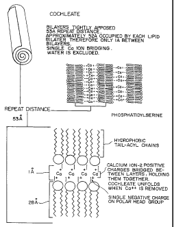
Plain lipid cochleates (Figure 1) have been
described previously. Protein-cochleates or
peptide-cochleates have been described heretofore
and patented by the instant inventors, as
intermediate structures which can be converted to
protein-lipid vesicles (proteoliposomes) (Figure 2)
by the addition of calcium chelating agents (see
U.S. Pat. No. 4,663,161 and U.S. Pat. No.
4,871,488).
Freeze-fracture
electron micrographs of protein-cochleates
containing Sendai glycoproteins made by the DC
method show the rolled up lipid bilayer structures
with a "bumpy" surface. Plain phospholipid
cochleates are smooth in that type of preparation.
CA 02212382 1997-08-06
WO 96/25942 PCT/US96/01704
- 2 -
The proteoliposomes resulting from
polypeptide-cochleates have been shown to be
effective immunogens when administered to animals
by intraperitoneal and intramuscular routes of =
immunization (G. Goodman-Snitkoff, et al., J.
Immunol., Vol. 147, p.410 (1991); M.D. Miller, =
et al., J. Exp. Med., Vol. 176, p. 1739 (1992)).
Further, when the glycoproteins of Sendai or
influenza virus are reconstituted by that method,
the proteoliposomes are effective delivery vehicles
for encapsulated proteins and DNA to animals and to
cells in culture (R.J. Mannino and S.
Gould-Fogerite, Biotechniaues, Vol. 6, No. 1,
pp. 682-690 (1988); S. Gould-Fogerite et al., Gene,
Vol. 84, p. 429 (1989); M.D. Miller, et al., J.
Exp. Med., Vol. 176, p. 1739 (1992)).
It would be advantageous to provide a means
for stabilizing or preserving biologic molecules in
a form that is stable at room temperature, capable
of desiccation and is suitable for oral
administration. For example, it would be
beneficial to have a formulation for stabilizing
polynucleotides and which could be used for
delivering polynucleotides to a cell.
SUMMARIC OF THE INVENTION
Accordingly, it is an object of the instant
invention to provide a means for stabilizing
biologic molecules to yield a formulation with
prolonged shelf life, which can be made into powder
form and which later can be rehydrated to yield a
biologically active molecule.
It also is an object of the instant invention
to provide a formulation suitable for use as a
vehicle to administer a biologically active
molecule to a host. The formulation can be used to
CA 02212382 1997-08-06
WO 96/25942 PCT/US96101704
- 3 -
deliver a biologic molecule to the gut for
absorption or to a targeted organ, tissue or cell.
A suitable biologic molecule is a
polynucleotide.
Other suitable biologic molecules are
= polypeptides such as hormones and cytokines.
Yet other suitable biologic molecules are
bioactive compounds such as drugs.
Those and other objects have been obtained by
providing a cochleate formulation comprising the
following components:
a) a biologically relevant molecule
component to be stabilized or delivered,
b) a negatively charged lipid component,
and
c) a divalent cation component.
In a preferred embodiment, the cochleate
formulation is administered orally.
The instant invention further provides a
cochleate formulation containing a polynucleotide,
wherein said polynucleotide-cochleate comprises the
following components: _
a) a. polynucleotide component,
b) a negatively charged lipid component,
and
c) a divalent cation component.
The polynucleotide can be one which is
expressed tci yield a biologically active
polypeptide or polynucleotide. Thus, the
polypeptide may serve as an immunogen or, for
example, have enzymatic activity. The
= polynucleotidea may have catalytic activity, for
example, be a ribozyme, or may serve as an
= inhibitor of transcription or translation, that is,
be an antisense molecule. If expressed, the
polynucleotide would include the necessary
CA 02212382 1997-08-06
WO 96/25942 PCT/US96/01704
- 4 -
regulatory elements, such as a promoter, as known
in the art.
The instant invention further provides a
cochleate formulation containing a polypeptide, 5 wherein said polypeptide-
cochleate comprises the
following components: =
a) a polypeptide component,
b) a negatively charged lipid component,
and
c) a divalent cation component.
A specific example is an insulin cochleate.
The advantages of cochleates are numerous.
The cochleates have a nonaqueous structure while
not having an internal aqueous space, and therefore
cochleates:
(a) are more stable than liposomes because
the lipids in cochleates are less susceptible to
oxidation;
(b) can be stored lyophilized which provides
the potential to be stored for long periods of time
at room temperatures, which would be advantageous
for worldwide shipping and storage prior to
administration;
(c) maintain structure even after
lyophilization, whereas liposome structures are
destroyed by lyophilization;
(d) exhibit efficient incorporation of
biological molecules, particularly with hydrophobic
moieties into the lipid bilayer of the cochleate
structure;
(e) have the potential for slow or timed
release of the biologic molecule in vivo as
cochleates slowly unwind or otherwise dissociate;
(f) have a lipid bilayer matrix which serves
as a carrier and is composed of simple lipids which =
are found in animal and plant cell membranes, so
CA 02212382 1997-08-06
WO 96125942 PCT/I7S96/01704
- 5 -
that the lipids> are non-toxic, non-immunogenic and
non-inflammatory;
(g) contain high concentration of divalent
= cation, such as, calcium, an essential mineral;
(h) are ,safe, the cochleates are non-living
subunit formulations, and as a result the
cochleates have none of the risks associated with
use of live vaccines, or with vectors containing
transforming sequences, such as life threatening
infections in immunocompromised individuals or
reversion to wild type infectivity which poses a
danger to even healthy people;
(i) are produced easily and safely; and
(j) can be produced as defined formulations
composed of predetermined amounts and ratios of
biologically relevant molecules, including
polypeptides, carbohydrates and polynucleotides,
such. as DNA.
The advantages of oral administration also are
numerous. An oral route has been chosen by the WHO
Children's Vaccine Initiative because of ease of
administration. Oral vaccines are less expensive
and much safcar to administer than parenterally
(intramuscular or subcutaneous) administered
vaccines. The: use of needles adds to the cost, and
also, unfortunately, in the field, needles are
often reused.
BRIEF DESCRIPTION OF THE DRAWINGS
Figure 1 is a schematic representation of a
plain lipid cochleate.
Figure 2 shows the structure of
polypeptide-lipid vesicles with integrated membrane
proteins.
Figure 3 summarizes the various alternative
procedures for the preparation of cochleates.
CA 02212382 1997-08-06
WO 96/25942 PCTIUS96/01704
- 6 -
Figures 4(A) and 4(B) show serum antibody
titers in mice following oral administration of
influenza polypeptide-cochleates.
Figure 5 is a graph showing the results of
oral administration of polypeptide-cochleates when
challenged with live virus.
Figure 6 is a graphic representation of serum
antibody titers in mice following oral
administration of Sendai-cochleates.
Figure 7 is a graph depicting the induction of
antigen-specific cytotoxic splenocytes following
oral administration of Sendai cochleates.
Figure 8 provides a series of bar graphs
depicting serum glucose levels before and after
oral insulin adiministration.
DETAILED DESCRIPTION OF THE INVENTION
The instant inventors have now found
surprisingly and have demonstrated that cochleates
themselves be used as means for stabilizing and
delivering biologic molecules. The cochleates
survive the harsh acid environment of the stomach,
protecting the susceptible biologic molecules
immersed therein, probably by virtue of their
unique multilayered precipitate structure. It is
likely that cochleates then are taken up by
microfold cells (M cells) in the small intestine.
The instant inventors have demonstrated that
oral administration by drinking cochleates
containing the glycoproteins and viral lipids from
the surface of influenza or Sendai viruses plus
phosphatidylserine and cholesterol, stimulate both
mucosal and circulating antibody responses. In
addition, strong helper cell (proliferative) and
killer (cytotoxic) cell responses also are
generated. Perhaps most impressively, oral
CA 02212382 1997-08-06
WO 96/25942 PCT/US96101704
- 7 -
admiriistration of the influenza cochleates protects
agairist intranasal challenge with live virus.
Those results are unexpected for a number of
reasons.
It was not known and was not expected that the
cochleates would survive the stomach and protect
the polypeptides associated with them from the acid
environment and degradative enzymes. It is known
that without the presence of at least 3 mM calcium,
the cochleates begin to unwind and form liposomes.
It was possible, in fact likely, that the
cochleates would not remain intact during the
transit from the mouth, down the esophagus and
through the stomach. If cochleates did come apart,
they would be digested as food.
Also, having survived the stomach, that the
cochleates would interact in an effective way with
the mucosal and circulating immune systems was
unknown and unexpected. Everyone ingests large
quan;tities of proteins, fats and sugars on a daily
basis which simply get digested and used as fuel,
without stimulating any kind of mucosal or
circulating immune responses. Thus, the cochleates
deliver molecules which retain biologic activity at
the delivery site within the host.
As used herein, the term "immune response"
means either antibody, cellular, proliferative or
cytotoxic activities, or secretion of cytokines.
Also, as used herein, the term "antigen" is
meant to indicate the polypeptide to which an
immune resporise is directed or an expressible
polynucleotide encoding that polypeptide.
"Polynuc:leotide" includes DNA or RNA, as well
as antisense and enzymatically active molecules.
Thus the biologically relevant molecule can be the
polynucleotide itself, the transcript thereof or
the translated polypeptide encoded thereby.
CA 02212382 1997-08-06
WO 96/25942 PCT/US96/01704
- 8 -
"Polypeptide" is any oligomer or polymer of
amino acids. The amino acids can be L-amino acids
or D-amino acids.
A "biologically relevant molecule" is one that
has a role in the life processes of a living
organism. The molecule may be organic or
inorganic, a monomer or a polymer, endogenous to a
host organism or not, naturally occurring or
synthesized in vitro and the like. Thus, examples
include, vitamins, minerals, amino acids, toxins,
microbicides, microbistats, co-factors, enzymes,
polypeptides, polypeptide aggregates,
polynucleotides, lipids, carbohydrates,
nucleotides, starches, pigments, fatty acids,
hormones, cytokines, viruses, organelles, steroids
and other multi-ring structures, saccharides,
metals, metabolic poisons, drugs and the like.
The instant invention also can be practiced
using whole cells other subcellular replicative
entities, such as viruses and viroids. Hence,
bacteria, yeasts, cell lines, viruses and the like
can be mixed with the relevant lipid solution,
caused to precipitate to yield structures wherein
the cells and the like are fixed within the
cochleate structure.
Polypeptides are suitable molecules to be
incorporated with cochleates. The procedure for
preparing cochleates is set forth in greater detail
hereinbelow. The polypeptide is suspended in a
suitable aqueous buffer. The lipids are dried to
form a thin film. Then the aqueous buffer is added
to the lipid film. The vessel is vortexed and then
the sample dialyzed against a cation-containing
buffer.
In that way, for example, cochleates carrying
insulin can be obtained. The insulin cochleates
were made with a 1 mg/ml solution of insulin, but
CA 02212382 1997-08-06
WO 96/25942 PCT/US96/O1704
- 9 -
various other beginning concentrations of insulin
can be used to obtain cochleates loaded with
varying concentrations of insulin.
Recent studies indicate that the direct
injection of DNA plasmids can lead to the
expression of the proteins encoded by those
plasmids resulting in humoral and cell mediated
immuzle responses, see, for example, Wang et al.,
Procõ Natl Acad. Sci. 90: 4156-4160 (1993);
Zhu et al., Science 261: 209-211 (1993). Those
studies indicate that DNA vaccines could provide a
safe and effective alternative for human
vaccination. Those studies also suggest that DNA
vaccines could benefit from simple, more efficient
delivery systems.
The use ojE lipids to facilitate the delivery,
entry and exprE:ssion of DNA in animal cells is well
documented, see, for example, Philip et al., Mol.
Cell Biol. 14: 2411-2418 (1994). Indeed, DNA-lipid
complexes curriantly form the basis for a number of
human gene therapy protocols.
Because cochleates are stable structures which
can withstand a variety of physiologic conditions,
cochleates are suitable means for delivering
biologic molecules, such as, polypeptides or
polynucleotides, to a selected site in a host. The
polypeptide or polynucleotide is incorporated into
and integral with the cochleate structure. Thus
the polypeptide or polynucleotide, which may need
to be expressed, are protected from degrading
proteases and nucleases.
' The cochleates used in the instant invention
can be prepared by known methods such as those
described in U.S. Patent No. 4,663,161, filed
22 April 1985,, U.S. Patent No. 4,871,488, filed
13 April 1987, S. Gould-Fogerite et al., Analytical
Biochemistry, Vol. 148, pages 15-25 (1985);
CA 02212382 1997-08-06
WO 96/25942 PCT/US96/01704
- 10 -
S. Gould-Fogerite et al., Advances in Membrane
Biochemistry and Bioenergetics, edited by Kim,
C.H., Tedeschi, T., Diwan, J.J., and Salerno, J.C.,
Plenum Press, New York, pages 569-586 (1988);
S. Gould-Fogerite et al., Gene, Vol. 84,
pages 429-438 (1989); Liposome Technology, 2nd
Edition, Vol. I, Liposome Preparation and Related
Techniques, Vol. II, Entrapment of Drugs and Other
Materials, and Vol. III, Interactions of Liposomes
with the Biological Milieu, all edited by Gregory
Gregoriadis (CRC Press, Boca Raton, Ann Arbor,
London, Tokyo), Chapter 4, pp 69-80, Chapter 10,
pp 167-184, and Chapter 17, pp. 261-276 (1993); and
R.J. Mannino and S. Gould-Fogerite, Liposome
Mediated Gene Transfer, Biotechniaues, Vol. 6,
No. 1 (1988), pp. 682-690.
The polynucleotide can be one which expresses
a polypeptide, that is, pathogen membrane
polypeptides, aberrant or atypical cell
polypeptides, viral polypeptides and the like,
which are known or which are suitable targets for
host immune system recognition in the development
of immunity thereto.
The polynucleotide may express a polypeptide
which is biologically active, such as, an enzyme or
structural or housekeeping protein.
Also, the polynucleotide may be one which
necessarily is not expressed as a polypeptide but
nevertheless exerts a biologic effect. Examples
are antisense molecules and RNA's with catalytic
activity. Thus, the expressed sequence may on
transcription produce an RNA which is complementary
to a message which, if inactivated, would negate an
undesired phenotype, or produce an RNA which
recognizes specific nucleic acid sequences and
cleaves same at or about that site and again, the
CA 02212382 1997-08-06
WO 96/25942 PCT/US96/01704
- 11 -
non-e.xpression of which would negate an undesired
phenotype.
The polynucleotide need not be expressed but
may bs used as is. Thus, the polynucleotide may be
an antisense molecule or a ribozyme. Also, the
polynucleotide -may be an immunogen.
Thus, for polynucleotides, the relevant coding
sequence is subcloned downstream from a suitable
promoter, othEer regulatory sequences can bia
incorporated as needed, in a vector which is
expanded in an appropriate host, practicing methods
and using materials known and available in the art.
For example, two plasmids, pDOLHIVenv (AIDS
Research and Reference Reagent Program, Jan. 1991
catalog p. 113; Freed et al. J. Virol. 63: 4670
(1989) ) and pCM'iIHIVLenv (Dr. Eric Freed, Laboratory
of Molecular Immunology, NJAID, NIH) are suitable
expression plasmids for use in
polynucleotide-cochleates.
The plasmids contain the open reading frames
for the env, tat and rev coding regions of HIV-1
(LAV strain).
pDOLHIVenv was constructed by introducing the
SalI-XhoI fragment from the full length infectious
molecular clonea pNL4-3 into the SalI site of the
retrovirus vector, pDOL (Korman et al. Proc. Natl.
Acad., Sci. 84: 2150 (1987)). Expression is from
the Moloney mui-ine virus LTR.
pCMVHIVLenv was constructed by cloning the
same SalI-XhoI fragment into the XhoI site of the
cytomegalovirus (CMV) -based expression vector p763.
The polynucleotide can be configured to encode
mult:tple epitopes or epitopes conjugated to a known
immunogenic peptide to enhance immune system
recognition, particularly if an epitope is only a
few amino acids in size.
CA 02212382 1997-08-06
96/D170~
IPEA/US03 CT1996
- 12 -
To form cochleate precipitates, a majority of
the lipid present should be negatively charged.
One type of lipid can be used or a mixture of
lipids can be used. Phosphatidylserine or
phosphatidylglycerol generally have been used.
Phosphatidylinositol also forms a precipitate which
converts to liposomes on contact with EDTA. A
substantial proportion of the lipid can, however,
be neutral or positively charged. The instant
inventors have included up to 40 mol% cholesterol
based on total lipid present and routinely make
polypeptide=-lipid or polynucleotide-lipid
cochleates which contain 10 mol% cholesterol and
2 0% v i r a 1 m e m b r a n e 1 i p i d s.
Phosphatidylethanolamine, plain or cross-linked to
polypeptides, also can be incorporated into
cochleates.
While negatively charged lipid can be used, a
negatively charged phospholipid is preferred, and
of those phosphatidylserine, phosphatidylinositol,
phosphatidic acid and phosphatidylglycerol are most
preferred.
One skilled in the art can determine readily
how much lipid must be negatively charged by
preparing a mixture with known concentrations of
negative and non-negative lipids and by any of the
pi_-ocedures described herein, determining whether
precipitates form.
There are several known procedures for making
the cochleates of the instant invention and those
are schematized in Figure 3.
A suiteible procedure for making cochleates is
one wherein a negatively charged lipid such as
phosphatidylserine, phosphatidylinositol,
phosphatidic acid or phosphatidylglycerol in the
absence or presence of cholesterol (up to 3:1,
preferably 9:1 w/w) are utilized to produce a
ANIENOEO SHEEf
CA 02212382 1997-08-06
WO 96/25942 PCT/US96/01704
- 13 -
suspension of multilamellar lipid vesicles
containing or surrounded by a biologically relevant
molecule (polypeptide, polysaccharide or
polynucleotide, such as DNA) which are converted to
small. unilame:Llar protein lipid vesicles by
sonication under nitrogen. Alternatively, to avoid
damage, the biologically relevant molecule can be
added to the solution following sonication. The
vesicles are dialyzed at room temperature against
buffered divalent cation, e.g., calcium chloride,
resulting in the formation of an insoluble
precipitate which may be presented in a form
referred to as a cochleate cylinder. After
centrifugation, the resulting pellet can be taken
up in buffer to yield the cochleate solution
utilized in the instant invention.
In an alternative and preferred embodiment, an
amount of negatively charged lipid, e.g.,
phosphatidylserine and cholesterol in the same
proportions as above and equal to from about 1 to
10 times the weight, preferably equal to four times
the weight of the viral or_other additional lipids
are utilized to prepare the cochleates. Either a
polypeptide, mineral, vitamin, carbohydrate or
polynucleotideõ such as DNA, is added to the
solution. Tha-t solution then is dialyzed against
buffered divalant cation, e.g., calcium chloride,
to produce a p:recipitate which can be called a DC
(for direct ca:tcium dialysis) cochleate.
An add:i.tional, related method for
recoinstituting cochleates has been developed and is
calliad the LC method (liposomes before cochleates) .
The initial steps involving addition of extracted
polypeptide, polysaccharide,polynucleotide, such as
DNA or combinations thereof, to dried down
nega=tively charged lipid and cholesterol are the
same as for the DC method. However, the solution
CA 02212382 1997-08-06
WO 96/25942 PCT/1JS96/01704
- 14 -
next is dialyzed against buffer (e.g., 2 mM TES,
2 mM L-histidine, 100 mM NaCl, pH 7.4) to form
small liposomes containing the polypeptide,
polynucleotide, such as DNA, and/or polysaccharide.
A divalent cation, e.g., calcium, then is added
either directly or by dialysis to form a
precipitate which can consist of cochleates.
In the above procedures for making the
cochleates of the instant invention, the divalent
cation can be any divalent cation that can induce
the formation of a cochleate or other insoluble
lipid-antigen structures. Examples of suitable
divalent cations include Ca+2, Mg+Z, Ba+Z, and Zn+Z or
other elements capable of forming divalent ions or
other structures having multiple positive charges
capable of chelating and bridging negatively
charged lipids.
Cochleates made with different cations have
different structures and convert to liposomes at
different rates. Because of those structural
differences, the rate of release of the
biologically relevant molecules contained therewith
varies. Accordingly, by combining cochleates made
with different cations, formulations which will
release the biologically relevant molecule over a
protracted period of time are obtainable.
The amount of biologically relevant molecule
incorporated into the cochleates can vary. Because
of the advantageous properties of cochleates
generally, lesser amounts of biologically relevant
molecule can be used to achieve the same end result
as compared to using known delivery means.
An artisan can determine without undue
experimentation the optimal lipid:biologically
relevant molecule ratio for the targeted purposes.
Various ratios are configured and the progress of
precipitation of each sample is monitored visually
CA 02212382 1997-08-06
WO 96/25942 PCTIUS96101704
- 15 -
under a phase contrast microscope. Precipitation
to form, for example, cochleates, is monitored
readily. Then, the precipitates can be
admiriistered to the targeted host to ascertain the
nature and tenor of the biologic response to the
admiriistered cochleates.
It should be evident that the optimized ratio
for any one use may range from a high ratio, for
example, to mir.iimize the use of a rare biologically
relevant molectile, to a low ratio to obtain maximal
amount of biologically relevant molecule in the
cochleates.
Cochleates can be lyophilized and stored at
room temperatu're indefinitely or can be stored in
a divalent cation-containing buffer at 4 C for at
least six mont:hs.
The cochleate formulations also can be
prepared both with and without fusogenic molecules,
such as Sendai virus envelope polypeptides. Prior
studies with proteoliposomes have demonstrated that
cytoplasmic delivery of liposome contents requires
a fusogenic liposome bilayer. The exact role of
Sendai virus envelope polypeptides in facilitating
the immune response to polypeptide-cochleates as
yet is not cle.ar.
It is preferred to use cochleates without
fusogenic molecules over fusogenic molecule
cochleates because of a more simple structure and
ease of preparation favors eventual use in humans.
Because polynucleotides are hydrophilic
molecules and cochleates are hydrophobic molecules
that do not contain an internal aqueous space, it
is surprising polynucleotides can be integrated
into cochleates. The polynucleotides are not
exposed on the surface of the cochleates because
the polynucleotides are resistant to nucleases.
CA 02212382 1997-08-06
WO 96125942 PCT/US96/01704
- 16 -
In the case of polynucleotide cochleates,
considerations for dosage parallel the standard
methodologies regarding vaccines as known in the
art. Also, methods for using polynucleotides in
liposomes and the "naked DNA" are available to
serve as a baseline for empirically determining a
suitable dosing regimen, practicing known methods.
For example, a suitable scheme for determining
dosing is as follows.
The initial dose of polynucleotides in
cochleates administered by injection to animals is
selected to be about 50 g, although it is know
that as little as 2 g of tested plasmids is
effective. That dose is proposed to maximize the
probability of observing a positive response
following a single administration of a cochleate.
Any formulations which do not elicit a response at
that dose are to be considered ineffective but
retained for further study.
Developing formulations which can be
administered easily and non-invasively is
desirable. Thus, PO administration of cochleates
will be targeted and higher doses will be tried
initially (100 g/animal and 200 g/animal).
However, lower doses are required for parenteral
routes.
Then graded doses will be used to develop a
dose response curve for each formulation. Thus,
cochleates containing 50 g, 10 g, 2 g, 0.4 and
0 g polynucleotide/animal will be inoculated with
at least 10 animals per group.
Immune response or enzymatic activity are
responses easily monitored when expression of the
polynucleotide is required. Altered phenotype is
another response for tracking efficacy of antisense
or ribozyme type molecules. In the case of immune
system monitoring, T cell proliferation, CTL and
CA 02212382 1997-08-06
WO 96125942 PCT/US96101704
- 17 -
antibody preseince at specific body sites can be
evaluated, using known methods, to assess the state
of specific immune response.
To determine the duration of activity of
cochleate formu.lations, groups which have responded
to a single immunization are monitored periodically
for up to a year or more to determine the effective
life of a coch].eate on administration.
Animals which fail to develop a detectable
response on first exposure can be re-inoculated
(boosted) to p:rovide insights into the ability of
the low dose formulations to prime the immune
system for later stimulation.
Pharmaceutical formulations can be of solid
form including tablets, capsules, pills, bulk or
unit dose powders and granules or of liquid form
including solutions, fluid emulsions, fluid
suspensions, semisolids and the like. In addition
to the active ingredient, the formulation would
comprise suitable art-recognized diluents,
carriers, fillers, binders, emulsifiers,
surfactants, water-soluble vehicles, buffers,
solubilizers and preservatives.
An advantage of the cochleates is the
stability of the composition. Thus, cochleates can
be administe:red orally, topically or by
instillation without concern, as well as by the
more traditional routes, such as subcutaneous,
intradermal, intramuscular and the like. Direct
application to mucosal surfaces is an attractive
delivery means made possible with cochleates.
The skilled artisan can determine the most
efficacious and therapeutic means for effecting
treatment practicing the instant invention.
Reference can also be made to any of numerous
authorities anid references including, for example,
"Goodman & Gilman's, The Pharmaceutical Basis for
CA 02212382 1997-08-06
WO 96/25942 PCTIUS96/01704
- 18 -
Therapeutics", (6th Ed., Goodman, et al., eds.,
MacMillan Publ. Co., New York, 1980).
The cochleates of the instant invention can be
used as a means to transfect cells with an efficacy
greater than using currently known delivery means,
such as liposomes. Hence, the polynucleotide
cochleates of the instant invention provide a
superior delivery means for the various avenue of
gene therapy, Mulligan, Science 260: 926-931
(1993). As Mulligan noted, the many possibilities
of treating disease by gene-based methods will be
enhanced by improved methods of gene delivery.
The cochleates of the instant invention also
serve as excellent means for delivering other
biologically relevant molecules to a host. Such
biologically relevant molecules include nutrients,
vitamins, co-factors, enzymes and the like.
Because the biologically relevant molecule is
contained within the cochleate, in a non-aqueous
environment, the biologically relevant molecule
essentially is stabilized and preserved. As
described hereinabove, the biologically relevant
molecule is added to the lipid solution and
processed to form a precipitated structure
comprising lipid and biologically relevant
molecule. As demonstrated herein, hydrophilic
molecules can be "cochleated", that is, can be made
part of the cochleate structure, with little
difficulty.
Also, suitable lipophilic biologically
relevant molecules, such as drugs and other
therapeutic compounds, are amenable to cochleation.
For example, lipophilic drugs such as cyclosporin,
ivermectin and amphotericin are readily cochleated.
The instant invention now will be described by
means of specific examples which are not meant to
limit the invention.
CA 02212382 2006-09-27
WO 96125942 PCT/US96/01704
- 19 -
ERAMPLE 1
Bovine brain phosphatidylserine in chloroform
was purchased from Avanti Polar Lipids, Birmingham,
Alabama in glass ampules and stored under nitrogen
at -20 C. Cholesterol (porcine liver) grade I,
O-D-octyl-glucopyranoside (OCG), fluorescein
isothiocyanate (FITC)-dextran (average mol. wt.
67,000), metrizamide grade I, and chemicals for
buffers and protein and phosphate determinations,
were obtained from Sicqma Chemical Company, St.
Louis, Missouri. Organic solvents were purchased
from Fisher Scientific Co., Fairlawn, New Jersey.
Reagents for polyacrylamide gel electrophoresis
were from BioRad Laboratories, Richmond,
*
California. S1000 Sephacryl Superfine was obtained
from Pharmacia, Piscataway, New Jersey. Thick
walled polycarbonate centrifuge tubes (10 ml
capacity) from Beckman Instruments, Palo Alto,
California, were used for vesicle preparations,
washes, and gradients. A bath type sonicator,
Model G112SP1G, from Laboratory Supplies Company,
Hicksville, New York was used for sonications.
Virus was grown and purified essentially as
described by M.C. Hsu et al., Virology, Vol. 95,
page 476 (1979). Sendai (parainfluenza type I) and
influenza (A/PR8/34) viruses were propagated in the
allantoic sac of 10 or 11 day old embryonated
chicken eggs. Eggs were inoculated with 1-100 egg
infectious doses (103 to 105 viral particles as
determined by HA titer) in 0.1 ml of phosphate
buffered saline (0.2 gm/L KC1, 0.2 gm/L KH2P04,
8.0 gm/L NaCl, 1.14 gm/L NaZH-P041 0.1 gm/L CaC12,
0.1 gm/L MgC126H2O (pH 7.2)). Eggs were incubated
at 37 C for 48 to 72 hours, followed by incubation
at 4 C for 24 to 48 hours. Allantoic fluid was
collected and clarified at 2,000 rpm for 20 minutes
* Trade-mark
CA 02212382 2006-09-27
WO 96/25942 PCT/US96/01704
- 20 -
~
at ~j z'"' ~,n a n;~, M
?~ ,' =T,?~-j ce-tatrifuge, The
supernatant was then centrifuged at 13,000 rpm for
60 minutes. This and all subsequent
centrifugations were performed in a sorvall* RC2-B
centrifuge at 5 C using a GG rotor. The pellets
were resuspended in phosphate buffered saline
(pH 7.2) by vortexing and sonicating, followed by
centrifugation at 5,000 rpm for 20 minutes. The
pellet was resuspended by vortexing and sonicating,
diluting, and centrifuging again at 5,000 rpm for
minutes. The two 5,000 rpm supernatants were
combined and centrifuged at 13,000 rpm for 60
minutes. The resulting pellets were resuspended in
phosphate-buffered saline by vortexing and
15 sonicating, aliquoted, and stored at -70 C.
Sterile technique and materials were used
throughout viral inoculation, isolation, and
purification.
Virus stored at -70 C was thawed, transferred
20 to sterile thick-walled polycarbonate tubes and
diluted with buffer A (2 mM TES, 2 mM L-histidine,
100 mM NaCl (pH 7.4)). . Virus was pelleted at
30,000 rpm for 1 hour at 5 C in a Beckman TY65
rotor. The supernatant was removed and the pellet
resuspended to a concentration of 2 mg viral
protein per ml of extraction buffer (EB) (2 M NaCl,
0.02 M sodium phosphate buffer (pH 7.4)) by
vortexing and sonicating. The nonionic detergent
fl-D-octyl-glucopyranoside was then added to a
concentration of 2% (w/v). The suspension was
mixed, sonicated for 5 seconds and placed in a 37 C
water bath for 45 minutes. At 15, 30 and 45 minute
incubation times, the suspension was removed
briefly for mixing and sonication. Nucleocapsids
were pelleted by centrifugation at 30,000 rpm for
minutes in a TY65 rotor. The resulting clear
supernatant was removed and used in the formation
* Trade-mark
CA 02212382 1997-08-06
WO 96/25942 PCT/US96/01704
- 21 -
of viral glycoprotein-containing cochleates. Some
modification of the above procedure may have to be
employed with other membrane proteins. Such
modifications are well known to those skilled in
the art.
EXAMPLE 2
A. DC Cochleates.
An amount of phosphatidylserine and
cholesterol (9:1 wt ratio) in extraction buffer and
non-ionic dete:rgent as described hereinabove was
mixed with a pre-selected concentration of
polynucleotide and the solution was vortexed for
5 minutes. T:he clear, colorless solution which
resulted was d'.ialyzed at room temperature against
three changes (minimum 4 hours per change) of
buffer A (2 mlvi TES N-Tris[hydroxymethyl]-methyl-2
aminoethane sulfonic acid, 2 mM L-histidine, 100 mM
NaCl, pH 7.4, also identified as TES buffer)
containing 3 mr2 CaCl.. The final dialysis routinely
used is 6 mM CaZ`, although 3 mM Ca2+ is sufficient
and other concentrations may be compatible with
cochleate formation. The ratio of dialyzate to
buffer for eaclz change was a minimum of 1:100. The
resulting white calcium-phospholipid precipitates
have been termied DC cochleates. When examined by
light microscopy (x 1000, phase contrast, oil), the
suspension contains numerous particulate structures
up to several. microns in diameter, as well as
needle-like structures.
B. LC Cochleates.
An amount of phosphatidylserine and
cholesterol (9:1 wt ratio) in extraction buffer and
non-ionic detergent as described hereinabove was
mixed with a pre-selected concentration of
CA 02212382 1997-08-06
WO 96/25942 PCT/US96101704
- 22 -
polynucleotide and the solution was vortexed for
minutes. The solution first was dialyzed
overnight using a maximum ratio of 1:200 (v/v) of
dialysate to buffer A without divalent cations,
5 followed by three additional changes of buffer
leading to the formation of small protein lipid
vesicles. The vesicles were converted to a
cochleate precipitate, either by the direct
addition of Ca2+ ions, or by dialysis against two
changes of buffer A containing 3 mM Ca2+ ions,
followed by one containing buffer A with 6 mM Ca2+.
E%AMPLE 3
IIrIISLTNE RESPONSES TO ORALLY
DELIVERED PROTEIN-COC$ILEATE VACCINES
To make the vaccine, influenza virus was
grown, purified, and the glycoproteins and lipids
extracted and isolated as described in Example 1.
Protein-cochleates were made according to the "LC
cochleate" procedure described above.
Cochleate vaccines containing the
glycoproteins and lipids from the envelope of
influenza virus and phosphatidylserine and
cholesterol were given to mice by gradually
dispensing 0.1 ml liquid into the mouth and
allowing it to be comfortably swallowed.
Figures 4(A) (from Experiment A) and 4(B) (from
Experiment B) show resulting total circulating
antibody levels specific for influenza
glycoproteins, as determined by ELISA. Antibody
titer is defined as the highest dilution that still
gives the optimal density of the negative control.
In Experiment A that generated the data shown
in Figure 4(A), initial vaccine doses of 50, 25,
12.5 or 6.25 g of glycoproteins (groups 1 through
4 respectively) were administered at 0 and 3 weeks.
CA 02212382 1997-08-06
WO 96/25942 PCT/US96101704
- 23 -
The third and fourth immunizations (6 and 19 weeks)
were at one fc-urth 'the dose used for the initial
two immunizations. Bleed 1 - Bleed 6 occurred at
0, 3, 6, 9, 19, and 21 weeks. The data demonstrate
that high circulating antibody titers can be
achieved by s;imply drinking cochleate vaccines
containing viral glycoproteins. The response is
boostable, increasing with repeated administration,
and is directly related to the amount of
glycoprotein iin the vaccine.
Those observations were confirmed and extended
in Experiment B that generated the data shown in
Figure 4(B). The dose range was expanded to
include 100 g and 3.1 g initial doses. Vaccine
was given at 0, 3 and 15 weeks, with the third
immuinization at one fourth the dose of the initial
two. Bleed 1 to Bleed 6 occurred at 0, 3, 6, 15
and 16 weeks. Circulating influenza
glycoprotein-specific responses were detectable
after a singlie administration for the top five
doses, and for all groups after two feedings. The
data shown is jEor pooled sera from each group, but
all mice given the four highest doses, and four of
five mice in groups five and six, responded to the
vaccine with circulating antibody titers ranging
from 100 to 102,400. Group seven, which received
no vaccine, had titers less than 50 for all mice at
all =time points.
The antibody response is long lived. Titers
13 weeks after the third immunization (Figure 4(A) ,
bleed 5) and 12 weeks after the second immunization
(Figure 4(B), bleed 4) remained the same or within
one dilution higher or lower than seen at 3 weeks
after the previous boost.
To determine whether oral administration of
the subunit vaccine described in Example 2 could
lead to protective immunity in the respiratory
CA 02212382 1997-08-06
WO 96/25942 PCTIUS96/01704
- 24 -
tract, the mice described in Experiment B of
Example 2 were immunized with cochleates at 0, 3
and 15 weeks. The immunized mice were challenged
by intranasal application of 2.5 x 10⁹ particles of
influenza virus at 16 weeks. Three days after
viral challenge, mice were sacrificed, and lungs
and trachea were obtained. The entire lung or
trachea was triturated and sonicated, and aliquots
were injected into embryonated chicken eggs to
allow amplification of any virus present. After
three days at 37 C, allantoic fluid was obtained
from individual eggs and hemagglutination (HA)
titers were performed.
Mice were al--:o challenged with live influenza
intranasally following oral cochleate
administration in Experiment A of Example 2. Lungs
were obtained three days later and cultured to
detect presence of virus.
The combined data for the two experiments is
giveri in Table 1. The results also are shown
graphically in Figure 5.
CA 02212382 1997-08-06
WO 96125942 PCT/17S96/01704
- 25 -
TABLE 1
Vaccine Trachea' Lungsz Lungs3
Dose # Infected/Total # Infected/Total # Infected/Total
g Protein
100 0/5 0/5 0/5
50 2/5 0/5 2/10
25 0/5 0/5 1/10
1.25 1/5 0/5 1/10
6.25 0/5 5/5 6/10
3.12 4/5 5/5 5/5
0 5/5 5/5 9/10
1. Mice from Experiment B.
2. Mice from Experiment B.
3. Mice from Experiments A and B.
The data in Table 1 shows that all five of the
unvaccinated mice had sufficient virus in the
trachea to infect the embryonated chicken eggs
(greater than 103 particles per trachea or at least
one egg infectious dose (EID) per 0.1 ml of
suspE:nsion). In contrast, the oral vaccine
provided a high degree of protection from viral
replication in the trachea. All mice in groups 1,
3 and 5 of Experiment B were negative for virus.
Two mice in group 2, 1 in group 4, and 4 in group
6(the lowest vaccine dose) of Experiment B had
sufficient virius to test positive in this very
sensitive assay used to detect presence of virus.
The oral protein cochleate vaccine also
provided protection against viral replication in
the lungs. All twenty mice which received the four
highest doses of vaccine were negative for virus
when lung suspensions were cultured in embryonated
chicken eggs (Table 1). All mice in the groups
immunized with 16.25 g and 3.1 g glycoproteins and
CA 02212382 1997-08-06
WO 96/25942 PCT/US96/01704
- 26 -
all mice in the unvaccinated control were positive
for virus.
Even in the lowest two vaccine doses, there
was some inhibition of viral replication. When
lung suspensions were diluted 1/10 and inoculated
into eggs, only one animal in the groups immunized
with 6.25 g was positive, as compared to three in
the groups immunized with 3.12 g and three in the
unvaccinated control. Culturing of 1/100 dilutions
resulted in one positive animal in each of the
groups immunized with 6.25 and 3.12 g, but 3 of 5
remained positive in the unvaccinated group. In
addition, for the two animals in the group that was
immunized with 3.12 g, but which were negative at
1/100, only 50% of the eggs'were infected at 1/10
and had low HA titers. In contrast, for the
unvaccinated group, all eggs were infected and
produced maximal amounts of virus at 1/10 and 1/100
dilutions.
C57BL/6 mice were given cochleates containing
Sendai virus glycoproteins orally at 0 and 3 weeks.
They were bled at 0 (bleed 1), 3 (bleed 2), and 6
(bleed 3) weeks. Group 1 received approximately
50 g protein, Group 2 about 25 g, Group 3 about
12.5 g, Group 4 about 6.25 g, and Group 5
(negative control) received 0 g protein. The
levels of Sendai specific antibodies in the serum
pooled from 5 mice in each dose group were
determined by ELISA. The results are shown in
Figure 6. It can be seen that strong antibody
responses were generated, that the magnitude of the
response was directly related to the immunizing
dose, and that the magnitude of the response
increased (boosted) after a second immunization.
The response was extremely long-lived. The
response is predominantly IgG, indicative of the
involvement in T cell -help and establishment of
CA 02212382 1997-08-06
WO 96/25942 PCT/US96101704
- 27 -
long--term memoi:y cells associated with a secondary
immune response. Surprisingly, the lowest dose
which initially had the lowest response, now had
the highest circulating antibody levels. This may
be due to the immune system's down regulation of
the very high responses originally but allowing the
low response to slowly climb. This may also
indicate a persistence and slow release of antigen.
It is also interesting and consistent with the use
of the oral rotite of immunization that significant
IgA titers are generated and maintained.
A 50 g protein dose of Sendai
glycoprotein-containing cochleates was given
orally. Two weeks later the animal (BALB/c mouse)
was sacrifice:d and spleen cells obtained.
Cytolytic activity of the spleen cells was measured
by their ability to cause the release of
chromium-51 from target cells presenting Sendai
antigens. The non-immunized mouse did not kill
Sendai virus (SV) pulsed cells with in culture
restimulation (N/SV/SV) or non-Sendai presenting
cells (N/N/N). (Figure 7) In contrast, Sendai
cochleate immunized mice killed SV pulsed targets
to a very high degree and non-pulsed targets to a
lesser degree. Cytolytic activity is crucial to
clearance of cells infected with viruses, or
intracellular lparasites or to cancer cells. It is
a highly desirable activity for a vaccine to
induce, but classically has not been seen with most
non-living vaccines. This is an important feature
of protein-coclhleate vaccines.
E%AMPLE 4
Eight week old BALB/c female mice were
immunized IM twice with various
polynucleotide-cochleate formulations,
CA 02212382 1997-08-06
WO 96/25942 PCT/US96/01704
- 28 -
polynucleotide alone and controls and then
splenocytes from the mice were tested for the
ability to proliferate in response to a protein
encoded by the polynucleotide.
Cochleates with and without fusogenic Sendai
virus protein were prepared as described
hereinabove. The polynucleotide used was the
pCMVHIVLenv plasmid. The solution containing lipid
and extracted Sendai virus envelop proteins as
described hereinabove and polynucleotide were mixed
at a 10:1 (w/w) ratio and 50:1 (w/w) ratio. That
protocol yielded four groups, cochleate/DNA, 10:1;
cochleate/DNA, 50:1; SV-cochleate/DNA, 10:1; and
SV-cochleate/DNA, 50:1. Naked DNA was used at a
rate of 10 g/mouse and 50 g/mouse. The control
was buffer alone. Mice were immunized twice,
15 days apart at 50 l/mouse.
Splenocytes were obtained and tested in a
T-cell proliferation assay using tritiated
thymidine, as known in the art. Control cultures
contained no antigen or con A. The antigen used
was p18 peptide, at 1 M, 3 M and 6 M. Cells
were harvested at days 2, 4 and 6 following
preparation of the splenocyte cultures.
The naked DNA provided a marginal response
above background. All four cochleate preparations
yielded a p18-spec}fic response which increased
over time. At six days, the response was about
four times above background.
The DNA concentration range at the 10:1 ratio
was about 120-170 g/ml. At the 50:1 (w/w) ratio;
the DNA concentration was about 25-35 g/ml.
The polynucleotide-cochleates were exposed to
micrococcal nuclease and little or no nucleic acid
degradation was observed.
The polynucleotide encapsulation efficiency
was found to be about 50% based on quantification
CA 02212382 1997-08-06
WO 96/25942 3PCTlIJS96/02 704
- 29 -
of free DNA from lipid, that is present in the
supernatant following a precipitation reaction.
After washing the precipitate and opening the
structures by removing cation about 35% of the DNA
was recovered.
EXAMPLE 5
In similar fashion, splenocytes from animals
immunized as described in Example 4, were tested
for antigen specific cytotoxic activity using a
chrornium release assay using labelled H-2
compatible target cells known to express an HIV
protein, such as gp160. The responder cells can be
stimulated by brief exposure to purified HIV
peptides.
On prest:imulation, animals exposed to
polyriucleotide cochleates demonstrated specific
cytotoxic splenocytes directed to gp160, with
nearly 100% cytotoxicity observed at an
effector: target: ratio of 100.
ERAMPLE 6
Fifteen mg of insulin were added to 15 ml of
extraction bufiEer (EB) in a 50 ml plastic tube.
Then 300 mg of OCG were added to the mixture. The
resulting suspension was colloidal and not clear at
pH 7.4. The solution was titrated with 1 N NaOH to
pH 8.5, resulting in a clear solution.
In a separate vessel, 6.8 ml of a 10 mg/ml
solution of phosphatidylserine and 1.5 ml of a
5 mg/ml solution of cholesterol were mixed and then
dried to yield a thin film. The insulin solution
was added to the vessel yielding a colloidal
suspension. The suspension was vortexed for seven
minutes and then set on ice for one hour. The pH
CA 02212382 1997-08-06
WO 96/25942 PCT/US96/01704
- 30 -
of the solution was adjusted to 9-9.5 with 1 N
NaOH, the sample was filter sterilized and placed
in dialysis tubing at about 2 ml per bag.
Two different dialysis schedules were used.
A. DC cochleates:
1. 100 ml overnight 1 x TES pH 9.0 co~tain
ng
3 ~i Ca+ , Zn or
Mg
2. 250 ml 4h 1 x TES pH 8.5 coptai~ng
3~i Ca , Zn or
Mg
3. 250 ml 4h 1 x TES pH 8.0 co~itainzing
3 ~I Ca , Zn or
Mg
4. 250 ml 4h 1xTES pH 7.4 coptain~ing
6 a ~ i Ca+ , Zn+ or
M +
g
B. LC cochleates:
1. 100 ml overnight 1 x TES, pH 9.0
2. 250 ml 4h, 1 x TES, pH 9.0
3. 250 ml 4h 1 x TES, pH 9.0
4. 100 ml overnight 1 x TES, pH 9+g co~tainin~
3 mM Ca , Zn or Mg
5. 250 ml 4h 1 x TES, pH 8.5 containing
+2 +2
3 mMCa+, Zn orMg
6. 250 ml 4h 1 x TES, pH 7.4 contai~ing +2 +2
6 mM Ca , Zn or Mg
Following dialysis, the resulting precipitate
was found to comprise numerous cochleates.
E7CAMPLE 7
Mice were given insulin cochleate samples
orally. Serum glucose levels were measured at 0
time, (prior to cochleate administration), 30 min.
and 60 min. post administration using standard
methods. Cochleate formulations of Example 6 with
CA 02212382 1997-08-06
WO 96125942 PCT/US96101704
- 31 -
a starting concentration of 1 mg insulin/ml
solution were used. Each mouse was administered
100 ul or 200 ul of the designated preparations as
indicated. For comparison, one mouse was given the
standard commercial human insulin, Humulin R, by
intraperitoneal administration.
Sample Volume Serum Glucose
Given mg/dl
0 Time 30 min. 60 min.
LC Ca++ 200 ul 100 49.12 43
LC Ca++ 200 ul 102.9 252.4 61.9
Humulin R 200 ul 88.8 66 48.5
Oral administration of insulin affected serum
glucose levels.
EXAMPLE 8
Insulin cochleates as produced in Example 6
were fed orally to three-month-old female BALB/c
mice made diabetic through intraperitoneal
injection of streptozotocin, practicing known
methods. Two days after exposure to
streptozotocin, the mice were allocated into groups
of five and administered with oral insulin
cochleates at 200 pl per mouse. Other mice were
injected with 2 IU of Humulin R.
Serum samples were obtained at time 0, prior
to insulin dosing, and two hours post insulin
administration. Glucose levels were measured using
a kit from Sigma (St. Louis). Control animals were
untreated, thzit is, received no streptozotocin or
insulin. Representative data are set forth in
Figure 8. Orally administered insulin, simply by
SUBSTITUTE SHEET (RULE 26)
CA 02212382 2006-09-27
WO 96/25942 PCT/US96/01704
- 32 -
Y'1sducing blood glucose
levels. No reduction in blood glucose was observed
in control animals.
While the invention has been described in
detail and with reference to specific embodiments
thereof, it will be apparent to one skilled in the
art that various changes and modifications can be
made therein without departing from the spirit and
scope thereof.